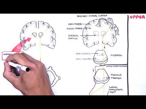
0:00 In this video, we're going to look at the motore pathways. 0:14 If you have not watched the video on the introduction to the descending and 0:18 ascending tracts, 0:19 please watch that beforehand. 0:22 There is also a sensory motor pathway video if you are interested. 0:25 So in the brain, there are two important cortexes, one in front of the central 0:32 gyrus known as 0:33 the motor cortex and the other at the back, which is the somatosensory cortex. 0:39 Now these two cortexes are important, one controls motor movements and the 0:45 other sensation. 0:47 We will be focusing on the motor cortex. 0:49 So let us take a cross section of the motore cortex here. 0:56 So here is a motor, the cortex of the brain. 0:59 It is made up of the outer gray matter where synapses are and then there is the 1:04 inner white 1:05 matter which contains a myelinated axons. 1:09 On each side of the cortex, right and left, there are areas that represent a 1:15 particular 1:16 region in our body. 1:18 So here I am drawing on the left side and we can see, here we have represented 1:25 particular 1:26 areas of the body such as the hands, the face, the legs. 1:33 And these areas, these regions make up the motor cortex and essentially signals 1:37 will arise 1:38 from one of these areas which will then travel to that particular area of the 1:45 body to perform 1:46 that movement or action. 1:50 So hope that made sense and if it does it, we will look at some examples soon 1:55 enough. 1:56 And of course in the cortex itself, there is a region called the thalamus which 2:00 is the 2:00 relay station and this is quite important to know. 2:04 So the motor pathways can be divided into two main pathways, the lateral 2:10 pathway and the 2:11 anterior medial pathway. 2:14 We will first focus on the lateral pathway which is responsible for voluntary 2:19 movements 2:20 so we control these movements and there are three main tracks involved in the 2:25 lateral pathway. 2:29 And these are the lateral corticospinal tract which control distal muscles such 2:34 as our forearm. 2:36 There is also the anterior corticospinal tract which control the proximal 2:41 muscles so it would 2:42 be the arm for example. 2:45 The anterior corticospinal tract I highlighted here because it is voluntary but 2:51 not part 2:52 of the lateral pathway itself. 2:55 Regardless, the lateral and anterior corticospinal tracts are very important in 3:01 voluntary muscle 3:02 control. 3:04 The third tract that we will talk about is the rubrospinal tract. 3:10 Before looking at these tracts, let us learn some other important structures 3:14 involved. 3:15 So here I am drawing the midbrain. 3:18 Below the midbrain is the pons which I have not drawn, then we have the medulla 3:22 here and 3:23 here is a section of a spinal cord. 3:25 Let us just say it's a cervical segment of the spinal cord and here is a 3:31 skeletal muscle 3:33 for movement of the arm or forearm for example. 3:40 Now within sections of the spinal cord, like within the spinal cord, there are 3:46 designated 3:47 tracts. 3:48 For example, there are designated tracts for the ones I mentioned earlier. 3:53 So here we have a tract for the lateral corticospinal pathway, the anterior 3:59 corticospinal pathway 4:01 and then the rubrospinal tract is here. 4:04 And there are many other tracts which we will look at later in this video. 4:10 Let us first focus on the corticospinal tract. 4:14 So let us say we want to move our hands, so distal muscle. 4:19 So a neuron will arise from the motor cortex which control hand movements here. 4:26 It will pass through the thalamus, pass through the seribri of the midbrain and 4:32 the pyramids 4:33 of the medulla where it will then cross over and land on the lateral corticosp 4:40 inal tract 4:41 before synapsing with a second neuron on the ventral horn of the spinal cord. 4:49 The first neuron coming down is therefore part of the lateral corticospinal 4:54 pathway. 4:55 The second neuron coming from the ventral horn of the spinal cord will then 5:00 target the skeletal 5:01 muscle which has to be the distal muscle. 5:04 So for example, muscles of the hands. 5:08 The second neuron is known as the lower motor neuron and the first neuron 5:13 arising from the 5:14 cortex is the upper motor neuron. 5:18 Of course, if this neuron was part of the anterior corticospinal tract, it 5:24 would pass 5:25 through the anterior corticospinal tract here before synapsing with the lower 5:31 motor neuron. 5:33 Okay, so we know about the lateral and anterior corticospinal tract. 5:41 Now let us learn about the rubrospinal tract. 5:47 The rubrospinal tract is for voluntary control of big muscles. 5:52 Basically, it is important. 5:55 There is an area in the midbrain known as the red nucleus where the rubrospinal 6:00 pathway 6:00 begins. 6:02 It essentially descends down and crosses over passing the pawns, the medulla, 6:08 before landing 6:09 on the rubrospinal tract of the spinal cord and then synapsing with the lower 6:15 motor neuron. 6:23 With the corticospinal lesion, there can be paralysis on the contralateral side 6:29 , the 6:31 opposite side. 6:32 The function of the muscle can be recovered by the rubrospinal tract if the rub 6:38 rospinal 6:39 tract is intact. 6:41 Now it is important to understand that the left side of the brain controls 6:45 movements of 6:46 the right side of the body and vice versa. 6:48 So if we damage the left side of the brain corticospinal pathway, there will be 6:54 paralysis 6:55 on the right side, on the opposite side. 6:58 And this is known as the contralateral side. 7:06 So I hope you understood the lateral pathways which are made up of the lateral 7:13 corticospinal 7:14 tract and the rubrospinal tract but we also added onto this section the 7:20 anterior corticospinal 7:22 tract because it is voluntary control. 7:26 But now let's just recap the descending motor tracts that we've learned where 7:30 it's located 7:31 and some of the new ones. 7:32 So here is a spinal cord section and here I'm drawing, I'm coloring in yellow 7:37 the descending 7:37 motor tract which we have learned so far are the lateral motor pathways which 7:42 are part 7:42 of our voluntary control. 7:45 These are the lateral corticospinal tract and here is the rubrospinal tract and 7:49 here in the 7:50 front the anterior, the ventral side is the anterior corticospinal tract. 7:57 Some new tracts which we will now investigate are the tectospinal, vestibular 8:04 spinal and 8:05 reticular spinal tract. 8:07 Let us look at these tracts and briefly look at their functions. 8:11 You won't however go to too much detail. 8:15 All these tracts are part of the anterior medial pathway. 8:21 These pathways control axial muscles such as the spine muscles and the rib 8:27 muscles which 8:29 are responsible for posture and balance and the course control of axial and 8:35 proximal muscles. 8:39 Note the proximal muscles. 8:41 So the anterior medial pathway include the vestibular spinal tract which also 8:48 include 8:49 the medial and lateral vestibular spinal tract. 8:52 There is also the reticular spinal tract which include the pontine and medull 8:57 ary tracts and 8:58 there is also the tectospinal tract. 9:02 And part of this category is the anterior corticospinal tract which we already 9:08 talked 9:09 about and which is voluntary and controls proximal muscles and so I highlighted 9:16 it. 9:16 So basically the anterior medial pathway mainly controls the axial muscles for 9:27 posture and 9:28 balance except for the anterior corticospinal tract which is voluntary control 9:33 of proximal 9:34 muscles. 9:35 Again here we are drawing some important structures. 9:40 The brain, the cortex, the thalamus, the motor cortex here. 9:44 We have the midbrain, pons which I have not drawn, the medulla and there is the 9:50 cerebellum 9:51 here which is important and it actually connects with all these motor pathways 9:56 and is responsible 9:57 for coordination essentially as well as posture and balance. 10:02 Then of course we have the spinal cord and here are some skeletal muscles which 10:08 we will 10:08 target. 10:11 On the spinal cord level we can find the tract. 10:15 Here is the tectospinal tract and the medullary reticular spinal tract. 10:23 We will first focus on the vestibular spinal pathway which originates from the 10:28 medulla's 10:29 vestibular nucleus. 10:33 It descends down and synapses with a second neuron in the spinal cord but we 10:37 won't look 10:37 at that. 10:39 Just know that the vestibular spinal tract is responsible for head, maintaining 10:44 head balance 10:45 and turning. 10:49 Now the reticular spinal tract can either originate from the medulla or the p 10:55 ons so it 10:55 can be medullary or pontine. 10:58 Here I am drawing the reticular spinal tract which originates from the medulla, 11:03 an area 11:03 known as the medullary or the medullary reticular formation. 11:09 It travels down this neuron that arises from here and lands on the medullary 11:13 reticular spinal 11:14 tract before it synapses with a second order neuron on the ventral horn and 11:19 then targets 11:20 an axial muscle responsible for posture and balance. 11:25 If the reticular spinal tract is damaged a harmless stimulus can elicit a flex 11:34 or reflex. 11:35 The last tract we will look at is the tectospinal tract which originates from 11:40 the midbrain here. 11:42 The area known as the superior coliculus. 11:49 The tectospinal pathway descends down and then crosses over, descends again and 11:54 lands 11:55 on the tectospinal tract before synapsing with a second neuron which will 11:59 target an axial 12:00 muscle. 12:01 Now the tectospinal tract is responsible in the orientation response so orient 12:09 ating yourself 12:10 in the world essentially. 12:12 So I hope that made sense so the motor pathways again can be divided into the 12:17 lateral pathways 12:19 or the anterior medial pathway. 12:22 The corticospinal tract is probably the most important because it's voluntary 12:26 control anyway. 12:27 So anyway I hope you enjoyed this video thanks for watching, bye.