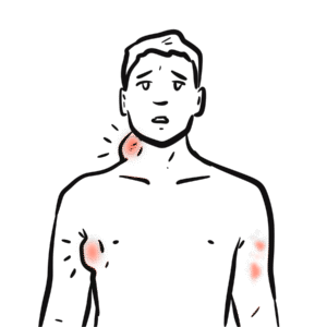
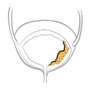
0:00 Hello, in this video we are going to talk about the oncological emergency 0:09 spinal cord compression. 0:12 Spinal cord compression, more specifically metastatic spinal cord compression, 0:16 is one 0:16 of the omnis causes of back pain where there is a compression of the fecal sac 0:22 and its 0:22 components by tumor mass. 0:25 The spinal cord is the continuation of the brain and brainstem which all make 0:30 up your 0:30 central nervous system. 0:32 It is surrounded and protected by the vertebrae, the bones. 0:37 The spinal cord ends at about the L2 vertebral level where it has fibers 0:41 continuing down called 0:43 the corda equana or the horse's tail. 0:46 Making up the spinal cord are many nerve fibers relaying motor information down 0:51 to the periphery 0:52 and sensory information from the periphery up to the brain. 0:57 Spinal nerve roots emerge from either side of the spinal cord giving rise to 1:03 the spinal 1:04 nerves. 1:09 For example, here is a superior view of the thoracic spinal cord. 1:15 It has a thoracic spinal root emerging giving rise to the spinal nerves on 1:19 either side. 1:21 The spinal nerves contain the sensory and motor nerve fibers. 1:28 Because of its delicate important nature, the spinal cord is first protected by 1:33 the vertebrae, 1:34 the bone, and then also further protected by the meninges made up of three 1:41 layers. 1:42 The innermost meningial layer is the piamata. 1:46 Then you have the arachnoid matter and then the dura matter. 1:56 Above the dura matter is what's called the epigural space. 2:00 We can find the venous plexus here which drains blood from the spinal cord as 2:05 well as this 2:07 area contains fat and arteries which supply the spinal cord with blood. 2:15 Separating the arachnoid matter and the piamata is the cerebrospinal fluid. 2:21 Separating the arachnoid matter and the piamata is the cerebrospinal fluid. 2:31 The spinal cord is protected in the sac filled with cerebrospinal fluid which 2:38 we can call 2:39 the dural sac or the fecal sac because the dural matter is the most outer men 2:47 ingial layer. 2:49 Let's look at it at a different plane, a median plane. 2:54 Here you can see the terminal portion of the spinal cord at the L2 vertebral 2:59 level known 3:00 as the conus medallaris. 3:03 The spinal cord is protected by the vertebrae, the bone, and further protected 3:07 by the meninges 3:08 made up of three layers. 3:11 The innermost layer is the piamata, then the arachnoid matter, then the dura 3:20 matter. 3:21 Separating the arachnoid matter and the piamata is the cerebrospinal fluid. 3:27 The spinal dura is composed of a single layer and serves as the innermost 3:32 barrier of the 3:33 fecal sac. You can imagine the spinal cord sitting in the sac with this 3:39 illustration. 3:40 It's the same sort of principle. 3:51 The epidural space contains the venous plexus, fat and other blood vessels and 3:57 lies between 3:58 the spinal dura and the bony ring. 4:06 Spinal cord compression develops in approximately 2.5% of patients with metast 4:11 atic cancer and 4:12 may be the first presentation of someone with cancer. 4:16 Metastatic cancer, of course, originates from a primary source, primary tumor, 4:21 and this could 4:21 be the lungs or the breast, for example. 4:26 These cancers will metastasize into the spine. 4:31 Metastatic tumors in the osteous vertebral columns account for 85% of cases of 4:36 spinal 4:37 cord compression. 4:42 As tumor grows in the epidural space, it generally takes the path of least 4:46 resistance and encircles 4:48 and compresses the fecal sac. 4:53 As the epidural venous plexus becomes obstructed, basogenic edema may develop 4:58 in the white matter 5:00 and eventually the gray matter of the spinal cord. 5:03 And this will eventually lead to spinal cord infarction. 5:13 Another mechanism of spinal cord compression is through destruction of the 5:16 cortical bone 5:17 resulting in vertebral body collapse, with displacement of bone fragments into 5:28 the epidural 5:29 space which can compress the fecal sac. 5:40 Another mechanism is paraspinal tumors or paraortic nodal masses may invade the 5:47 spinal 5:48 cord through the neural foramen causing spinal cord compression. 6:01 Intra-medullary metastasis can produce internal compression of the spinal cord 6:07 and thus spinal 6:08 cord compression. 6:19 The primary tumors that are mostly involved with the spinal cord compressions 6:24 and metastasis 6:25 to the spine include lung cancers, breast cancers, prostate and lymphoma. 6:33 Most frequently the vertebral bodies become involved by hematogenous spread. 6:40 Primary bone tumors such as multiple myeloma and osteogenic sarcoma may also 6:46 lead to spinal 6:46 cord compression. 6:51 Spinal cord compression has a propensity for certain regions of the spine. 6:55 The most common level of spinal cord compression involvement is in the thoracic 7:02 spine between 7:03 60-80% and this is followed by the lumbus spine, roughly 25% and then the 7:08 cervical spine. 7:11 Multiple spinal levels are involved in up to 50% of patients with metastatic 7:17 cancer to 7:17 the bones. 7:19 If the pressure on the spinal cord is not relieved quickly it may result in 7:23 irreversible 7:24 loss of neurologic function. 7:27 The most important prognostic factor for functional outcome is neurologic 7:32 function before treatment. 7:34 Hence, any delay could result in poorer functional outcome and decrease quality 7:38 of life with 7:39 increased dependence on healthcare resources. 7:44 Majority of patients will have symptoms of back pain weeks before neurological 7:47 symptoms 7:48 come up and the back pain is usually worse with recumbency. 7:53 60% will have symptoms of weakness on presentation. 7:59 Radicular pain which may develop due to nerve root compression by the tumor or 8:06 secondary 8:06 to vertebral collapse may occur. 8:16 Other features include sensory changes and urinary and bowel dysfunction. 8:29 Spinal cord compression is an oncological emergency. 8:32 MRI should be performed in suspected individuals for acute diagnosis. 8:37 This is whole spine and even brain MRI. 8:43 Spinal cord compression needs to be treated immediately with steroids and this 8:47 is followed 8:48 by radiation therapy which aims to decompress the spinal cord and nerve roots 8:54 via cytoreduction 8:55 of tumors, prevention of progressive neurologic symptoms, pain relief and 9:00 durable local control. 9:02 Surgery is another option if radiation fails. 9:06 Surgery is also useful to relieve pressure within the area through decomp 9:12 ression. 9:13 An neurological status is the most important predictor of outcome in patients 9:16 with spinal 9:17 cord compression. 9:19 Ambulatory patients tend to remain ambulatory whereas non-ambulatory patients 9:23 typically 9:24 do not regain the ability to walk. 9:29 Thank you for watching this video. 9:30 In summary, spinal cord compression is an oncological emergency. 9:34 MRI needs to be ordered right away and commencement of steroids to reduce edema 9:39 is recommended. 9:43 [BLANK_AUDIO]