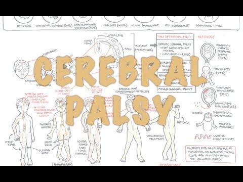
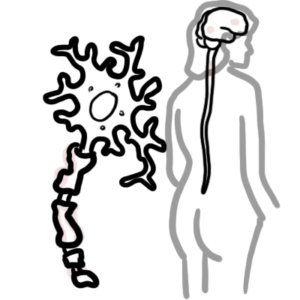
0:00 The basal ganglia, also known as the basal nucleus, or the extra-paramidal 0:09 nuclei, are 0:11 a collection of sub-cortical structures located deep within the white matter of 0:16 the brain. 0:17 It forms part of the extra-paramidal motor system, which has functions such as 0:21 regulation 0:22 of involuntary movements, so movements not under conscious control, the fine- 0:27 tuning 0:27 of voluntary movements, and the maintenance of posture. 0:34 The basal ganglia is made up of five pairs of nuclei, the cortine nucleus, the 0:40 putamen, 0:41 the globus pallidus, the subthalamic nucleus, and the sub-stanchor nigra. 0:47 Grouped into broader clusters, these nuclei are known as the striatum, the glob 0:52 us pallidus, 0:53 the subthalamic nuclei and the sub-stanchor nigra. 0:59 Let's talk about each of these nuclei in a bit more detail, and their function. 1:06 The striatum is found inside the insular lobe, and is made up of the dorsal 1:11 striatum 1:11 and the ventral striatum. 1:13 However, the ventral striatum is actually considered part of the limbic system, 1:17 rather 1:18 than the basal ganglia, it is the dorsal striatum, which is part of the basal 1:24 ganglia. 1:25 The dorsal striatum is made up of the cortine nucleus and the putamen. 1:31 During development, the cortine nucleus is separated from the putamen by 1:36 descending 1:37 white matte fibers, which at this level are known as the internal capsule. 1:43 The striatum is the primary input unit of the basal ganglia. 1:48 It receives input from the cerebral cortex. 1:53 It receives excitatory glutamatergic neurons from the cerebral cortex. 2:01 Glutamatergic neurons release glutamate, a stimulatory neurotransmitter. 2:10 The substance of the striatum itself is mainly composed of projection neurons 2:16 and interneurons. 2:17 Functionally, they are inhibitory neurons called GABAergic neurons that release 2:23 inhibitory 2:23 neurotransmitters called GABA. 2:27 The axons of these neurons form the direct and indirect pathways of the basal 2:33 ganglia, 2:33 which project into the globus pallidus and the subthalamic nuclei. 2:42 The next nuclei to talk about is the globus pallidus, which is made up of an 2:46 internal 2:47 segment, GPI, and an external segment, GPE. 2:53 The globus pallidus is composed mainly of inhibitory GABAergic projection 2:59 neurons, which 2:59 fire spontaneously and irregularly at high frequencies. 3:04 GABAergic neurons inhibit things, they release the neurotransmitter GABA. 3:10 Neurons that project from the striatum to the internal part of the globus pall 3:14 idus are 3:14 part of what's called the direct pathway of the modalup. 3:19 Meanwhile neurons that project from the striatum to the external part of the 3:23 globus pallidus 3:24 are part of the indirect pathway. 3:28 The direct pathway basically means it goes from the striatum straight to thal 3:37 amus. 3:38 Both of these segments play an essential role to the modulation of motor 3:42 pathways, in particular 3:44 the creation of smooth and precise motor actions, and also inhibitory actions, 3:50 to balance the 3:52 excitatory component coming from the cerebral cortex. 3:59 Next we have the subthalamic nuclei, which are biconcave-paid structures found 4:05 within 4:05 the subthalamus. 4:07 They are not an anatomical part of the basal ganglia, however due to their 4:11 functional 4:12 connection they are considered a part of the basal ganglia. 4:15 The function of the subthalamic nucleus is not well known, however some 4:20 theories suggest 4:21 it has a crucial role related to modulation of planned motor movement. 4:26 They receive input and also give output to the globus pallidus and the nuclei 4:36 contains 4:38 glutamatergic neurons, which are the excitatory neurons. 4:44 Lastly, we have the substantia nigra, which is a small motor nucleus found in 4:51 the anterior 4:52 part of the midbrain. 4:54 Even though it is located in the midbrain, which is part of the brainstem, 4:59 functionally 5:00 the substantia nigra is considered part of the basal ganglia. 5:04 The substantia nigra is made up of the past compactor and the past reticulata. 5:12 The past compactor supplies the striatum with dopamine, a neurotransmitter, 5:20 acting as an 5:22 input to the basal ganglia, area, the striatum. 5:27 The past reticulata serves as an output, as it conveys signals from the basal 5:32 ganglia 5:32 to the thalamus. 5:38 So putting all these different nuclei of the basal ganglia together, all these 5:44 connections 5:45 allow for a regulation of involuntary movements. 5:50 It allows for the fine-tuning of voluntary movements and the maintenance of 5:54 posture via 5:55 skeletal muscles. 5:59 The main efferent nerve fibers, or output of the basal ganglia, are made up of 6:05 neurons 6:06 that are directed towards the thalamus and the brainstem. 6:10 These originate from the internal part of the globus pallidus and the reticular 6:15 part of 6:16 the substantia nigra. 6:21 There are many afferents or inputs to the basal ganglia. 6:26 And these include the cerebral cortex through the corticosterietal pathway. 6:34 This is the largest afferent connection to the basal ganglia. 6:39 The second input to the basal ganglia is from the substantia nigra, because 6:45 there are fibers 6:46 here that play an important role, specifically from the compacta, as they 6:51 supply the striatum 6:52 with dopamine, enabling the regulation of movement initiation, termination and 6:58 modulation. 6:59 The third input to the basal ganglia is from the thalamus. 7:02 These fibers are glutamatergic and are responsible for excitatory effects on 7:08 the cerebral cortex 7:10 and brainstem, and then it sort of loops back around to the striatum. 7:14 This is what we call the thalamo striatal pathway. 7:23 The functions of the basal ganglia are not fully understood, however its known 7:27 primary 7:28 function is to fine-tune voluntary movements. 7:32 It receives impulses for the upcoming movement from the cerebral cortex, 7:37 processes these impulses, 7:41 and then conveys instructions to the thalamus, which then sends this 7:46 information back to 7:47 the cortex. 7:49 The instructions of the fine-tune movements is then sent to the skeletal muscle 7:54 via the 7:54 pyramidal motor system. 7:57 Much of this involves preventing unwanted movements to start, and reducing the 8:02 excitatory 8:02 input from the cerebral cortex, preventing excessive and exaggerated movements. 8:10 Other functions that have been established include the reward processing and 8:16 motivation, 8:17 decision making, working memory and eye movements. 8:25 Some clinical anatomy now. 8:27 Parkinson's disease is the most well-known disease of the basal ganglia. 8:33 Classic signs and symptoms of Parkinson's disease include bradykinesia, which 8:37 is slowness 8:37 of movement, arresting tremor, postural instability, which is the inability to 8:43 basically stand 8:45 upright, and shuffling gait. 8:48 This disease is a result of neurodegeneration of dopaminergic neurons, 8:54 specifically dopaminergic 8:56 neurons which are found in the substantia nigra past compactor. 9:02 Communication of the substantia nigra results in decreased dopaminergic input 9:07 to the striatum, 9:09 resulting in the classic features of Parkinson's disease, bradykinesia, resting 9:15 tremor, rigidity 9:16 and postural instability. 9:18 Basically, it is a hypokinetic movement disorder. 9:26 Next is Huntington's disease, unlike Parkinson's disease, which is a hypokin 9:32 etic movement 9:33 disorder, Huntington's disease is a hyperkinetic movement disorder, meaning it 9:38 is characterized 9:39 by excessive abnormal and involuntary movements. 9:43 Classic signs and symptoms include involuntary movements, cognitive degener 9:47 ation and psychiatric 9:48 dysfunction. 9:50 The cause of Huntington's disease is a genetic error, specifically it is caused 9:57 by a CAG 9:58 repeat sequence on chromosome 4P on the HTT gene, the Huntington's D gene. 10:05 The resultant abnormally long Huntington gene leads to neuronal death in the 10:10 chordate 10:11 and putamen of the basal ganglia. 10:18 There is no cure for Huntington's disease, however, a drug called tetrabenazine 10:24 helps 10:24 reduce disease symptoms. 10:27 Tetrabenazine inhibits uptake of monoamine such as serotonin, dopamine and 10:32 adrenaline 10:33 in two synaptic vessels, resulting in reduced stimulation of the striatum. 10:42 The next case we will discuss is hemibolism, which is derived from the Greek to 10:48 throw. 10:49 Hemibolism is used to describe hyperkinetic involuntary, forceful movements of 10:56 the ipsilateral 10:58 arm and leg. 11:00 The most common cause of hemibolism is a lesion in the contralateral subthal 11:07 amic nuclei, such 11:08 as from a stroke, traumatic brain injury, neoplasm, vascular malformation, 11:14 amongst many 11:15 other causes. 11:18 The last clinical case we will discuss is Tourette syndrome, which presents a 11:22 sudden 11:22 repetitive uncontrolled movements and vocalization called tics. 11:28 These tics are associated with dysfunction of projection from the striatum and 11:32 results 11:32 in increased dopaminergic activity, similar to Huntington's disease and hemibol 11:42 ism. 11:43 So in summary, in this video we talked about the basal ganglia, which is a 11:47 collection of 11:48 subcortical structures located deep within the white matter of the brain. 11:53 We discussed the five pairs of nuclei that make up the basal ganglia. 11:57 The cortine nucleus, the putamen, the globus palitis, the subthalamic nucleus 12:01 and the 12:02 substantial nigra, and the fact that they can be grouped into broader clusters. 12:07 We then talked about the function of each nuclei and the function of the basal 12:12 ganglia 12:13 broadly. 12:14 We finished off discussing four relevant clinical anatomy cases, which were 12:21 Parkinson's disease, 12:22 Huntington's disease, hemibolism, and Tourette syndrome. 12:26 Thank you.