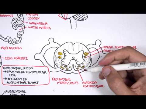
0:00 The brainstem is a stalk-like projection found in the distal part of the brain, 0:11 made up 0:11 of the midbrain, pons and medulla oblongata. 0:15 Each component of the brainstem has its own unique features and function. 0:19 As a whole structure, the brainstem allows communication between the cerebrum, 0:23 cerebellum 0:24 and spinal cord and contains the cardiovascular, respiratory, vomiting and vas 0:29 omotor centers 0:30 which regulate heart rate, breathing and blood pressure. 0:33 The brainstem is responsible for these important functions because it is a pass 0:37 ageway for many 0:37 neural pathways and is home to cranial nerve nuclei. 0:48 The brainstem begins at the level of the cerebral peduncles, anteriorly and cor 0:55 poria, quadrogeminal, 0:57 quadrogeminal plate and tectal plate posteriorly. 1:01 It ends at the level of the foramen magnum of the skull at the decastation of 1:13 the pyramids. 1:16 The midbrain is the most superior and widest part of the brainstem. 1:22 Below it is the pons and the medulla oblongata is the most distal and narrowest 1:27 part of the 1:28 brainstem. 1:31 The parts of the brainstem will be focused on in more detail in separate videos 1:39 . 1:39 Let's focus on the blood supply of the brainstem. 1:43 The blood supply of the brainstem is derived from vertebral and basilar 1:51 arteries. 1:52 The vascular supply of the midbrain is from the basilar artery and its branches 1:56 . 1:56 The major vessels are the posterior cerebral artery and its peduncular branches 2:02 , the superior 2:03 cerebellar artery, the posterior coroidal artery and the interpandicular 2:09 branches of the basilar 2:10 artery. 2:13 The majority of the vascular supply of the pons is supplied by the pontine 2:21 arteries, 2:22 which are branches of the basilar artery and a smaller part comes from the 2:28 anterior inferior 2:30 cerebellar artery and the superior cerebellar artery. 2:39 The vascular supply of the medulla is complex and it depends on the level being 2:44 viewed. 2:45 In general, the vessels that supply the medulla are the spinal artery, the 2:50 posterior inferior 2:52 cerebellar artery, the anterior inferior cerebellar artery and the vertebral 3:05 arteries. 3:07 The rule of four for the brainstem, this is a simplified method that is used to 3:13 understand 3:14 brainstem anatomy and brainstem vascular syndromes. 3:18 So the rules of four is basically everything to do with fours. 3:23 The first rule is that there are four structures in the midline beginning with 3:29 M and these 3:30 are, so, medially in the brainstem, these are the motor pathway, which is the 3:36 corticospinal 3:37 tract. 3:38 You have the medial lemaniscal pathway and this is your touch, basically, 3:46 sensation. 3:46 You have the medial longitudinal fasciculus, which is basically the connection 3:50 between cranial 3:51 nerve number six and cranial nerve number three. 3:56 And then the last structure or thing that is medial is the motor nucleus and 4:02 the nerves. 4:03 So, medially, all these nuclei here would be your motor nuclei or the cranial 4:12 nerves. 4:14 The second rule is that there are four structures in the sides, or the lateral 4:19 aspect of the 4:20 brainstem, beginning with S. And these are the spinal cerebellar pathways, the 4:27 spinal 4:27 thalamic pathway, which is your pain, the sensory nuclei of cranial nerve five, 4:33 and lastly, 4:35 the sympathetic pathway. 4:41 The third rule of the rules of fours is that there are four cranial nerves in 4:47 the medulla. 4:48 There are four cranial nerve nuclei in the pons, and then there are four cran 4:53 ial nerve 4:54 nuclei that are above the pons, and so two are in the midbrain. 5:00 Above the pons, you have cranial nerves one, two, and then cranial nerves three 5:06 and four 5:06 are in the midbrain. 5:08 In the pons, you have cranial nerves five, six, seven, and eight, and they're 5:13 nuclei. 5:14 In the medulla, you have cranial nerves nine, ten, eleven, and twelve, so fours 5:21 . 5:21 The last rule of rules of four is that the four motor cranial nerve nuclei that 5:28 are in 5:28 the midline are those that divide equally into twelve, except one and two. 5:34 So the four motor cranial nerves are three, four, six, and twelve. 5:40 Cranial nerve five, seven, nine, and eleven are in the lateral brainstem. 5:46 So again, medially, midline, these are your motor nerve nuclei. 5:57 So let's try to incorporate the rules of four by looking at some examples. 6:03 So let's just say there is an old man that comes in with sudden hornet syndrome 6:07 on the 6:08 right. 6:09 They also have ataxia on the right. 6:12 They have loss of facial sensation on the right, loss of temperature and pain, 6:20 sensation on 6:21 the contralateral side of the body, in the arms and in the legs, and also they 6:26 have dysarthri 6:27 and dysphagia. 6:31 Well these constellations of symptoms and signs are caused by loss of blood 6:35 supply to 6:36 the posterior inferior cerebellar artery, causing what's called Wallenberg 6:40 syndrome or 6:41 lateral medallory syndrome. 6:43 In this example, specifically, it is an in fact caused by the right posterior 6:51 inferior 6:51 cerebellar artery, and it causes the following things, ipsilateral hornet 6:56 syndrome due to 6:57 the sympathetic pathway lesion, ipsilateral ataxia due to spinocerebellar 7:04 pathway, ipsilateral 7:06 loss of sensation of the face due to trigeminal nuclei, which actually extends 7:10 to the medallar 7:11 laterally, contralateral loss of temperature and pain due to spinothalamic 7:18 involvement, 7:19 and then you have dysarthria and dysphagia due to disruption of cranial nerve 9 7:24 and 10. 7:25 And so it's important, this syndrome is lateral because all the lateral 7:28 structures are affected 7:30 hence the constellation of symptoms. 7:36 So in summary, today we looked at the brainstem, which is a stalk-like 7:40 projection found 7:41 in the distal part of the brain. 7:43 We briefly talked about the anatomy and the three components of the brainstem, 7:46 the midbrain, 7:48 pons, and medalloblongata, and discussed the blood supply to these areas. 7:52 We then spent some time discussing the rule of four, which is a method that is 7:55 used to 7:56 understand brainstem anatomy and brainstem vascular syndromes, and we used a 8:00 patient 8:00 scenario to demonstrate how it can be applied clinically. 8:04 In the upcoming videos, we will further discuss the brainstem by going to 8:07 detail about its 8:08 three components, the midbrain, pons, and medalloblongata. 8:12 Thank you.