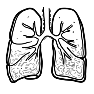
0:00 Hello, in this video we're going to talk about pulmonary circulation and we 0:10 will also talk 0:11 about ventilation and perfusion. 0:15 So first of all, ventilation is essentially the amount of air traveling into 0:21 your alveoli 0:22 ready for gas exchange, perfusion is the amount of blood flow going into the al 0:32 veoli 0:33 and thus prepared for gas exchange as well. 0:38 Ventilation is abbreviated V and perfusion is abbreviated Q. 0:44 A simple concept to understand is that the lung is divided into three zones. 0:50 The so-called ventilation perfusion ratio is higher at the apex of the lung on 0:56 the top. 0:58 Towards the base of the lung, the ventilation perfusion ratio is decreased. 1:07 The ventilation here again is the amount of air coming into the lungs 1:12 specifically into 1:13 the alveoli ready for gas exchange. 1:17 The perfusion is a blood flow to the lungs, to the alveoli, and thus ready for 1:24 gas exchange. 1:25 The average ventilation perfusion ratio is 0.8. 1:30 0.8 really means that there is more perfusion, more blood flow to the lungs to 1:36 the alveoli 1:37 than there is ventilation. 1:41 But fun fact, through the lungs, perfusion and ventilation differ. 1:47 Different lung diseases can also further affect the ventilation perfusion ratio 1:52 , also known 1:52 as the VQ ratio. 1:56 Let's now try to understand the concept of ventilation and perfusion and why 2:00 they differ 2:01 in different zones of the lungs. 2:03 So here we have two lungs. 2:05 The hot pumps deoxygenated blood through the pulmonary artery into the lungs. 2:12 In the upright position, the upper portion of the lungs are well above the 2:17 level of the 2:18 hot, and the base of the lungs are at or below it. 2:23 This has important implications on the perfusion as well as the ventilation. 2:29 Because as you can imagine, when standing up gravity will affect how much blood 2:33 goes 2:34 to different areas in the lungs. 2:36 So as mentioned, the lungs can be divided into three zones. 2:39 As you can imagine, blood traveling to the zone on the top at the apex of the 2:43 lung will 2:44 be decreased because of gravity. 2:48 There is decreased perfusion at the apex. 2:51 Perfusion here is represented by Q, remember. 2:55 This means that the blood flow to the base of the lungs will have increase in 3:00 perfusion. 3:01 So the Q is increased. 3:03 Thus we can say that at the apex of the lung we have wasted ventilation. 3:08 All the gas that goes to the alveoli is not exchanged efficiently because there 3:12 is less 3:13 perfusion. 3:15 Whereas at the base of the lungs we have wasted perfusion. 3:18 There is a lot of blood flow to this area but not as much ventilation, not as 3:22 much gas 3:23 going into these alveoli. 3:25 Fun fact, like perfusion, ventilation is actually higher at the base of the 3:29 lungs than 3:30 at the apex of the lungs. 3:32 It's important to keep note that both ventilation and perfusion is actually 3:36 greater at the base 3:37 of the lung. 3:39 So let's look at another representation here. 3:42 Because the apex of the lungs sits well above the heart, you have larger alve 3:46 oli. 3:46 There is reduced pulmonary intravascular pressure because of less blood flow to 3:51 this 3:51 area. 3:52 Less blood flow means less perfusion. 3:55 In the apex, you also have less ventilation occurring because of your large al 4:00 veoli. 4:01 But despite the reduced ventilation, the perfusion is far more reduced here. 4:06 And so you actually have wasted ventilation. 4:09 Which means you have a lot of gas, a lot of oxygen to offer but not enough red 4:12 blood 4:12 cells around. 4:14 As you go to the base of the lungs, your alveoli becomes smaller. 4:18 The size difference is in alveoli from the apex to the base of the lung is 4:22 attributed 4:23 to the difference in intraplural pressure which we won't actually talk about 4:27 here. 4:28 You have more blood flow to the base of the lungs as mentioned. 4:31 And so you have more perfusion. 4:33 At the base of the lungs, you also have more ventilation because the small alve 4:36 oli are 4:37 able to expand more. 4:39 At the base, the ventilation increase is not as much as the increase in perf 4:44 usion. 4:44 And thus, the ventilation perfusion ratio is lower here. 4:49 Thus we can create a ventilation perfusion ratio using this concept. 4:53 So at the base of the lung, you have good ventilation and great perfusion. 4:58 For the apex, you have not so good ventilation and pretty bad perfusion. 5:05 This is again in an upright position and these differences in VQ ratio is 5:11 thought to be primarily 5:12 due to gravity but also other factors play a role including different diseases. 5:18 If this concept of different ventilation and perfusion is still confusing, let 5:21 's take another 5:22 look at it and introduce pressures. 5:24 Again, you have two lungs, each lung can be divided into three zones. 5:30 The pulmonary artery brings blood to the lungs. 5:33 The pulmonary artery pressure P small a here brings blood to the alveoli. 5:41 The alveoli have their own pressure represented as P capital A. Gas exchange 5:47 occurs between 5:48 the pulmonary vessels and the alveoli. 5:52 Then the pulmonary vein will return this newly oxygenated blood to the heart. 5:56 The pulmonary vein pressure here is represented by P small V. 6:04 So now let's introduce the three zones again. 6:07 At the apex of the lung, you have large alveoli. 6:09 You have a large alveoli pressure, blood flow to the apex is less so pulmonary 6:13 arterial 6:13 pressure here is normally just sufficient to maintain perfusion. 6:18 But if pulmonary artery pressure is reduced or if alveoli pressure is increased 6:23 , some 6:23 of these capillaries collapse. 6:25 Under these circumstances, no gas exchange actually takes place in the affected 6:30 alveoli 6:30 and they become part of the physiological dead space. 6:35 Easy to remember that in zone one, alveoli pressure is highest followed by 6:41 pulmonary 6:42 artery pressure, but normally in healthy adults, pulmonary artery pressure will 6:47 be just higher 6:49 in order to maintain the blood flow. 6:51 Then pulmonary vein pressure is obviously lowest, thus in zone one, perfusion 6:57 is crap. 6:58 In the middle portion of the lungs, the pulmonary artery pressure exceeds the 7:02 alveoli pressure. 7:04 Perfusion is good and so ventilation is good. 7:09 And towards the base of the lung, alveolar pressure decreases and pulmonary 7:12 blood flow 7:13 increases as the arterial pressure increases. 7:17 In the lower portion of the lungs, alveolar pressure is lowest in the pressure 7:22 in all 7:22 parts of the pulmonary circulation. 7:26 Again recapping the apex of the heart, focusing on the pulmonary artery 7:30 pressure. 7:31 If this drops, no gas exchange takes place. 7:35 You thus have physiological dead space. 7:38 Let's look at the pulmonary blood flow in more detail. 7:42 Here is the right side of the heart, which pumps deoxygenated blood to the 7:46 lungs. 7:46 The lungs can be divided into three zones. 7:49 Gas exchange occurs in the lungs, then the pulmonary vein will return the newly 7:53 oxygenated 7:54 blood to the left side of the heart. 7:57 To the middle zone of the lung, perfusion is good, ventilation is good. 8:00 However, remember, perfusion to the apex of the lung is poor. 8:04 You have decrease in Q. 8:07 Perfusion is lower at the apex of the lung, but not as bad as perfusion at the 8:11 apex of 8:11 the lung. 8:12 Thus, VQ ratio is high here. 8:18 As you go to the base of the lung, ventilation increases, but perfusion 8:22 increases a lot more, 8:24 so your VQ ratio will decrease at the base of the lung. 8:30 So in summary, your ventilation, the amount of gas you breathe in, increases 8:35 from the 8:35 apex of the lung to the base of the lung, but your perfusion, the amount of 8:39 blood flow 8:40 going to your lungs, going to your avioli, increases a lot more from the apex 8:44 of the 8:45 lung to the base of the lung. 8:47 And so your VQ ratio decreases from the apex of the lung to the base of the 8:53 lung. 8:54 So I just want to talk about one more thing about venous return to the left 8:58 side of the 8:59 heart. 9:00 I wanted to introduce the waterfall effect whereby the height does not actually 9:05 influence 9:06 blood flow. 9:08 So here, looking at an example, distance as in the height, as in towards the 9:13 apex and 9:13 blood flow, we would assume that blood flow coming from the apex would be 9:19 highest. 9:20 But just like the waterfall, the height of the waterfall has no influence on 9:30 flow. 9:31 So in summary, your ventilation, the amount of gas you breathe in, increases 9:35 from the 9:36 apex of the lung to the base of the lung, but your perfusion, the amount of 9:40 blood flow 9:40 going to your lungs, going to your avioli, increases a lot more from the apex 9:45 of the 9:46 lung to the base of the lung, and so your VQ ratio decreases from the apex of 9:51 the lung 9:52 to the base of the lung. 9:54 Again ventilation is the amount of gas oxygen coming into your avioli, ready 10:00 for gas exchange. 10:01 And perfusion is the amount of flow to the lungs, the red blood cells to the 10:06 lungs, ready 10:07 for gas exchange. 10:10 The average VQ ratio through the whole lung is about 0.8. 10:15 The VQ ratio can change drastically depending on diseases. 10:20 So let's take a look at some examples here. 10:23 So here again is your heart. 10:24 The right side of your heart will pump blood to your lungs through the 10:27 pulmonary arteries 10:28 here in blue. 10:29 The blood will go to the avioli eventually, gas exchange occurs and new oxygen 10:33 ated blood 10:33 will go back to the heart via the pulmonary veins colored here in red. 10:38 Now in different diseases, ventilation and perfusion gets messed up. 10:42 Imagine you breathe air in to your avioli. 10:45 This is ventilation. 10:47 Normal VQ ratio is about 0.8, which really means you have a higher perfusion 10:52 than ventilation 10:53 on average. 10:55 In pneumonia for example, where you have consolidation, you have decreased in 11:00 ventilation, you don't 11:01 get enough gas moving into your avioli as a consequence, your ventilation perf 11:05 usion ratio 11:06 is low. 11:10 Similarly, if you have mucous thickening and build up along the airways, like 11:17 in COPD, 11:17 you get reduced gas coming into your avioli and so you have reduced ventilation 11:22 . 11:22 Thus, your VQ ratio is decreased. 11:28 Another example of a decrease in VQ ratio is in pulmonary edema. 11:33 When you have fluid overload in your lungs, this results in reduced ventilation 11:38 . 11:38 There is reduced gas traveling into your avioli, where gas exchange should take 11:44 place. 11:45 And so you can see spectrum of VQ mismatch, whereby on the left side, your 11:50 ventilation 11:51 is decreased, so does your VQ ratio. 11:58 On the very end of the spectrum on the left side, you can have one area of the 12:02 lung where 12:03 the ventilation is zero, which would mean that in this area, the VQ ratio is 12:09 zero. 12:10 When this happens, this is called absolute physiological shunt, or absolute 12:15 pulmonary 12:15 shunt, where you have perfusion without ventilation. 12:23 With this scenario, you can imagine no oxygen is coming into the avioli, so you 12:27 get net 12:27 low oxygen levels leaving the pulmonary system, and you still have all that 12:32 carbon dioxide 12:33 because you cannot breathe it out. 12:35 You have perfusion, but no ventilation. 12:39 Now back to COPD, we mentioned how mucus buildup causes reduced ventilation. 12:44 But in COPD, you can also get destruction of the pulmonary capillaries 12:48 supplying the 12:49 avioli, and so when this happens, you get a reduced perfusion because you are 12:54 destroying 12:55 the blood flow to the avioli. 12:57 So I mean in COPD, like in many other lung diseases, it's a fine balance of 13:01 reduced ventilation 13:03 and reduced perfusion. 13:05 Reduced perfusion in COPD occurs more in the late stages of the disease. 13:11 As the VQ ratio spectrum moves more to the right, your VQ ratio increases. 13:18 It increases until the middle when you get a ventilation and perfusion ratio 13:23 about one, 13:24 which is ideal. 13:26 Here you get enough air entering the avioli, and you are getting enough blood 13:30 flow to the 13:30 avioli. 13:32 In real life, the average humans, as mentioned, have a VQ ratio of 0.8. 13:37 However, as a spectrum of VQ ratio moves to the right, you have an increase in 13:44 VQ ratio, 13:45 which means you really have a decrease in perfusion. 13:49 You have a decrease in blood flow to the avioli. 13:53 The example of this is the damage to the capillaries as seen in late COPD. 13:58 Another good example is pulmonary embolism, where a clot includes the pulmonary 14:02 capillary 14:03 or artery causing reduced perfusion, which will subsequently increase the VQ 14:09 ratio causing 14:10 a mismatch. 14:13 When the perfusion to an area of the lung is so low, the number could really be 14:17 anything, 14:18 because it will depend on the top value, which is ventilation. 14:22 When perfusion is zero to an area of the lung, the area is called an absolute 14:27 dead space, 14:28 which really means that gas is actually entering the avioli, but does not 14:32 participate in gas 14:33 exchange. 14:34 And so this space is dead space. 14:38 The trachea, for example, is dead space because air flows through here, but the 14:42 trachea does 14:43 not participate in gas exchange. 14:45 So I hope this video on ventilation perfusion makes sense. 14:50 Thank you for watching. 14:52 [BLANK_AUDIO]