

Chronic Recurrent Multifocal Osteomyelitis
Overview
Chronic Recurrent Multifocal Osteomyelitis (CRMO), also referred to as Chronic Non-bacterial Osteomyelitis (CNO), is a rare, autoinflammatory bone disease primarily affecting children and adolescents. It is characterized by sterile, non-infectious, inflammatory bone lesions that follow a chronic, waxing-and-waning clinical course.
The disease results from an innate immune dysregulation featuring unbalanced pro-inflammatory cytokine production, leading to localized bone destruction (osteolysis) and reactive hyperostosis. Because CRMO clinically and radiologically mimics infectious osteomyelitis and bone malignancies (e.g., Ewing sarcoma, osteosarcoma, and leukemia), it remains a diagnosis of exclusion. Early recognition is vital to avoid unnecessary repeated courses of antibiotics, invasive surgical debridements, and long-term skeletal deformities.
CRMO is a sterile autoinflammatory disease, not an infection. Bone cultures are characteristically negative, and patients do not respond to antibiotics.
Definition
Chronic Non-bacterial Osteomyelitis (CNO): The umbrella term describing non-infectious inflammatory bone disease across all age groups and severity spectrums.
Chronic Recurrent Multifocal Osteomyelitis (CRMO): The severe, pediatric-onset, multifocal, and relapsing phenotype of CNO.
Whole-Body MRI (WBMRI): The gold-standard imaging modality using sensitive short tau inversion recovery (STIR) sequences to detect active bone inflammation, including subclinical (“silent”) lesions.
SAPHO Syndrome: An acronym for Synovitis, Acne, Pustulosis, Hyperostosis, and Osteitis; considered the adult spectrum variant of CNO/CRMO.
Hyperostosis: Excessive bone growth and cortical thickening resulting from chronic inflammatory osteoblast activation following osteolytic destruction.
Why is Whole-Body MRI essential in CRMO?
Patients with CRMO often present with pain at a single anatomical site, but up to 80% have additional asymptomatic (“silent”) inflammatory bone lesions elsewhere in the skeleton. WBMRI identifies the true multifocal extent of the disease without ionizing radiation, confirming the diagnosis and preventing unnecessary localized biopsies.
Classification
CRMO/CNO exists on a spectrum ranging from mild, isolated monostotic bone lesions to complex, syndromic autoinflammatory conditions.
Non-Syndromic CNO / CRMO
- Monostotic CNO
- Single bone lesion without recurrence
- Often difficult to distinguish initially from acute bacterial osteomyelitis or bone tumors.
- Classic Polyostotic / Multifocal CRMO:
- Multiple bone lesions occurring simultaneously or sequentially over time
- Characteristic periods of exacerbation and remission.
Syndromic & Monogenic Autoinflammatory Variants
- Majeed Syndrome: Autosomal recessive disorder caused by mutations in LPIN2; presents with early-onset CRMO, congenital dyserythropoietic anemia, and neutrophilic dermatosis.
- DIRA (Deficiency of IL-1 Receptor Antagonist): Autosomal recessive mutation in IL1RN; presents in neonates with severe multifocal osteomyelitis, periostitis, and pustulosis.
- SAPHO Syndrome: Associated with prominent dermatological manifestations (palmoplantar pustulosis, severe acne vulgaris) and anterior chest wall hyperostosis in young adults.
Although CRMO was historically considered an idiopathic condition, the discovery of monogenic syndromes like Majeed syndrome and DIRA helped pinpoint dysregulated interleukin-1 (IL-1) signaling as the central driver of non-bacterial bone inflammation.
Aetiology & Risk Factors
Pathogenic Triggers & Genetics
- Autoinflammatory Etiology: Driven by aberrant innate immune activation rather than autoimmune antibody formation or external microbial infection.
- Genetic Susceptibility: Polygenic in most sporadic cases, with linked susceptibility loci on chromosome 18q21. Polymorphisms in genes regulating cytokine expression (such as IL10, IL1B, and NLRP3) are heavily implicated.
Demographics & Co-Existing Conditions
- Age at Onset: Peak incidence occurs between 10 and 12 years of age (rarely presents under 2 years old unless monogenic).
- Sex Ratio: Female predominance (~2:1 female-to-male ratio).
- Associated Conditions: Strongly associated with other inflammatory and autoinflammatory disorders:
- Dermatological: Palmoplantar pustulosis, psoriasis, severe acne, and pyoderma gangrenosum.
- Gastrointestinal: Inflammatory Bowel Disease (Crohn’s disease and Ulcerative Colitis occur in 10–15% of patients).
- Rheumatological: Seronegative Spondyloarthropathies and Juvenile Idiopathic Arthritis (JIA).
Pathophysiology
- Cytokine Imbalance: Monocytes from CRMO patients exhibit defective p38 MAPK pathway signaling, leading to reduced expression of the anti-inflammatory cytokines Interleukin-10 (IL-10) and Interleukin-19 (IL-19).
- Inflammasome Activation: Unopposed pro-inflammatory signaling triggers NLRP3 inflammasome activation, causing massive production of Interleukin-1 beta (IL-1β), Tumor Necrosis Factor-alpha (TNF-α), and Interleukin-6 (IL-6).
- Uncoupled Bone Remodeling: High local levels of IL-1β and TNF-α upregulate RANKL on osteoblasts, leading to unchecked osteoclastogenesis and focal bone resorption. As lesions age, reactive osteoblasts lay down unorganized woven bone, causing sclerosis and hyperostosis.
The Histological Progression of CRMO
- – Early Stage: Dense infiltration of neutrophils and osteoclasts causing focal bone erosion and marrow destruction (mimicking acute bacterial osteomyelitis).
- – Intermediate Stage: Infiltration shifts to lymphocytes, monocytes, and plasma cells.
- – Late Stage: Fibrosis, osteosclerosis, and hyperostosis dominate, with an absence of inflammatory cells.Cultures remain consistently sterile across all stages.
Clinical Manifestations
Primary Musculoskeletal Features
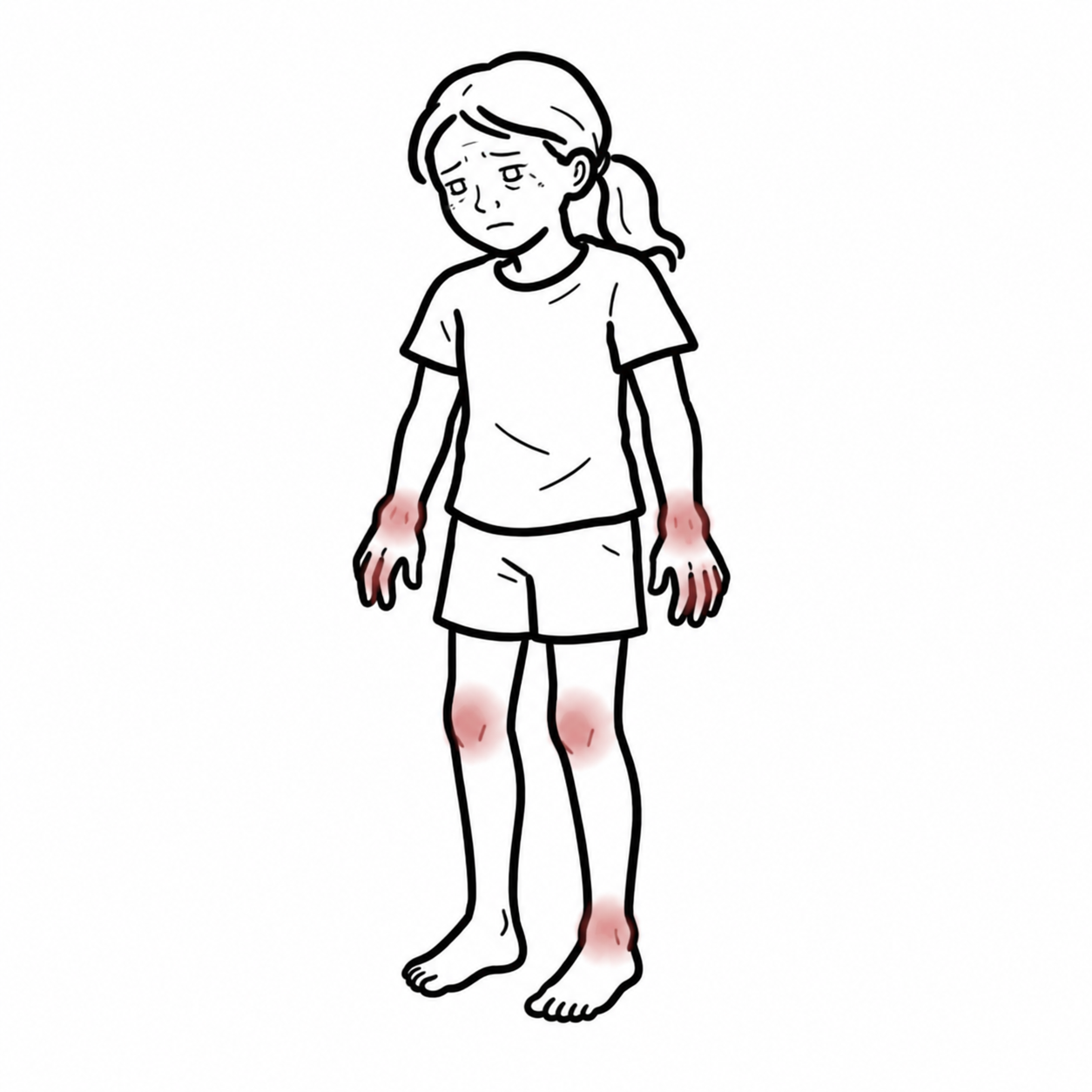
- Bone Pain: Insidious onset of deep, aching bone pain, frequently worsening at night and during activity.
- Localized Signs: Local tenderness, warmth, and soft tissue swelling over affected bones. Joint effusions may occur adjacent to metaphyseal lesions.
- Functional Deficits: Antalgic gait or limp (when lower limbs/pelvis are involved); restricted spinal mobility.
Anatomical Site Predilection
Lesions are typically bilateral and asymmetric, with a strong predilection for the metaphyses of long bones:
| Skeletal Region | Affected Anatomical Sites | Clinical Significance |
| Lower Extremities (40–50%) | Distal femur, proximal/distal tibia, fibula | Most common site; causes limp and localized limb swelling. |
| Clavicle (20%) | Medial or lateral clavicle | Highly specific for CRMO; rare site for bacterial osteomyelitis. |
| Pelvis (20%) | Ilium, ischium, pubic rami | Often presenting with deep buttock, hip, or groin pain. |
| Spine (10–15%) | Thoracic and lumbar vertebral bodies | High-risk site; can lead to vertebra plana, collapse, and kyphosis. |
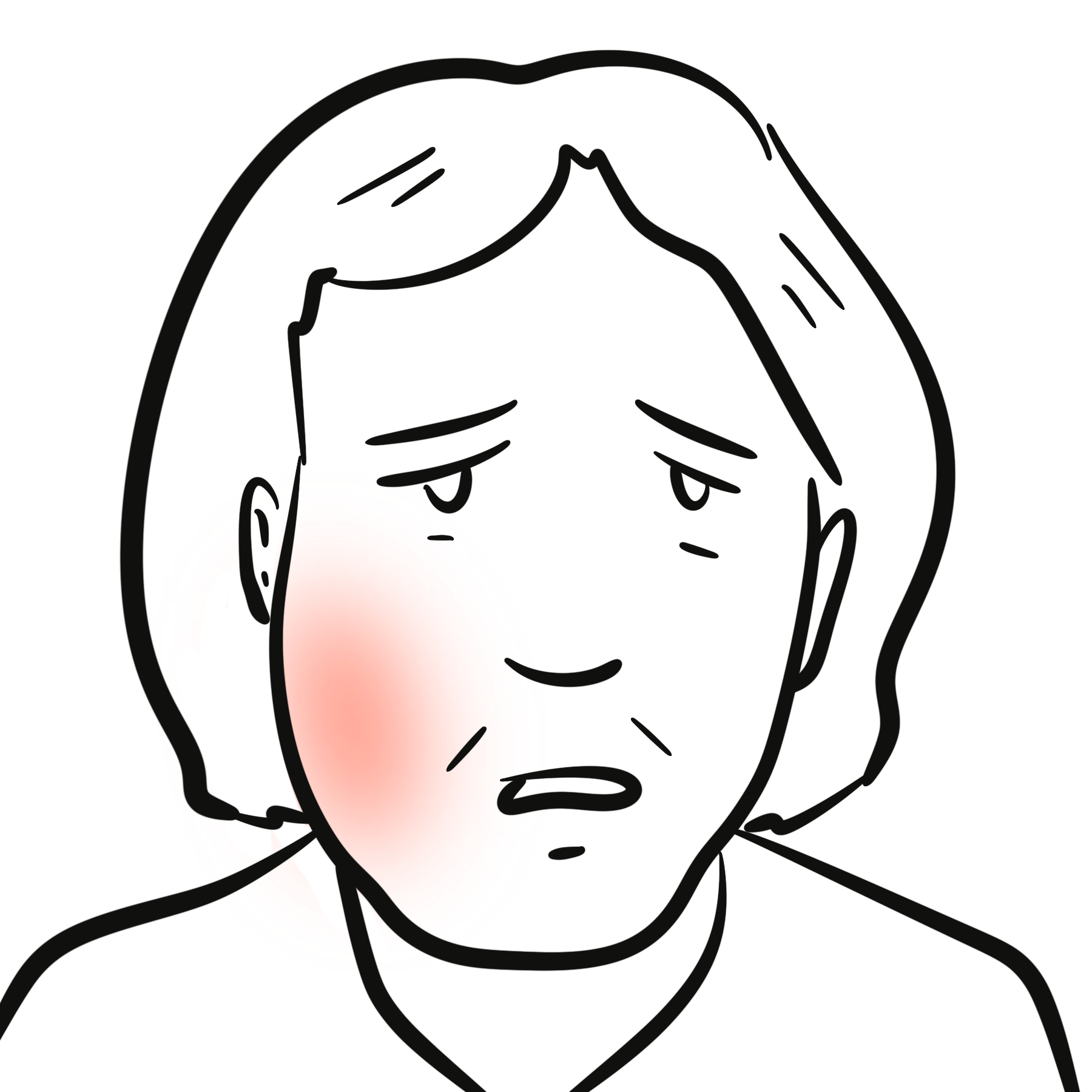
| Mandible (5%) | Body and angle of mandible | Causes facial asymmetry, trismus, and dental malocclusion. |
Clavicular osteomyelitis in a child or adolescent without prior trauma or central venous catheterization is CRMO until proven otherwise.
Diagnosis
CRMO is a diagnosis of exclusion. Diagnostic workup focuses on ruling out bacterial osteomyelitis, septic arthritis, primary bone tumors (Ewing sarcoma, osteosarcoma), hematological malignancies (leukemia, lymphoma), and Langerhans Cell Histiocytosis (LCH).
Laboratory Evaluation
- ESR and CRP elevated
- FBC: Usually normal; severe leukocytosis, marked anemia, or blast cells point toward infection or hematological malignancy.
- Microbiology: Blood cultures and serologies are negative.
Imaging Studies (Essential for Diagnosis)
- Whole-Body MRI (WBMRI with STIR sequences): Gold standard. Demonstrates hyperintense bone marrow edema, periosteal reaction, soft tissue inflammation, and uncovers “silent” asymptomatic lesions.
- Plain Radiography:
- Early: Radiolucent / osteolytic lesions in the metaphysis adjacent to the epiphyseal growth plate.
- Late: Osteosclerosis, cortical thickening, hyperostosis, and widening of affected bones.
- CT Scan: Useful for evaluating complex bony anatomy (e.g., spinal collapse or mandibular cortical erosion).
Diagnostic Criteria (Bristol Criteria)
Diagnosis can be made without bone biopsy if typical clinical and radiological features are present:
- Presence of typical osteolytic/sclerotic bone lesions on imaging.
- Multifocal bone involvement (especially if clavicular or symmetrical).
- Absence of local or systemic infectious signs (no purulent discharge, negative blood cultures).
- Mild or moderately elevated inflammatory markers.
- Lack of significant response to antibiotic therapy.
When is a Bone Biopsy Mandatory in Suspected CRMO?
A biopsy is required if any “red flags” exist: unifocal solitary lesion, severe constitutional B-symptoms (unexplained weight loss, night sweats), presence of an associated soft tissue mass, atypical imaging features (e.g., sunburst periosteal reaction), or cytopenias on CBC.
Treatment
The goal of treatment is to relieve bone pain, achieve clinical and radiological remission, prevent growth plate damage, and protect against vertebral compression fractures.
First-Line Pharmacotherapy
- Non-Steroidal Anti-Inflammatory Drugs (NSAIDs): First-line treatment for all patients. High-dose NSAIDs (e.g., Naproxen 10–15 mg/kg BD, Indomethacin, or Celecoxib) act as disease-modifying agents in CRMO by inhibiting prostaglandin synthesis and downregulating osteoclast activity.
- Response Rate: Up to 50–60% of patients achieve clinical remission within 1 to 3 months of continuous NSAID therapy.
Second-Line & Advanced Therapies
Indicated for NSAID non-responders, active spinal lesions (due to risk of collapse), or co-existing IBD/psoriasis:
- Bisphosphonates (Intravenous Pamidronate or Zoledronic Acid): Highly effective for severe pain and refractory lesions, particularly spinal involvement. Bisphosphonates inhibit osteoclast-mediated resorption, promote bone mineralization, and possess potent anti-inflammatory properties.
- Anti-TNF Alpha Agents (Adalimumab, Infliximab, Etanercept): First-line second-line option when CRMO co-occurs with inflammatory bowel disease or severe cutaneous psoriasis.
- Disease-Modifying Anti-Rheumatic Drugs (DMARDs): Methotrexate or Sulfasalazine may be used as adjunctive steroid-sparing agents.
- Systemic Corticosteroids: Oral Prednisolone or IV Methylprednisolone pulses used strictly as a short-term bridge for rapid symptom relief during acute severe disease flares.
Intravenous Bisphosphonates (Pamidronate) are the treatment of choice for CRMO with vertebral/spinal lesions, as they rapidly reduce pain and halt progressive vertebral collapse.
Complications & Prognosis
- Vertebral Compression Fractures & Vertebra Plana: Severe damage to vertebral bodies leading to spinal deformity (thoracic kyphosis) or neurological compromise.
- Physeal Bridge & Growth Arrest: Involvement of the growth plate (epiphyseal plate) resulting in premature physeal closure, angular limb deformities, or leg length discrepancy.
- Hyperostosis & Chronic Remodeling: Permanent enlargement and thickening of affected bones (especially the clavicle and mandible).
- Chronic Pain & Functional Disability: Centralized pain amplification syndromes secondary to prolonged unmanaged bone pain.
- With early diagnosis and targeted NSAID or biologic therapy, the overall long-term physical prognosis is favorable.
- The clinical course is typically prolonged, lasting 3 to 10 years with unpredictable exacerbations and remissions, before eventual spontaneous resolution in early adulthood.
References
- Roderick MR, Shah R, Rogers V, Finn A, Ramanan AV. Chronic recurrent multifocal osteomyelitis (CRMO) – advancing the diagnosis. Pediatr Rheumatol Online J. 2016;14(1):47. doi:10.1186/s12969-016-0109-1
- Girschick H, Finetti M, Soto-Regaña F, et al. Diagnosis and management of chronic non-bacterial osteomyelitis (CNO)/chronic recurrent multifocal osteomyelitis (CRMO): a consensus statement. Rheumatology (Oxford). 2021;60(11):4945-4964. doi:10.1093/rheumatology/keab440
- Zhao Y, Wu EY, Oliver MS, et al. Consensus Treatment Plans for Chronic Nonbacterial Osteomyelitis Refractory to Nonsteroidal Anti-Inflammatory Drugs and/or With Active Spinal Lesions. Arthritis Care Res (Hoboken). 2018;70(8):1228-1237. doi:10.1002/acr.23462












Members only discussions coming soon…