

Pyoderma Gangrenosum
Overview
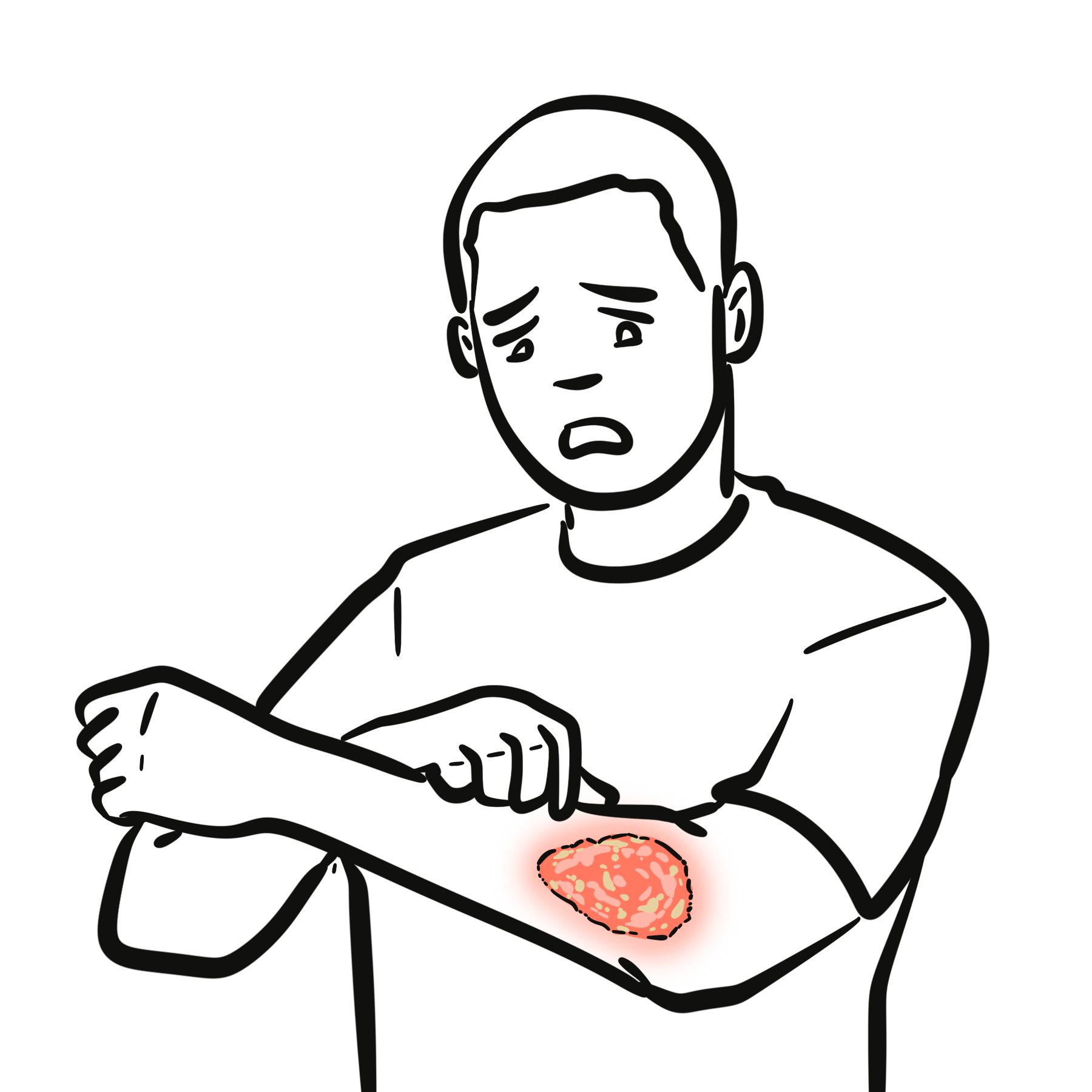
Pyoderma gangrenosum (PG) is a rare, severe, non-infectious inflammatory cutaneous disease belonging to the group of neutrophilic dermatoses. Despite its name, it is neither an infectious pyoderma nor a primary gangrenous necrosis; rather, it is driven by dysregulated innate immune activation leading to dermal neutrophilic infiltration and tissue destruction.
Characterized by rapidly expanding, excruciatingly painful cutaneous ulcers with gunmetal-colored or violaceous, undermined borders, pyoderma gangrenosum is associated with underlying systemic inflammatory diseases—such as Inflammatory Bowel Disease (IBD), inflammatory arthritis, and hematological malignancies—in approximately 50% of cases. Recognizing pyoderma gangrenosum is a critical clinical skill because surgical debridement is strictly contraindicated due to the phenomenon of pathergy.
Definition
Pathergy: The development of new skin lesions or the dramatic, rapid enlargement of existing ulcers following minor cutaneous trauma (e.g., surgical debridement, skin biopsy, venipuncture, or stoma placement).
Neutrophilic Dermatosis: An autoinflammatory skin disorder characterized histologically by a dense infiltrate of mature, non-infectious neutrophils in the dermis or subcutis.
Violaceous Undermined Border: The pathognomonic border of a classic pyoderma gangrenosum ulcer—a raised, purplish-red margin that overhangs the eroded necrotic ulcer bed.
Cribriform Scarring: A characteristic pitted, net-like, “cigarette paper” scar left behind following the slow healing of a pyoderma gangrenosum ulcer.
Aetiology & Risk Factors
Systemic Associations (~50% of Patients)
Pyoderma gangrenosum frequently serves as a cutaneous manifestation of an underlying systemic autoinflammatory condition:
- Inflammatory Bowel Disease (IBD): Ulcerative Colitis (more common) and Crohn’s Disease.
- Inflammatory Arthritis: Rheumatoid Arthritis, Seronegative Spondyloarthropathies, and Ankylosing Spondylitis.
- Hematological Malignancies & Dyscrasias: Acute Myeloid Leukemia (AML), Myelodysplastic Syndrome (MDS), Monoclonal Gammopathy of Undetermined Significance (MGUS), and Polycythemia Vera.
- Monogenic Autoinflammatory Syndromes: PAPA syndrome (Pyogenic Arthritis, Pyoderma gangrenosum, and Acne) and PASH syndrome (Pyoderma gangrenosum, Acne, and Suppurative Hidradenitis).
- Most commonly presents in adults aged 30 to 60 years.
- Triggers: Minor skin trauma, surgery, insect bites, or catheter insertion sites (via pathergy).
Approximately 50% of pyoderma gangrenosum cases are idiopathic, meaning a negative systemic workup does not rule out the diagnosis!
Pathophysiology
- Immune Dysfunction: Impaired regulation of the innate immune system leads to abnormal neutrophil chemotaxis, priming, and hyper-reactivity.
- Cytokine Cascade: Markedly elevated levels of interleukin-1 beta (IL-1β), interleukin-8 (IL-8), interleukin-17 (IL-17), and Tumor Necrosis Factor-alpha (TNF-α\alpha-α) drive continuous neutrophil recruitment to the skin.
- Tissue Destruction: Neutrophils release Matrix Metalloproteinases (MMPs) and reactive oxygen species, causing local microvascular damage, tissue infarction, and rapidly spreading ulceration.
Clinical Manifestations
- Initial Lesion: Small, painful, dusky-red nodule, pustule, or blood-filled bulla that breaks down within 24 to 48 hours.
- Ulcer Features:
- Border: Raised, violaceous (purple-gunmetal), overhang/undermined edges.
- Base: Purulent, fibrinous, or necrotic bed with focal hemorrhagic dots.
- Surrounding Skin: Erythematous, indurated halo expanding radially.
- Pain: Severe, excruciating pain that is strikingly disproportionate to the physical size of the lesion.
- Pathergy Sign: Lesion flares, expands rapidly, or develops new satellite ulcers following minor trauma or diagnostic procedures.
Diagnosis
Pyoderma gangrenosum is a diagnosis of exclusion. There are no specific diagnostic laboratory markers or pathognomonic histological features.
Diagnostic Criteria (PARACELSUS Score / Delphi Consensus)
Diagnosis requires ruling out other causes of cutaneous ulceration (e.g., vascular, infectious, vasculitic, or malignant etiologies).
- Major Criteria: Rapidly progressive, painful, necrotizing cutaneous ulcer with violaceous, undermined borders, and exclusion of relevant differential diagnoses.
- Minor Criteria: History of pathergy, systemic disease association (IBD/arthritis), prompt response to systemic corticosteroids, and dense neutrophilic infiltrate on skin biopsy.
Workup & Laboratory Investigations
- Skin Biopsy (Edge of Ulcer):
- Purpose: Performed carefully at the active, advancing border to rule out vasculitis, deep fungal infections, or malignancy.
- Histology: Demonstrates dense dermal neutrophilic infiltration and microabscesses without evidence of primary leukocytoclastic vasculitis.
- Microbiological Swabs & Tissue Cultures: Gram stain, bacterial, fungal, and atypical mycobacterial cultures to exclude primary infectious ulcers.
- Systemic Screening Workup:
- Gastrointestinal: Colonoscopy / Sigmoidoscopy to evaluate for occult IBD.
- Hematological: Complete Blood Count (CBC), peripheral blood smear, Serum Protein Electrophoresis (SPEP) to evaluate for leukemia, MDS, or MGUS.
- Rheumatological: ANCA, ANA, Rheumatoid Factor, and Anti-CCP to rule out systemic vasculitis (e.g., Granulomatosis with Polyangiitis) and Rheumatoid Arthritis.
CRITICAL WARNING: Skin biopsy should be performed with extreme caution. Always take a small, precise punch sample from the active border to minimize the risk of triggering pathergy and worsening the ulcer!
Treatment
General Measures & Wound Care
- AVOID SURGICAL DEBRIDEMENT: Absolute Rule. Surgical excision, debridement, or aggressive wound scraping triggers severe pathergic expansion of the ulcer.
- Non-Adherent Dressings: Gently apply non-adherent, moist, barrier dressings to protect against secondary infection and mechanical irritation.
- Mild / Localized Disease:
- Topical High-Potency Corticosteroids: Clobetasol propionate 0.05% ointment applied to the active violaceous border.
- Topical Calcineurin Inhibitors: Tacrolimus 0.1% ointment.
- Severe / Rapidly Progressive / Extensive Disease (First-Line Systemic):
- Systemic Corticosteroids: Oral Prednisone (1.0–2.0 mg/kg/day) or IV Methylprednisolone pulse therapy for rapid control.
- Cyclosporine: Oral Cyclosporine (3–5 mg/kg/day) is highly effective, either as monotherapy or combined with corticosteroids.
- Refractory / Second-Line Therapy (Biologics):
- TNF-Alpha Inhibitors: Infliximab (strongest evidence base) or Adalimumab—particularly useful in patients with co-existing Inflammatory Bowel Disease.
- Interleukin Inhibitors: Anakinra (IL-1 inhibitor), Ustekinumab (IL-12/23 inhibitor), or Secukinumab (IL-17 inhibitor).
Complications & Prognosis
- Iatrogenic Ulcer Worsening: Rapid expansion of the wound caused by mistaken diagnosis followed by surgical debridement (a common clinical diagnostic trap!).
- Secondary Bacterial Infection: Superinfection of the open ulcer bed by Staphylococcus aureus or Pseudomonas aeruginosa.
- Severe Scarring: Extensive cribriform or hypertrophic scarring causing cosmetic disfigurement and joint contractures when crossing flexor surfaces.
- Sepsis: Rare, secondary to severe systemic bacterial invasion through extensive open wounds.
- The clinical course is unpredictable and highly variable. While lesions can respond rapidly to systemic immunosuppression, complete healing often takes several months.
- Prognosis is closely tied to the control of any underlying systemic disorder (e.g., controlling IBD flares often leads to remission of peristomal pyoderma gangrenosum).
- Recurrence rates reach up to 30%, frequently triggered by minor trauma or systemic disease relapse.
References
- Su WP, Davis MD, Weenig RH, Powell FC, Perry HO. Pyoderma gangrenosum: clinicopathologic correlation and proposed diagnostic criteria. Int J Dermatol. 2004;43(11):790-800. doi:10.1111/j.1365-4632.2004.02128.x
- Maverakis E, Ma C, Shinkai K, et al. Diagnostic Criteria of Ulcerative Pyoderma Gangrenosum: A Delphi Consensus of International Experts. JAMA Dermatol. 2018;154(4):461-466. doi:10.1001/jamadermatol.2017.5980
- George C, Deroide F, Rustin M. Pyoderma gangrenosum – a guide to diagnosis and management. Clin Med (Lond). 2019;19(3):224-228. doi:10.7861/clinmedicine.19-3-224













Members only discussions coming soon…