Overview
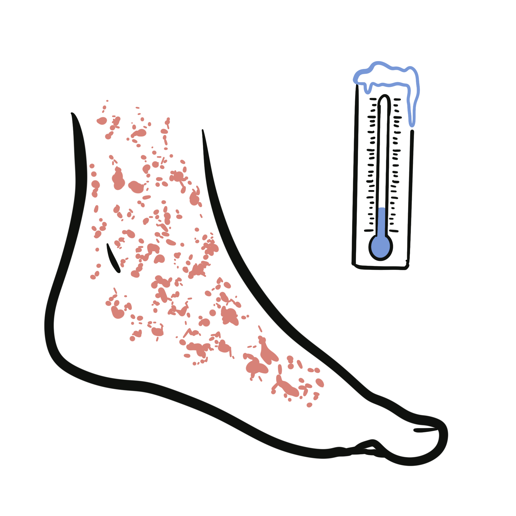
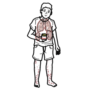
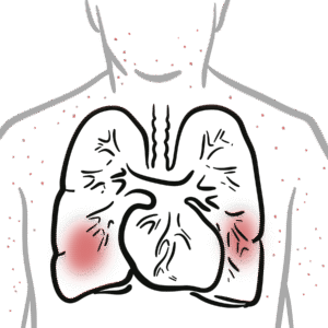
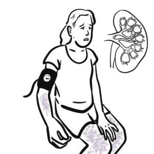
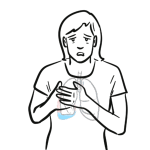
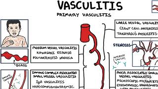
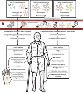
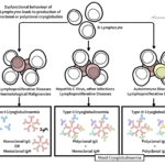
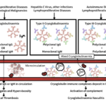
Cryoglobulinaemic vasculitis is an immune complex–mediated small to medium vessel vasculitis caused by circulating cryoglobulins—immunoglobulins that precipitate below 37 °C and redissolve on warming. These immune complexes deposit in vessel walls, activate complement (typically with marked C4 consumption), and drive leukocytoclastic vasculitis. “Mixed” CV (types II and III) is most often linked to hepatitis C virus (HCV), autoimmune diseases, or B-cell clonal disorders, while type I CV is monoclonal and associated with plasma-cell dyscrasias or lymphoproliferative disorders. Adults are most commonly affected.
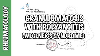
Definition
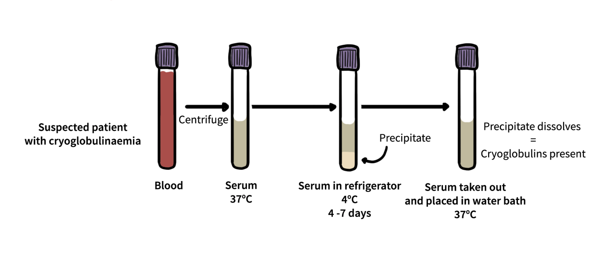
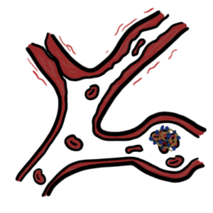
Cryoglobulins: Immunoglobulins that precipitate at <37 °C and dissolve on rewarming; quantified as cryocrit (%) and typed by immunofixation into Brouet types I–III.
Mixed cryoglobulinaemia: Type II (monoclonal IgM-RF + polyclonal IgG) or Type III (polyclonal IgM-RF + polyclonal IgG); RF activity drives immune complex formation.
Type I cryoglobulinaemia: Monoclonal immunoglobulin (IgM or IgG) from a haematologic clone; causes ischemia/hyperviscosity more than vasculitis.
Membranoproliferative glomerulonephritis (MPGN): Immune-complex GN typical of mixed CV renal disease.
Remember
Always keep blood samples at 37 °C until serum separation to avoid false-negative cryoglobulin results.
Aetiology & Risk Factors
Aetiology:
- HCV infection (leading global cause of mixed CV)
- Autoimmune diseases (e.g., Sjögren’s syndrome, SLE)
- B-cell clonal disorders (Waldenström macroglobulinaemia, multiple myeloma, MGUS)
- Less common: HBV, HIV, solid tumours
Risk Factors for Severe Disease:
- High cryocrit level
- Very low C4 and/or low C3
- Active HCV viremia or persistent monoclonal B-cell clone
- Renal or peripheral nerve involvement
- Extensive skin ulcers/necrosis
- Older age
Pathophysiology
- Chronic antigenic stimulation (e.g., HCV) or B-cell clone produces IgM with RF activity + IgG.
- Circulating immune complexes (cryoglobulins) form and precipitate in cooler environments.
- Immune complexes deposit in vessel walls → activate classical/lectin complement pathways → marked C4 consumption.
- Complement activation → neutrophil recruitment → leukocytoclastic vasculitis (skin/nerve) and immune-complex MPGN (kidney).
- Type I CV: Monoclonal proteins → hyperviscosity and vascular occlusion → ischemia.
Remember
Low C4 + high RF in the right setting is a strong clue to mixed CV.
Clinical Manifestations
- Skin: Palpable purpura (dependent areas), livedo, retiform purpura, ulcers/necrosis, acrocyanosis, Raynaud phenomenon.
- Musculoskeletal: Arthralgia, non-erosive arthritis, myalgia
- Mononeuritis multiplex.
- Membranoproliferative Glomerulonephritis pattern, possible rapidly progressive GN
- Type I CV may cause hyperviscosity (headache, visual changes, epistaxis) and digital ischemia.
Diagnosis
- Serum cryoglobulin positivity
- Must include clinical features: Purpura, low C4, positive RF, renal involvement, peripheral neuropathy, leukocytoclastic vasculitis on biopsy.
Investigations
- Cryoglobulin testing with proper handling (pre-warmed tubes, 37 °C transport until serum separation)
- Complement levels (very low C4, ± low C3)
- Rheumatoid factor (often high in mixed CV)
- HCV Ab + RNA, HBV serology, ± HIV testing
- Autoimmune serology (ANA, anti-SSA/SSB)
- Monoclonal screen (SPEP/UPEP, serum free light chains)
- Skin biopsy: leukocytoclastic vasculitis with IgM/IgG/C3 deposition
- Renal biopsy: immune-complex MPGN
- Nerve biopsy if diagnosis unclear
Differentials Diagnosis
- ANCA-associated vasculitis
- Infective endocarditis-related vasculitis
- Antiphospholipid syndrome
- Cholesterol embolism
- Cold agglutinin disease
Classification (Brouet Types)
Type | Immunochemistry | Typical associations | Key complications |
I | Monoclonal IgM/IgG | Waldenström, myeloma, MGUS | Hyperviscosity, ischemia |
II | Monoclonal IgM-RF + polyclonal IgG | HCV, autoimmune disease, B-cell clone | Mixed CV – purpura, neuropathy, MPGN |
III | Polyclonal IgM-RF + polyclonal IgG | Autoimmune disease, infections | Milder mixed CV, can evolve to type II |
Treatment
Treat underlying cause, control immune complex production, protect organs, avoid cold exposure, wound care.
Type I CV:
- Clone-directed therapy (e.g., BTK inhibitor, bortezomib-based regimen)
- Plasma exchange for hyperviscosity, rapidly progressive GN, or severe ischemia
HCV-associated mixed CV:
- First-line: DAAs (SVR >95%)
- Add rituximab ± short glucocorticoid course for moderate–severe or organ-threatening disease
- Avoid interferon-based therapy
Non-viral mixed CV:
- Rituximab + short glucocorticoid course first-line
- Cyclophosphamide for rituximab-refractory disease
Renal protection: RAAS blockade, BP <130/80 mmHg, avoid nephrotoxins.
Complications and Prognosis
Complications
- Skin ulceration/necrosis ± infection
- Peripheral neuropathy
- MPGN → CKD/ESKD
- Hyperviscosity syndrome (type I Cryoglobulinaemia)
- Digital ischemia/gangrene (type I Cryoglobulinaemia)
- Cardiovascular events
Prognosis
- Improved outcomes for HCV-associated CV in the DAA era
- Poor prognostic factors: renal involvement with creatinine rise, severe neuropathy, extensive ulcers/necrosis, cardiac involvement, older age, high cryocrit, very low C4, persistent HCV viremia or B-cell clone
- Relapse risk if underlying cause remains untreated
Discussion