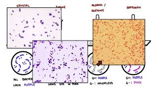
0:00 In this video we're going to talk about staining. 0:08 And so this would be a follow-up with the microscopy video. 0:16 So with staining, even with the best light microscope, which we learned about, 0:21 most bacteria are 0:22 nearly invisible because they are colorless and blend into their surroundings. 0:28 To truly study them, we need contrast, and that is where staining becomes 0:33 essential. 0:33 By adding dyes, we give microbes color and definition, allowing us to see their 0:38 shapes, 0:38 arrangements, and even special structures. 0:41 These staining methods are not just about making microbes visible. 0:47 They are the first steps in identifying pathogens, guiding treatment, and 0:52 uncovering the hidden 0:54 diversity of the microbial world. 1:02 So let's talk about some basic staining concepts, beginning with what's called 1:09 the gram stain. 1:11 Among all staining techniques, the gram stain stands as a cornerstone of 1:16 microbiology. 1:17 Developed by Hans Christian Graham in 1884, it was originally designed to 1:22 improve the 1:22 visibility of bacteria in infected tissue, and it ended up dividing the 1:28 bacteria world 1:28 into two major groups, gram positive and gram negative bacteria. 1:35 This distinction is based on their cell wall structure, and it is crucial 1:40 because it affects 1:41 how bacteria behave, how they cause disease, and which antibiotics work. 1:48 So how does gram staining work, let's look at the procedure and the reasoning. 1:53 The first step of gram staining is essentially when you have a sample of tissue 2:02 or whatever 2:04 you want to look at, you stain it with a crystal violet. 2:07 This is the primary stain. 2:09 All cells are flooded with the purple dye, which is called the crystal violet, 2:14 and at 2:14 this stage, all the bacteria look purple, regardless of the type. 2:20 The second part, iodine. 2:23 Iodine solution is then added, which binds with crystal violet inside the cells 2:29 . 2:30 This forms a large crystal violet iodine complex, locking the dye in place. 2:35 Still, all cells remain purple at this point. 2:41 The third step, using alcohol or acetone, a decolorizer, essentially. 2:47 This is a critical step. 2:48 So if it was gram positive cells, the thick peptidoglycan layer shrinks and it 2:56 traps the 2:57 dye complex. 2:58 So the purple color stays. 3:02 In gram negative cells, the alcohol dissolves lipids in the outer membrane, and 3:08 the thin 3:09 peptidoglycan cannot retain the dye, so the purple washes out. 3:16 Now gram positive cells are purple, gram negative cells are colorless. 3:21 And again, it's because of their peptidoglycan layer, one is thick and one is 3:27 thin. 3:28 The fourth part is adding something called saffronin, which is a counter stain. 3:35 Then a red counter stain is applied. 3:39 The gram positive cells remain purple because the purple is darker than the red 3:43 . 3:43 And remember, the gram positive cells are trapping that color, that purple 3:48 color in the 3:48 peptidoglycan, whereas gram negative cells, which were initially colorless, 3:54 take up the 3:55 red dye and then appear pink. 3:58 And so the results under the microscope, gram positive bacteria are purple. 4:03 Gram negative bacteria are pink or red. 4:07 So here are some example images of gram positive and gram negative stain. 4:12 Here you can see a gram positive cock eye in clusters. 4:17 This is Staphylococcus aureus. 4:21 Purple spherical bacteria arranged in grape-like clusters, characteristic of St 4:25 aphylococci. 4:27 Here is another gram positive because it's purple. 4:32 It does look like rod shape, but they're actually cock eye and they're sort of 4:36 two together 4:37 or in chains. 4:38 This purple round shade bacteria indicates, again, a thick peptidoglycan cell 4:46 wall. 4:47 And this is Staphylococcus pneumoniae. 4:50 Finally, we have gram negative bacilli. 4:56 This is E. coli. 4:58 It's pink, red, rod-shaped bacteria consistent with gram negative organism. 5:06 Why is it important? 5:07 Well, this is usually the first lab result that guides doctors in choosing 5:11 antibiotics. 5:12 Gram positive bacteria are often more sensitive to drugs like penicillin, while 5:17 gram negative 5:18 tends to be more resistant due to their extra outer membrane. 5:28 Another staining technique is the acid fast stain. 5:33 Some bacteria do not follow the rules of gram stain. 5:36 The most important example is mycobacterium tuberculosis, the cause of TB, the 5:41 condition 5:41 where you cough our blood, it causes lung infections. 5:45 Now, these bacteria have cell walls rich in mycolic acids, long waxy lipids 5:53 that make 5:54 the wolf thick, hydrophobic and resistant to many dyes and chemicals. 6:00 Because of this, they do not retain the gram stain well. 6:03 To see them clearly, we use the acid fast stain. 6:07 Why acid fast? 6:08 Well, the name comes from the fact that once stained, these bacteria resist dec 6:14 olorization 6:15 with acid alcohol. 6:16 In other words, the dye gets locked in their waxy walls and won't wash out, 6:21 even with strong 6:22 acid. 6:25 The acid fast stain procedure, also known as a Zeel Nielsen method, initially, 6:31 carbo fusine 6:32 is applied. 6:33 This is the primary stain. 6:35 It's a strong red dye. 6:38 Then with heat, it's used to help penetrate the waxy wall. 6:46 At this stage, all the cells look red. 6:49 The second part is the acid alcohol, the decolorizer. 6:53 This is, again, a key step. 6:55 Acid fast bacteria with mycolic acids hold on to the red dye. 7:01 Non acid fast bacteria lose the dye and become colorless. 7:07 The third step is methylene blue or brilliant green, which is a counter stain. 7:13 This contrasting color is added. 7:16 Acid fast bacteria stay red. 7:18 The non acid fast bacteria take up the counter stain and appear blue or green. 7:24 The result under the microscope, an acid fast bacteria will look red. 7:29 This includes mycobacterium tuberculosis, mycobacterium leprey, and some noch 7:34 adia. 7:36 Non acid fast bacteria will be blue or green, because this is the counter stain 7:43 . 7:43 The acid fast stain is a critical diagnostic tool for tuberculosis and leprosy, 7:49 because 7:49 mycobacterium grows very slowly, waiting for cultures takes weeks. 7:54 Microscopy and with acid fast staining provides a rapid answer, which can guide 7:59 early treatment 8:00 and infection control. 8:06 There are other stains used other than gram stain and the zeal Nielsen stain. 8:12 These other stains include endospor stain, which shows tough bacterial spores, 8:17 like bacillus 8:18 clostridium, which appear green against pink cells. 8:22 Capsule stain highlights protective capsules as clear halos, important in 8:25 organisms like 8:26 streptococcus pneumoniae, and the silver stain used for delicate organisms like 8:33 pneumocystis 8:34 uravicii. 8:37 So in summary, microscopy gives us the eye to see microbes, and staining gives 8:43 them color 8:44 and contrast. 8:45 The gram stain is the first and most important step in identifying bacteria, 8:50 while acid 8:50 fast stains pick out unique organisms like mycobacterium. 8:55 More advanced methods like fluorescence and electron microscopy add layers of 9:01 detail for 9:01 diagnosis and research, and this is touched on in the microscopy video. 9:05 Thank you.