

Mitral Stenosis
Overview
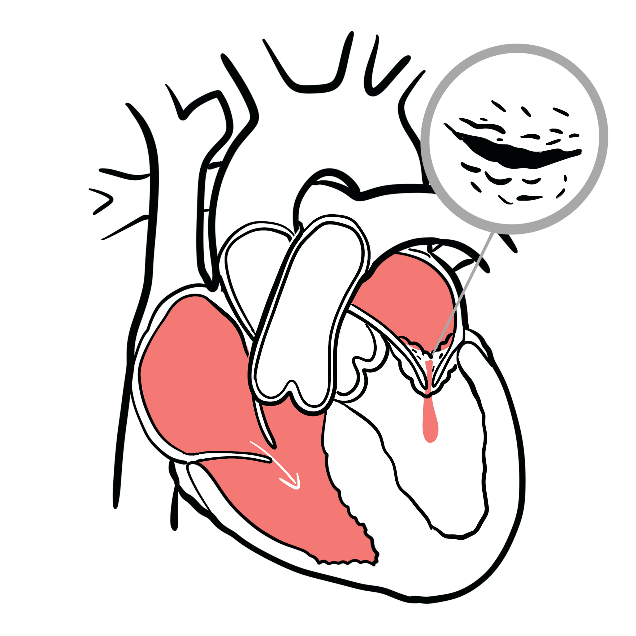
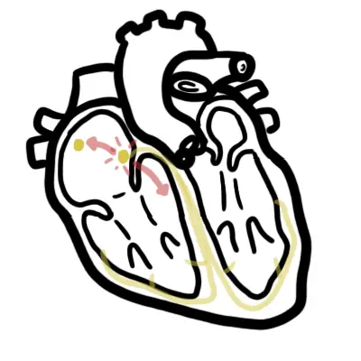
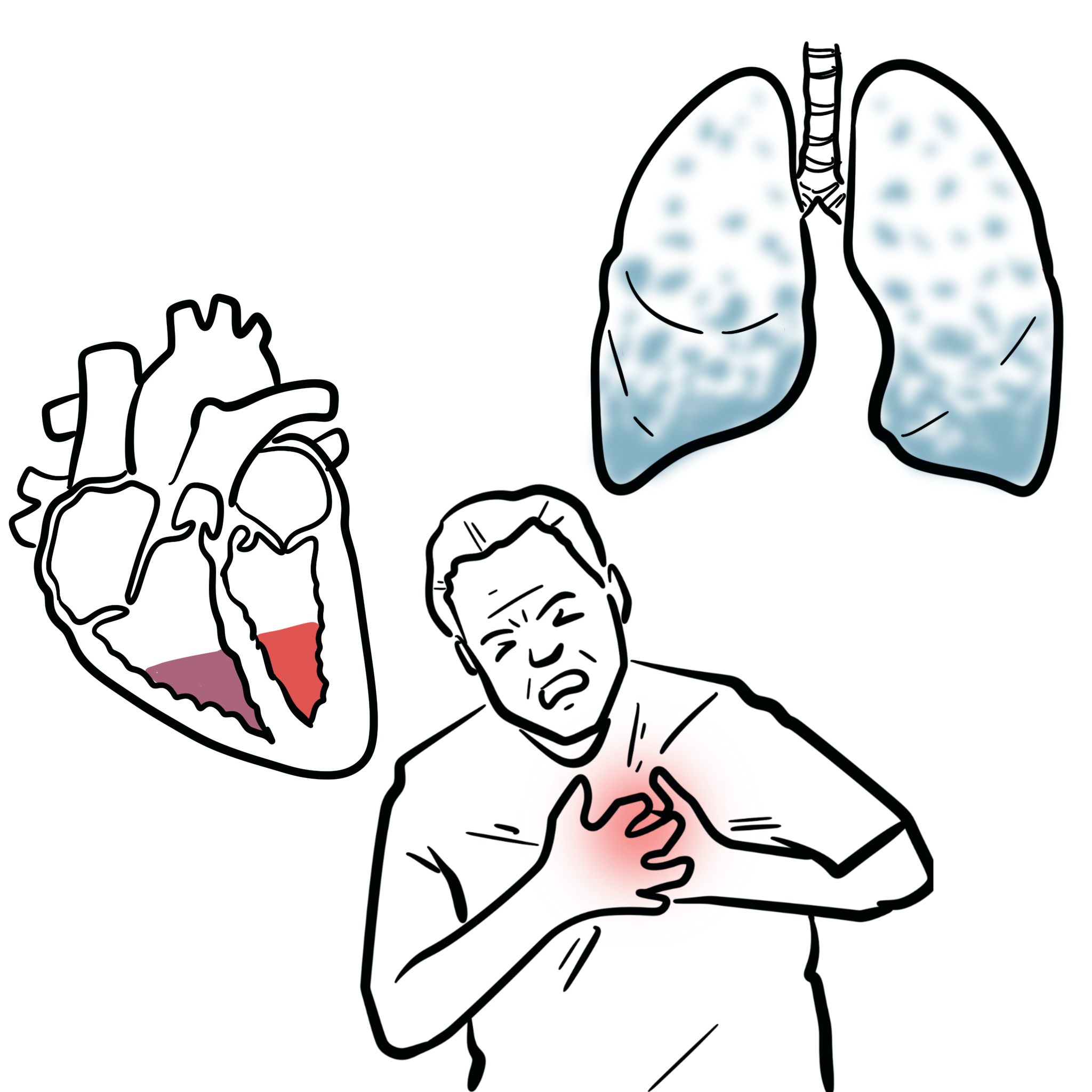
Mitral stenosis causes an obstruction to blood flow from the left atrium to left ventricle usually as a result of rheumatic heart disease. The stenosis results in increased pressure in the left atrium, pulmonary vasculature and right side of heart. Mitral valve disease is a frequent cause of heart failure and death.

Definition
Mitral valve area: area through which blood flows from the left atrium to the left ventricle; normal is approximately 4–6 cm².
Clinically significant/severe MS: mitral valve area ≤1.5 cm².
Very severe MS: mitral valve area ≤1.0 cm².
Commissural fusion: fusion of the leaflet edges, characteristic of rheumatic MS.
Percutaneous mitral balloon commissurotomy—PMBC: catheter-based balloon separation of fused mitral commissures; also called balloon mitral valvotomy.
Aetiology and Risk Factors
Aetiology
- Rheumatic fever leading to rheumatic heart disease (95% of cases)
- Congenital mitral stenosis
- Degenerative mitral-annular and leaflet calcification
- Systemic lupus erythematosus or antiphospholipid syndrome
- Left-atrial myxoma or large thrombus mimicking MS
- Rarely, carcinoid disease or infiltrative disorders
Pathophysiology
Narrowing of the mitral valve obstructs left-ventricular filling during diastole.Left-atrial pressure rises to maintain forward blood flow.Elevated left-atrial pressure causes:
- Left-atrial enlargement
- Pulmonary venous congestion
- Pulmonary oedema
Left-atrial enlargement predisposes to atrial fibrillation and thrombus formation.Chronic pulmonary venous hypertension causes pulmonary arterial hypertension.Progressive pulmonary hypertension may produce:
- Right-ventricular hypertrophy and failure
- Functional tricuspid regurgitation
- Peripheral oedema
Tachycardia shortens diastole, reducing filling time and markedly worsening symptoms.Left-ventricular systolic function is usually preserved unless another cardiac disorder is present.
Tachycardia worsens MS because it shortens diastolic filling time.
Clinical Manifestation
- Exertional dyspnoea
- Decreased excercise tolerance
- Haemoptysis
- Chest pain
- Fatigue
- History of rheumatic fever
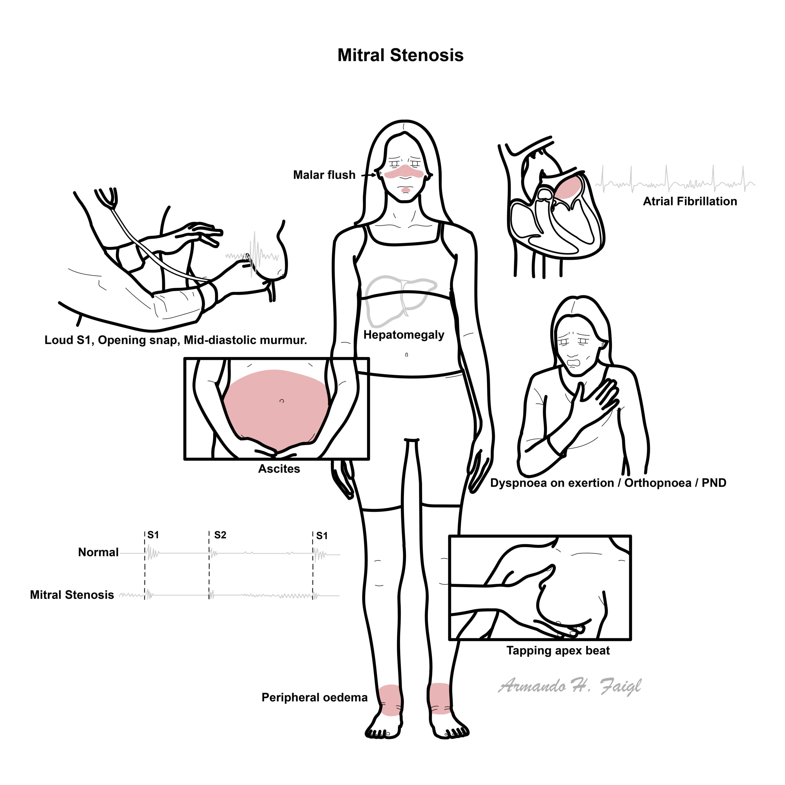
- Malar flush
- Signs of right-sided heart failure
- ascites
- raised JVP
- peripheral oedema
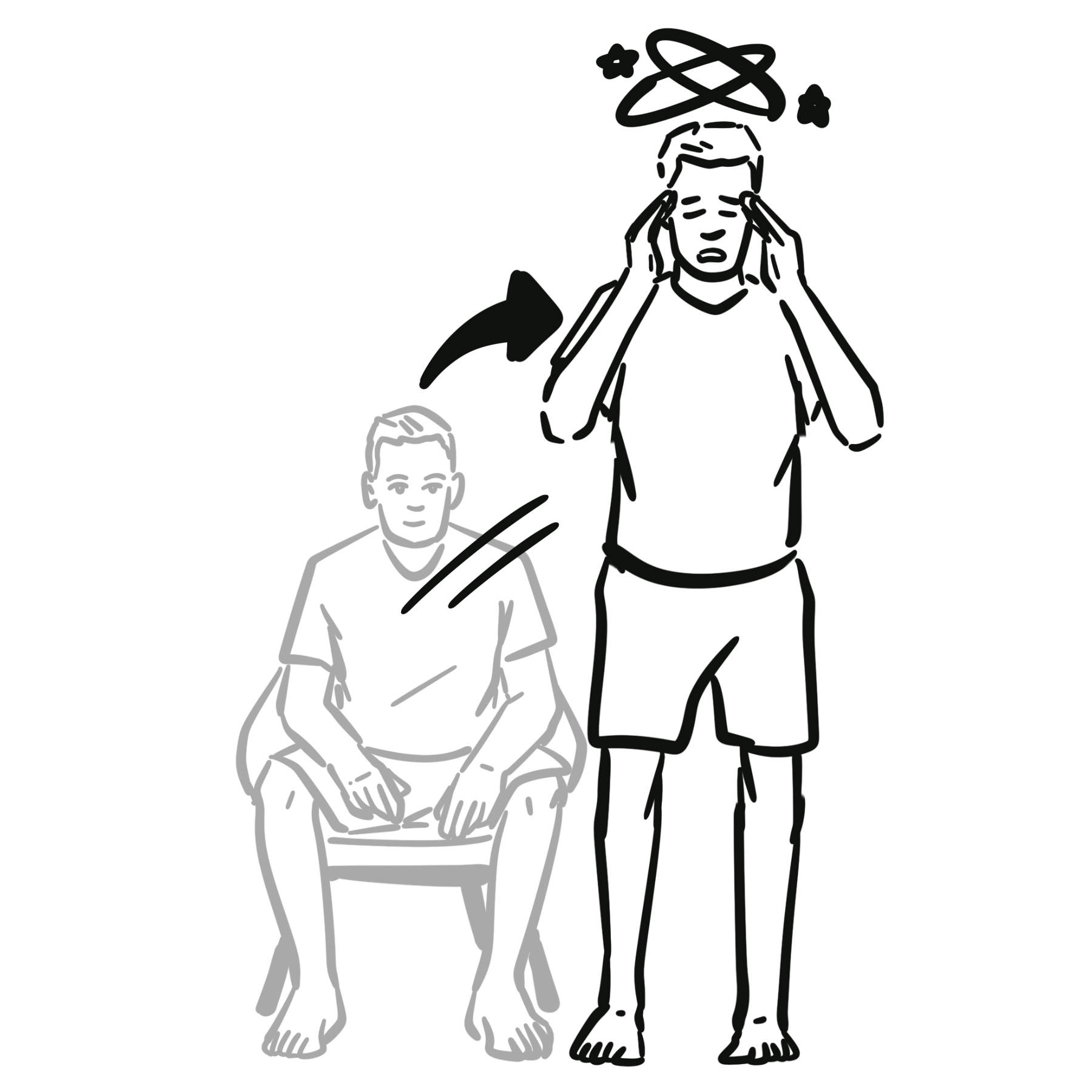
- Thromboembolic event “Stroke”
- Hoarseness (recurrent laryngeal nerve compressed)
- Dysphagia (esophagus compressed)
The classic murmur is a low-pitched mid-diastolic rumble at the apex with an opening snap.
- Malar flush
- Pulse
- Weak pulse due to reduced strove volume
- Atrial fibrillation
- Left parasternal heave (from right ventricular hypertrophy)
- Auscultation – Mitral valve (Apex – left 5th intercostal space mid-clavicular)
- Pre systolic murmur precedes S1, a result of increase blood flow from atrial contraction
- Opening snap of the mitral valve following S2 (closure of the aortic and pulmonic valves) is the opening of the stenotic mitral valve (SNAP)
- Long murmur during Diastole (longer in chronic mitral stenosis)
- Low-pitched diastolic rumble that is most prominent at the apex.
Early diastolic murmur (on inspiration) due to pulmonary regurgitation from pulmonary hypertension (Graham Steell murmur) may be heard rarely.
Diagnosis
- Transthoracic echocardiography—first-line
- Transoesophageal echocardiography
- ECG
- Atrial fibrillation
- Left atrial enlargement – P mitrale
- Right ventricular hypertrophy – Right axis deviation
- Chest X-ray
- Straight or convex L heart border
- Double shadow of LA behind RA
- Splaying of carina
- Dilated upper lobe veins
- Prominent pulmonary conus
- Pulmonary haemosiderosis
- Trans-thoracic echocardiography
- Transoesophageal echocardiography
- Cardiac catherization
Symptoms and signs similar to mitral stenosis
- left atrial myxoma
- prosthetic valve obstruction
- Cor tratriatum
Treatment
Mild or asymptomatic disease
- Regular clinical and echocardiographic surveillance.
- Treat hypertension and other cardiovascular risk factors.
Symptomatic medical treatment
Medical treatment reduces symptoms but does not relieve the valve obstruction.
- Diuretics: reduce pulmonary and peripheral congestion.
- Heart-rate control: increases diastolic filling time:
- Beta blocker
- Diltiazem or verapamil where appropriate
- Digoxin may assist ventricular-rate control in atrial fibrillation
- Treat precipitating factors such as infection, anaemia or hyperthyroidism.
Anticoagulation
A vitamin K antagonist such as warfarin is indicated in rheumatic MS with:
- Atrial fibrillation
- Previous systemic embolism
- Left-atrial thrombus
Direct oral anticoagulants are not recommended as substitutes for warfarin in patients with atrial fibrillation and clinically significant rheumatic MS.
Warfarin—not a DOAC—is used for atrial fibrillation associated with significant rheumatic MS
Percutaneous mitral balloon commissurotomy
Preferred intervention for symptomatic severe rheumatic MS when:
- Mitral valve area is ≤1.5 cm².
- Valve anatomy is suitable.
- There is no left-atrial thrombus.
- There is no more than mild mitral regurgitation.
It may also be considered in selected asymptomatic patients with pulmonary hypertension, very severe MS, high embolic risk or planned pregnancy.
| Contraindications to balloon commissurotomy |
| Left-atrial thrombus |
| More than mild mitral regurgitation |
| Severe or bicommissural calcification |
| Absence of commissural fusion |
| Significant associated valve or coronary disease requiring surgery |
| Unfavourable valve or subvalvular anatomy. |
Surgery
Mitral-valve repair, open commissurotomy or replacement is considered when:
- The patient is symptomatic with severe MS but unsuitable for balloon commissurotomy.
- Balloon commissurotomy has failed.
- Significant mitral regurgitation is also present.
- The valve is severely calcified.
- Other cardiac surgery is required.
A mechanical valve requires lifelong anticoagulation; a bioprosthetic valve has limited durability but usually avoids lifelong anticoagulation unless another indication exists.
Complications and Prognosis
- Atrial Fibrillation
- Stroke
- Warfarin-induced haemorrhage
- Systemic Embolism – due to thrombus formation in the right atrium
- Infective endocarditis
- Functional tricuspid reguritation
- Rheumatic MS often has a long asymptomatic period before symptoms develop.
- Prognosis worsens substantially after the development of:
- Atrial fibrillation
- Pulmonary hypertension
- Recurrent pulmonary oedema
- Right-sided heart failure
- Systemic embolisation
- Successful balloon commissurotomy or surgery can markedly improve symptoms and haemodynamics.
- Restenosis may occur over time, particularly with unfavourable anatomy or ongoing rheumatic disease.
References
- Praz F, Borger MA, Lanz J, et al. 2025 ESC/EACTS Guidelines for the management of valvular heart disease. Eur Heart J. 2025;46(44):4635–4736. doi:10.1093/eurheartj/ehaf194.
- Otto CM, Nishimura RA, Bonow RO, et al. 2020 ACC/AHA guideline for the management of patients with valvular heart disease. Circulation. 2021;143(5)–e227. doi:10.1161/CIR.0000000000000923.
- Vahanian A, Beyersdorf F, Praz F, et al. 2021 ESC/EACTS guidelines for the management of valvular heart disease. Eur Heart J. 2022;43(7):561–632. doi:10.1093/eurheartj/ehab395.
- Chandrashekhar Y, Westaby S, Narula J. Mitral stenosis. Lancet. 2009;374(9697):1271–1283. doi:10.1016/S0140-6736(09)60994-6.
- Karthikeyan G, Connolly SJ, Ntsekhe M, et al. Rivaroxaban in rheumatic heart disease-associated atrial fibrillation. N Engl J Med. 2022;387(11):978–988. doi:10.1056/NEJMoa2209051.


Members only discussions coming soon…