

Cerebral Aneurysm
Overview
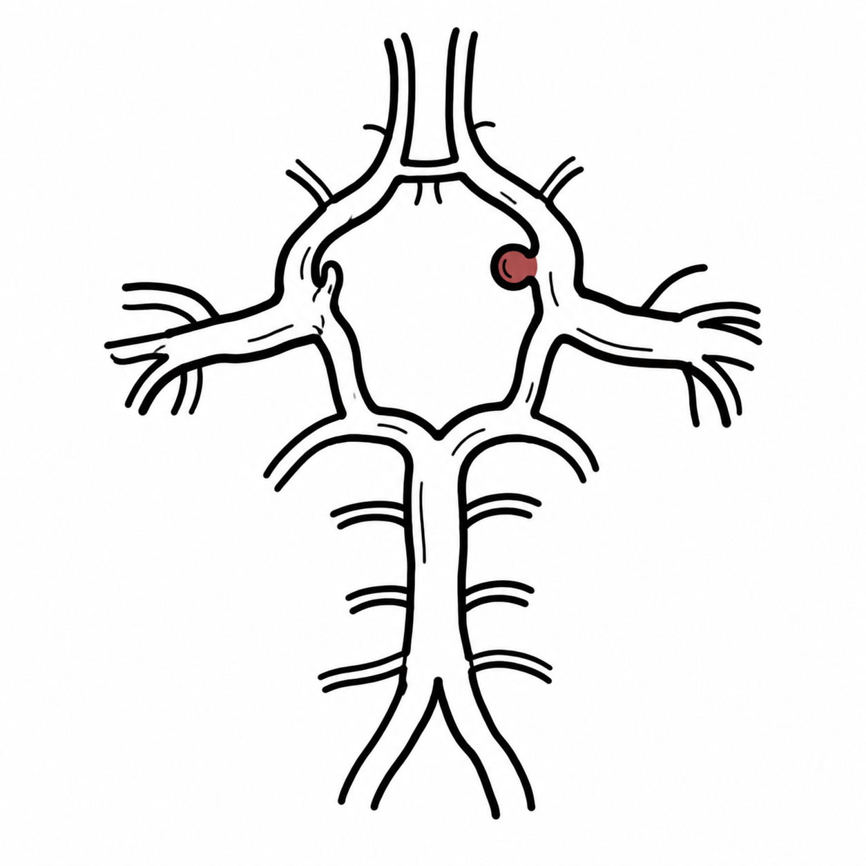
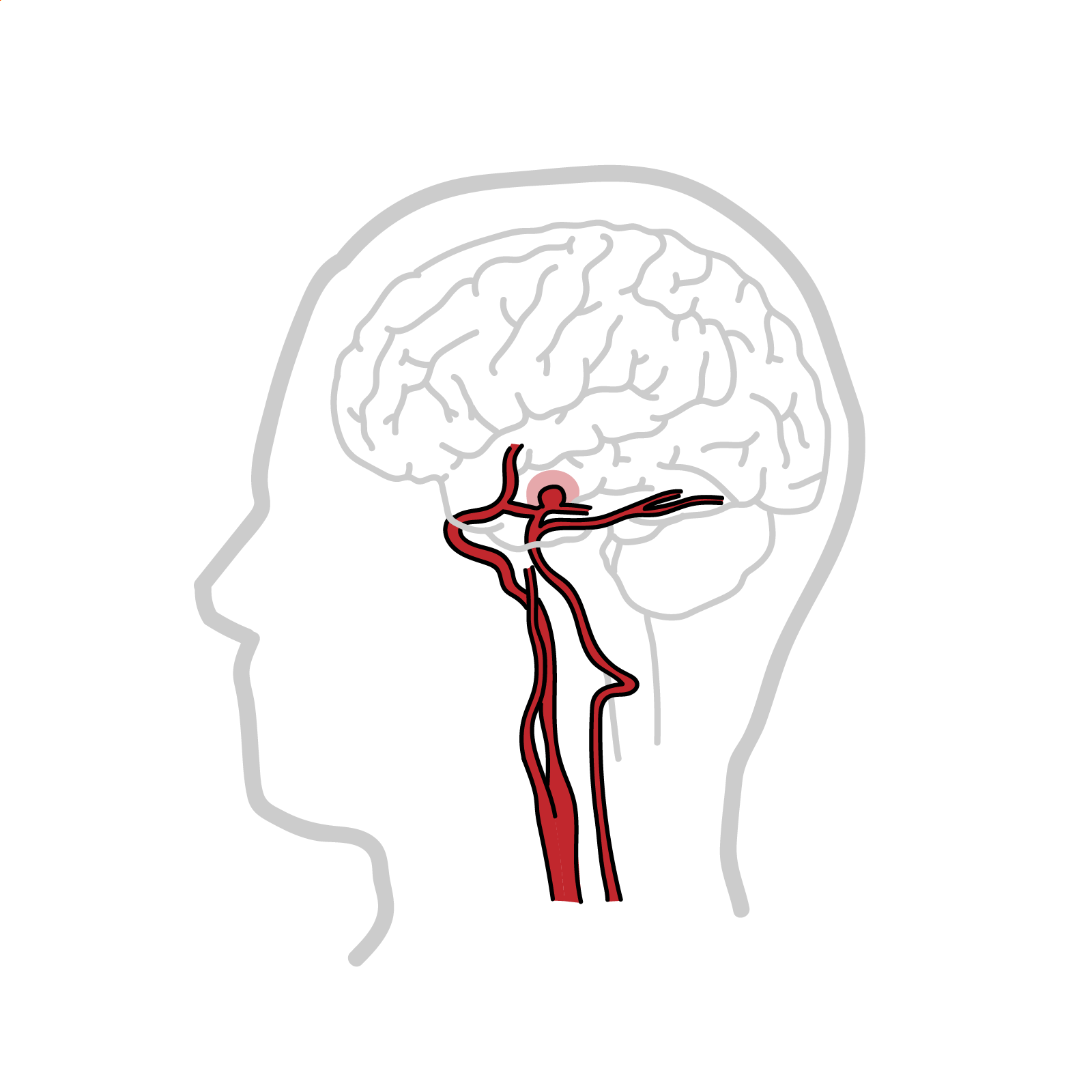
Cerebral (Intracranial) aneurysm is a focal dilation of an intracranial artery due to weakness of the arterial wall. It occurs in roughly 1-5% of adults and can lead to permanent disability. The most common type is a saccular “berry” aneurysm. Most common location is anterior communicating artery.
Cerebral aneurysms are typically asymptomatic until rupture, which leads to subarachnoid haemorrhage and presents with “thunderclap headache”.1
Definition
Aneurysm: an artery that has enlarged to greater than 1.5 times the expected diameter.
True aneurysm: the aneurysm is bound by all three layers of the vessel wall (tunica intima, media and adventitia). The wall may be attenuated.
False (pseudo) aneurysm: occurs when a blood vessel wall is injured and the blood is contained by the surrounding tissue, creating an apparent dilatation of the vessel.
Dissection: occurs when a tear in tunica intima of the vessel causes blood to pool between tunica intima and tunica media, forcing the layers apart.
Anatomy
Circle of Willis
Arterial ring located in the subarachnoid space and supplies majority of the brain
It connects the internal carotid arteries and the vertebral arteries
Vertebral arteries
- Arise from subclavian arteries
- Travel in vertebral foramina C1-C6
- Give the Posterior inferior cerebellar artery
- Combine to form basilar artery
Basilar artery
- Sits on the pons
- Branches:
- Pontine arteries
- Anterior inferior cerebellar artery
- Superior cerebellar artery
- Posterior cerebral artery
Internal carotids
- Arise from common carotid artery
- Travel in carotid sheath
- Branches:
- Ophthalmic artery
- Posterior communicating artery
- Middle cerebral artery
- Anterior cerebral artery
Communicating arteries
- Anterior communicating – connects the two Anterior Cerebral
- Most common location of Cerebral aneurysm
- Posterior communicating – connects Internal carotid to Posterior Cerebral
- Second most common location
Posterior communicating artery is closely related to Oculomotor nerve (CN III) – an aneurysm of this artery can cause compression of the nerve leading to: ptosis, diplopia, “down and out” eye position, and dilated pupil.
Large aneurysms of the Internal Carotid and Anterior Communicating can sometimes compress the Optic Nerve (CNII) leading to reduced visual acuity and visual field defects.
Subarachnoid haemorrhage is a common cause of hydrocephalus due to disruption of normal flow and absorption of CSF.
Classification
Morphology
- Saccular (berry): round pouch attached to the artery via a neck – most common
- Fusiform: symmetrical stretching of the artery
- Dissecting (pseudo-aneurysm): blood accumulates between the layers of the artery
- Mycotic: caused by an infection
Diameter
- Small: <7 mm
- Medium: between 7 and 12 mm
- Large: between 13 and 24 mm
- Very large: 25 mm or more1 2
Aetiology & Risk factors
Aetiology
Most intracranial aneurysms are acquired saccular (“berry”) aneurysms arising at arterial bifurcations in the Circle of Willis. They develop through:
- Haemodynamic shear stress at branch points
- Degeneration of the internal elastic lamina and tunica media
- Vascular inflammation, smooth-muscle-cell loss and extracellular-matrix breakdown
- Interaction between genetic susceptibility and acquired vascular injury
Risk factors for aneurysm formation
- Cigarette smoking—strongest modifiable association
- Uncontrolled hypertension
- Cocaine, amphetamine or other sympathomimetic use
- First-degree family history, especially ≥2 affected relatives
- Increasing age
- Previous intracranial aneurysm or aneurysmal subarachnoid haemorrhage
- Associated inherited/vascular disorders
- Autosomal dominant polycystic kidney disease
- Genetic connective tissue diseases: Vascular Ehlers–Danlos syndrome, Loeys–Dietz syndrome, Marfan syndrome
- Fibromuscular dysplasia
Features increasing rupture risk (not formation)
- Aneurysm growth on serial imaging
- Large aneurysm size
- Posterior circulation, anterior communicating artery or posterior communicating artery location
- Irregular shape or daughter sac
- Previous SAH from another aneurysm
- Ongoing smoking and uncontrolled hypertension
Clinical Manifestation
Unruptured cerebral aneurysm
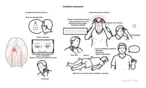
- Most are asymptomatic
- Potential symptoms – mainly due to compression:
- Visual acuity loss
- Oculomotor nerve palsy with pupil involvement
- Facial pain3
CNIII palsy with pupil involvement – think Posterior Communicating Aneurysm.
Ruptured cerebral aneurysm
- Sudden thunderclap headache
- “Worst headache of my life”
- Maximal intensity within seconds to minutes
- Nausea and vomiting
- Photophobia
- Neck pain
- Brief loss of consciousness, collapse or syncope
- Confusion or reduced consciousness
- Seizure
- Focal neurological symptoms, such as weakness, numbness or speech disturbance
- Possible preceding sentinel headache from a minor leak
- May occur during exertion, coughing, straining or sexual activity
Clinical examination
- Reduced GCS
- Meningism: Kernig’s sign and Brudzinski’s sign
- Cranial nerve palsy
- Especially a painful, pupil-involving third-nerve palsy
- Subhyaloid or retinal haemorrhage — Terson syndrome
- Hypertension and autonomic disturbance
- Raised ICP
- Papilloedema on fundoscopy
- Cushing’s triad: hypertension, bradycardia, irregular respiration
High-yield pattern: thunderclap headache with vomiting and meningism, with or without collapse, reduced consciousness, seizure or focal neurological deficits.
Leaking aneurysm – rare type of aneurysm that leaks small amount of blood (sentinel bleed) and causes headaches days to weeks before rupture.
bleed in the subarachnoid space irritates the two meningeal layers that surround it – pia and arachnoid mater (also known as leptomeninges), which leads to similar symptoms seen in meningitis.
Diagnosis
CT brain – first line
- Shows blood within the subarachnoid space
- Sensitivity decreases with time, meaning a delayed negative CT does not exclude haemorrhage
Lumbar puncture – required to exclude bleed for most patients with normal CT findings
- Elevated opening pressure
- Elevated RBC count that does not diminish between tubes
- Xanthochromia – represents haemoglobin degradation (pink or yellow tint)
Alternative imaging
- CT angiography
- MR angiography4
- Cerebral infection (e.g. meningitis)
- Cerebral venous thrombosis
- Intracerebral haemorrhage
- Ischaemic stroke
- Acute hypertensive crisis
- Cervical artery dissection4
Treatment
Unruptured aneurysm – either observe or operate; decision is made on case-by-case basis
- Conservative management
- Smoking cessation
- BP control
- Surveillance imaging
Ruptured aneurysm
- Surgery
- Microsurgical clipping
- Endovascular
- Coiling
- Flow diversion (stenting)
- Prevention of vasospasm
- Calcium channel blocker
- Blood pressure control
- Treat severe hypertension while avoiding hypotension
- Reduce rebleeding risk
- Ventricular shunt
- Used for acute hydrocephalus
Screening
- Considered in high risk groups
- Two or more first-degree relatives with intravenous aneurysm or aneurysmal SAH
- Autosomal dominant polycystic kidney disease with additional risk factors or family history
- Previous aneurysmal SAH with risk of additional aneurysms
- Either done with CTA or MRA1 5
Rebleeding of the aneurysm is associated with poor prognosis and usually occurs within the first 24 hours of the initial haemorrhage.
Complications
Rupture of cerebral aneurysm leads to subarachnoid haemorrhage
- Vasospasm
- Hyponatremia
- Recurrence
- Rebleeding
- Seizures
- Hydrocephalus1
References
- Eby B. Cerebral aneurysm. In: BMJ Best Practice [Internet]. 2023 [cited 2026 Jun 5]. Available from: BMJ Best Practice
- National Institute of Neurological Disorders and Stroke. Cerebral aneurysms [Internet]. Bethesda (MD): National Institute of Neurological Disorders and Stroke; [cited 2026 Jun 5]. Available from: https://www.ninds.nih.gov/health-information/disorders/cerebral-aneurysms
- Singer RJ, Ogilvy CS, Rordorf G. Unruptured intracranial aneurysms. In: Goddeau RP, editor. UpToDate [Internet]. [updated 2026 Feb 13; cited 2026 Jun 5]. Available from: UpToDate
- Singer RJ, Ogilvy CS, Rordorf G. Unruptured intracranial aneurysms. In: Goddeau RP, editor. UpToDate [Internet]. [updated 2026 Mar 6; cited 2026 Jun 5]. Available from: UpToDate
- Singer RJ, Ogilvy CS, Rordorf G. Treatment of cerebral aneurysms. In: Goddeau RP, editor. UpToDate [Internet]. [updated 2025 Jan 3; cited 2026 Jun 5]. Available from: UpToDate
Members only discussions coming soon…