

Acetabular Impingement Syndrome
Overview
Acetabular impingement syndrome is more commonly termed femoroacetabular impingement syndrome (FAIS). It is a motion-related clinical disorder in which symptomatic contact between the proximal femur and acetabular rim causes hip or groin pain, restricted movement and impaired function.
Cam and pincer morphology are common in people without hip pain. These findings describe bone shape; they constitute a syndrome only when they correspond with the patient’s symptoms and examination findings.
Definition
- Femoroacetabular impingement syndrome
- A motion-related clinical disorder involving symptoms, clinical signs and imaging findings associated with premature contact between the femur and acetabulum.
- Cam morphology
- Loss of the normal concavity or sphericity at the femoral head–neck junction, producing a bony prominence that may enter the acetabulum during hip movement.
- Pincer morphology
- Focal or global overcoverage of the femoral head by the acetabulum.
- Acetabular labrum
- A fibrocartilaginous ring attached to the acetabular rim that contributes to hip stability, load distribution and maintenance of the joint-fluid seal.
- FADIR test
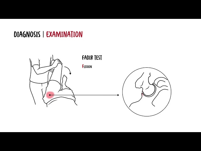
- Passive hip flexion, adduction and internal rotation used to reproduce symptoms associated with intra-articular hip pathology.
Anatomy & Physiology
The hip is a ball-and-socket synovial joint formed by the femoral head and acetabulum.
The acetabulum is formed by the ilium, ischium and pubis. Its articular surface is lined by hyaline cartilage, while the acetabular labrum attaches to its rim and deepens the socket.
The proximal femur consists of:
- The femoral head
- The femoral neck
- The greater and lesser trochanters
- The femoral head–neck junction
The normal femoral head is approximately spherical. The concave femoral head–neck junction provides clearance between the femur and acetabular rim during hip flexion and rotation.
The acetabular labrum:
- Deepens the acetabular socket
- Assists joint stability
- Distributes load around the acetabular rim
- Maintains a negative-pressure fluid seal
- Contributes to proprioception
Hip movement is controlled by coordinated activity of the gluteal muscles, deep external rotators, iliopsoas, adductors and trunk musculature. Pelvic position and femoral rotation influence how the femoral head–neck junction approaches the acetabular rim during movement.
Aetiology & Risk Factors
Aetiology
FAIS results from an interaction between hip morphology and movement. The morphology may be femoral, acetabular or mixed.
Cam morphology may develop during skeletal maturation, particularly in adolescents exposed to intensive loading through high-impact or multidirectional sport. The anterosuperior femoral head–neck junction becomes less concave, reducing clearance during hip flexion.
Pincer morphology results from acetabular overcoverage. This may be:
- Focal, such as with acetabular retroversion
- Global, with excessive coverage of the femoral head
Many individuals have combined cam and pincer features.
Morphology alone does not inevitably cause symptoms. The development of FAIS depends on the interaction between bone shape, activity demands, movement patterns, tissue sensitivity and associated labral or cartilage injury.
Risk Factors
Factors associated with FAIS include:
- Participation in high-impact sport during adolescence
- Sports involving repeated deep hip flexion, rotation or directional change
- Cam, pincer or mixed hip morphology
- Reduced hip internal rotation
- High sporting or occupational load
- Previous hip or groin symptoms
- Movement patterns that repeatedly reproduce impingement
- Associated acetabular labral or chondral pathology
- Participation in sports such as football, hockey, dancing and martial arts
Radiographic morphology is common among asymptomatic people, especially athletes.2 It should therefore be interpreted as a potential contributing factor rather than an isolated cause of pain.
Pathophysiology
During hip flexion and rotation, reduced clearance may cause the femoral head–neck junction to contact the acetabular rim.
In cam-type impingement, the non-spherical portion of the femoral head–neck junction enters the acetabulum. This can generate shear forces at the anterosuperior chondrolabral junction, producing:
- Separation between the labrum and adjacent cartilage
- Acetabular cartilage delamination
- Labral tearing
- Progressive chondral injury
In pincer-type impingement, acetabular overcoverage causes the femoral neck to contact the acetabular rim. This primarily compresses the labrum and may produce a lever effect with posteroinferior cartilage injury.
Repeated mechanical contact may lead to:
- Labral irritation or tearing
- Chondrolabral separation
- Articular cartilage damage
- Pain and movement restriction
- Progressive loss of hip function
FAIS is associated with the development of hip osteoarthritis, particularly when cam morphology and cartilage injury are present. However, progression is not inevitable, and the effect of treatment on the long-term risk of osteoarthritis remains uncertain.1
Why do deep squatting and prolonged low sitting commonly provoke symptoms?
Hip flexion brings the femoral head–neck junction closer to the acetabular rim. Adding adduction or internal rotation further reduces clearance, increasing contact at the anterosuperior joint in a susceptible hip.
Clinical Manifestations
Symptoms usually develop gradually and may be intermittent initially.
Common features include:
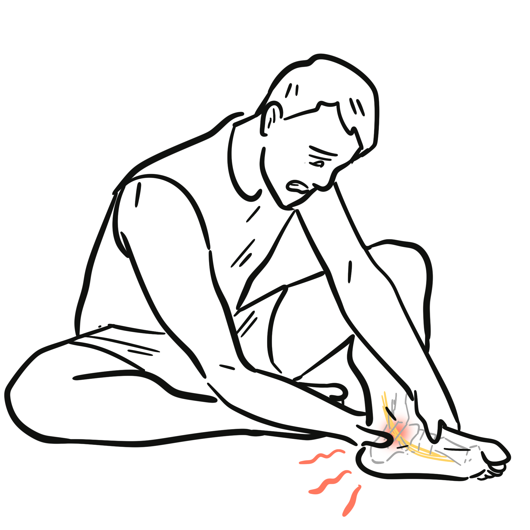
- Anterior hip or groin pain
- Pain described using a “C-sign”, with the hand cupped around the anterior and lateral hip
- Pain during deep squatting, lunging or pivoting
- Pain after prolonged sitting
- Pain when entering or leaving a car
- Symptoms during running, kicking or directional change
- Reduced sporting performance
- Hip stiffness or restricted movement
- Clicking, catching or locking
- Lateral hip, buttock, thigh or referred knee pain
Pain is often motion related or position related. Clicking and catching may suggest associated labral pathology but are not diagnostic by themselves.
Clinical Examination
- Reduced hip flexion
- Reduced internal rotation, particularly in flexion
- Pain with passive hip flexion
- Pain reproduced by the FADIR test
- Pain with the FABER test
- Reduced hip muscle strength
- Altered pelvic or lower-limb control
- Pain during a squat or single-leg squat
- Antalgic gait in more symptomatic disease
The FADIR test is commonly used to reproduce familiar symptoms. A negative test makes FAIS less likely, but a positive test is not specific and may occur with other intra-articular hip disorders.1
The lumbar spine, sacroiliac region, abdomen and surrounding hip structures should also be assessed when clinically indicated.
Diagnosis
FAIS is diagnosed when the patient has the recognised triad of symptoms, clinical signs and imaging findings.1 No single symptom, examination manoeuvre or radiographic measurement is independently diagnostic.
Cam or pincer morphology on imaging is not synonymous with FAIS.
The diagnosis requires concordant symptoms, clinical signs and imaging findings. Treating an asymptomatic radiographic shape is not supported.
Plain Radiography
Plain radiographs are commonly the first-line imaging investigation.
Radiographs assess:
- Cam morphology
- Acetabular overcoverage or retroversion
- Hip dysplasia
- Joint-space narrowing
- Osteophytes or other features of osteoarthritis
- Alternative bone or joint pathology
MRI and MR Arthrography
MRI provides further assessment of:
- Acetabular labral injury
- Articular cartilage damage
- Chondrolabral separation
- Bone marrow abnormality
- Synovitis
- Alternative soft-tissue pathology
CT
CT, including three-dimensional reconstruction, may be used when detailed assessment of femoral or acetabular morphology is required for operative planning. It is not routinely required for every patient.
Diagnostic Injection
An image-guided intra-articular local anaesthetic injection may help determine whether pain arises from the hip joint when the clinical picture is uncertain. A response supports an intra-articular pain source but does not independently establish FAIS.
Mechanical symptoms such as clicking or catching do not automatically indicate a labral tear requiring surgery. These symptoms must be interpreted alongside pain behaviour, examination findings and imaging.
Classification
FAIS is classified according to the predominant underlying morphology.
Cam Morphology
A bony prominence or reduced concavity at the femoral head–neck junction enters the acetabulum during movement. This pattern is particularly associated with shear injury at the anterosuperior chondrolabral junction.
Pincer Morphology
Focal or global acetabular overcoverage causes contact between the femoral neck and acetabular rim. Labral compression is more prominent.
Mixed Morphology
Cam and pincer features coexist. Mixed morphology is common in symptomatic clinical practice.
These categories describe morphology rather than symptom severity. Patients with similar radiographic appearances may have markedly different symptoms and functional limitations.
Treatment
Treatment aims to reduce pain, restore function and enable participation in desired activities. Management should be individualised according to symptom severity, functional goals, morphology, associated tissue injury and the presence of osteoarthritis.
Non-operative Management
Non-operative treatment is generally the initial approach and includes:
- Education about the condition
- Temporary modification of aggravating activities
- Avoidance of repeated painful end-range hip positions
- Progressive hip and lower-limb strengthening
- Trunk and lumbopelvic strengthening
- Movement-pattern retraining
- Improvement of balance and neuromuscular control
- Gradual return to sport or occupational activity
- Analgesia or NSAIDs when clinically appropriate
Manual therapy may be used as an adjunct when pain or soft-tissue and capsular restriction limits movement. It should not be used to force the hip repeatedly into painful impingement positions.
Injections
An intra-articular corticosteroid injection may provide temporary symptom relief in selected patients but does not correct the underlying morphology. Evidence supporting other injectables is variable, and injections should not replace an appropriate rehabilitation programme.
Surgical Management
Referral for specialist surgical assessment may be considered when:
- Symptoms remain functionally limiting
- The diagnosis is secure
- Imaging findings correspond with symptoms and clinical signs
- An adequate course of structured non-operative care has not produced acceptable improvement
- There is no advanced osteoarthritis that would make joint-preserving surgery inappropriate
Randomised trials in selected patients without established osteoarthritis have found that both structured physiotherapy and arthroscopic surgery can improve symptoms. Arthroscopy produced greater average short-term improvement in some trials, but not every patient benefited and surgery carries procedural risks.5,6 Shared decision-making should therefore consider symptom burden, prior rehabilitation, imaging, cartilage status, patient goals and the uncertainty around long-term disease modification.
Surgery treats symptomatic FAIS—not an incidental radiographic abnormality.
Appropriate surgical selection requires concordant symptoms, clinical signs and imaging findings, persistent functional limitation, and consideration of joint degeneration and previous non-operative treatment.
Complications & Prognosis
Complications
Potential consequences of untreated or persistent FAIS include:
- Recurrent hip or groin pain
- Reduced hip movement
- Impaired sporting or occupational function
- Acetabular labral injury
- Chondrolabral separation
- Progressive articular cartilage damage
- Hip osteoarthritis
Prognosis
Many patients improve with education, activity modification and a structured rehabilitation programme. Recovery may take several months and should be assessed through pain, function, strength, movement quality and participation rather than imaging appearance alone.
Patients undergoing appropriately selected arthroscopic treatment may experience meaningful improvement in pain and function.
Neither conservative treatment nor surgery has been conclusively shown to prevent future hip osteoarthritis.1 Early identification of cartilage damage and appropriate patient selection are important when considering joint-preserving surgery.
References
- Griffin DR, Dickenson EJ, O’Donnell J, Agricola R, Awan T, Beck M, et al. The Warwick Agreement on femoroacetabular impingement syndrome (FAI syndrome): an international consensus statement. Br J Sports Med. 2016;50(19):1169–1176.
- Frank JM, Harris JD, Erickson BJ, Slikker W 3rd, Bush-Joseph CA, Salata MJ, et al. Prevalence of femoroacetabular impingement imaging findings in asymptomatic volunteers: a systematic review. Arthroscopy. 2015;31(6):1199–1204.
- Enseki KR, Kohlrieser D, Lorenz DS, et al. Hip pain and movement dysfunction associated with nonarthritic hip joint pain: a revision. J Orthop Sports Phys Ther. 2023;53(7):CPG1–CPG70.
- Schmaranzer F, Kheterpal AB, Bredella MA. Best practices: hip femoroacetabular impingement. AJR Am J Roentgenol. 2021;216(3):585–598.
- Griffin DR, Dickenson EJ, Wall PDH, Achana F, Donovan JL, Griffin J, et al. Hip arthroscopy versus best conservative care for the treatment of femoroacetabular impingement syndrome: a multicentre randomised controlled trial. Lancet. 2018;391(10136):2225–2235.
- Palmer AJR, Ayyar Gupta V, Fernquest S, Rombach I, Dutton SJ, Mansour R, et al. Arthroscopic hip surgery compared with physiotherapy and activity modification for the treatment of symptomatic femoroacetabular impingement: multicentre randomised controlled trial. BMJ. 2019;364:l185.










Members only discussions coming soon…