

Lateral and Medial Elbow Tendinopathy (Epicondylitis)
Overview
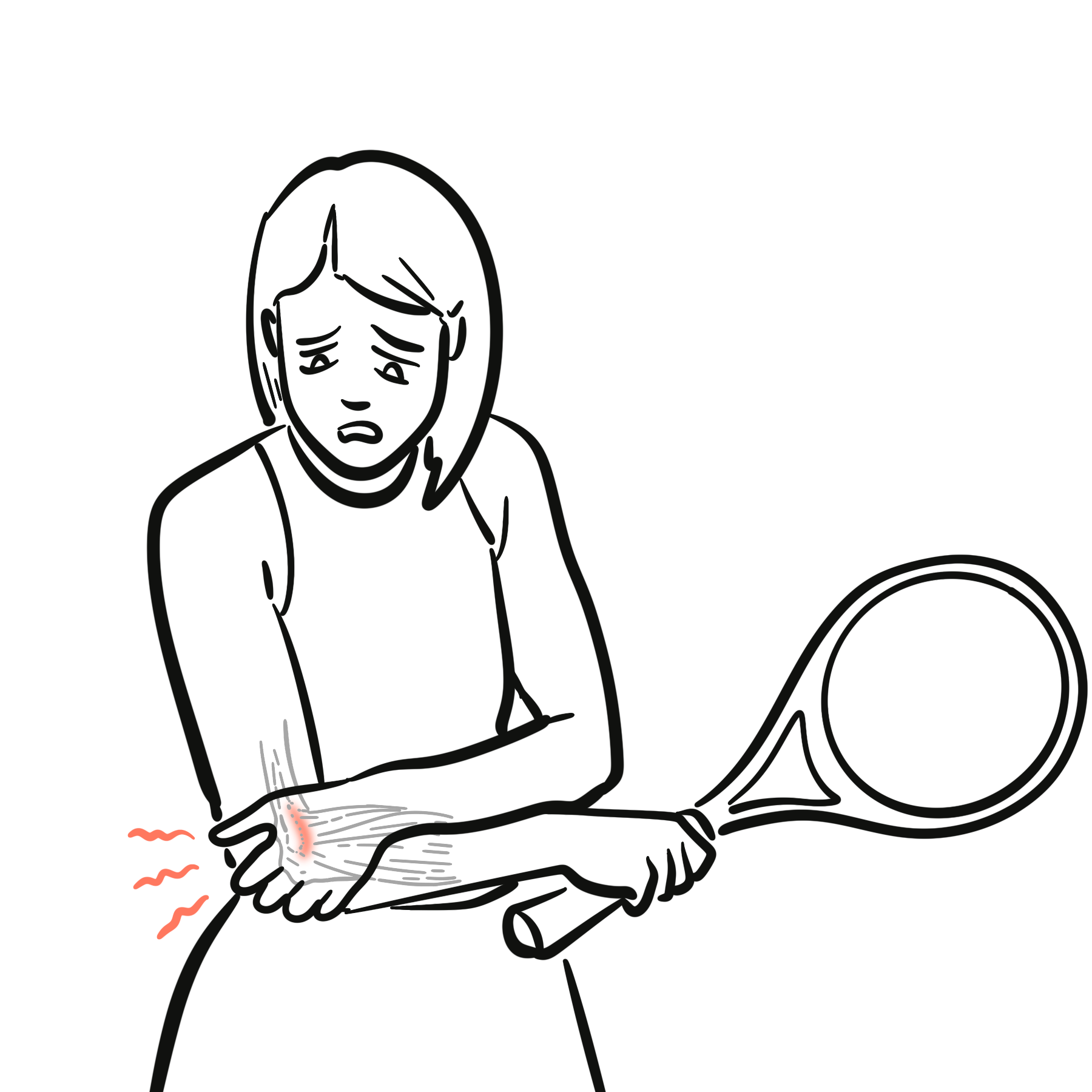
Medial and lateral elbow tendinopathies are common overuse injuries affecting the common flexor and extensor tendon origins at the elbow, respectively. Commonly known as Golfer’s Elbow (medial) and Tennis Elbow (lateral), these conditions are primarily degenerative (tendinosis) rather than inflammatory (tendinitis), resulting from repetitive microtrauma and failed healing responses.
Definition
Lateral Elbow Tendinopathy (Tennis Elbow / Lateral Epicondylitis): Degenerative tendinopathy involving the common extensor origin, primarily affecting the Extensor Carpi Radialis Brevis (ECRB).
Medial Elbow Tendinopathy (Golfer’s Elbow / Medial Epicondylitis): Degenerative tendinopathy involving the common flexor-pronator origin, primarily affecting the Pronator Teres and Flexor Carpi Radialis (FCR).
Angiofibroblastic Hyperplasia: The pathognomonic histological hallmark of tendinosis, characterized by hypervascularity, disorganized collagen bundles, hypercellularity, and an absence of classical acute inflammatory cells.
Anatomy & Comparison
| Feature | Lateral Elbow Tendinopathy (Tennis Elbow) | Medial Elbow Tendinopathy (Golfer’s Elbow) |
| Prevalence | High (~1–3% of adult population); 7–10× more common | Lower (~0.4% of population) |
| Anatomical Site | Common Extensor Origin (Lateral Epicondyle) | Common Flexor-Pronator Origin (Medial Epicondyle) |
| Primary Muscle Involved | Extensor Carpi Radialis Brevis (ECRB) | Pronator Teres & Flexor Carpi Radialis (FCR) |
| Primary Action Affected | Wrist extension & forearm supination | Wrist flexion & forearm pronation |
| Occupational/Sports Drivers | Racket sports, typing, manual labor (screwdriving) | Golf, baseball/throwing, heavy lifting, bowling |
| Associated Nerve Entrapment | Posterior Interosseous Nerve (PIN / Radial Tunnel) | Ulnar Nerve (Cubital Tunnel Syndrome in ~20%) |
Aetiology & Risk Factors
- Repetitive Microtrauma: Frequent eccentric loading, repetitive wrist flexion/extension, or pronation/supination under resistance.
- Age: Peak incidence between 30 and 50 years.
- Occupational Exposure: High physical workload, heavy lifting, high-vibration tool use, and repetitive manual tasks.
- Systemic Factors: Smoking, obesity, type 2 diabetes, and systemic metabolic disease.
Pathophysiology
- Tendinosis vs. Tendinitis: Historically termed “epicondylitis,” histological examination reveals an absence of acute inflammatory cells (e.g., polymorphonuclear leukocytes).
- Degenerative Cascade:
- Repetitive mechanical strain causes microtearing at the osteotendinous junction.
- Failed healing response leads to hypervascular invasion (neovascularization) and immature fibroblastic proliferation (angiofibroblastic hyperplasia).
- Normal parallel Type I collagen fibers are replaced by disorganized, non-functional Type III collagen, leading to altered biomechanics and localized mechanical pain.
Clinical Manifestations
- Lateral Tendinopathy: Insidious onset of burning or aching pain over the lateral epicondyle, radiating down the extensor surface of the forearm. Reduced grip strength due to pain, aggravated by lifting objects with an overhand grip (e.g., pouring a kettle).
- Medial Tendinopathy: Pain and tenderness localized to the medial epicondyle and proximal flexor muscle belly. Aggravated by resisted wrist flexion, pronation, or carrying heavy objects with an underhand grip.
Clinical Examination
- Lateral Elbow Tendinopathy (Tennis Elbow)
- Cozen’s Test: Patient actively extends and radially deviates the wrist against resistance with the elbow extended; positive if pain is elicited at the lateral epicondyle.
- Mill’s Test: Passive extension of the elbow while the wrist is fully flexed and the forearm pronated; positive if pain is reproduced at the lateral epicondyle.
- Maudsley’s Test: Resisted extension of the 3rd digit (middle finger) with the elbow extended; positive if pain occurs over the ECRB origin.
- Medial Elbow Tendinopathy (Golfer’s Elbow)
- Golfer’s Elbow Test: Passive extension of the elbow while the wrist and fingers are extended and forearm supinated; positive if pain is reproduced at the medial epicondyle.
- Resisted Wrist Flexion / Pronation: Resisted active flexion and pronation of the wrist with the elbow in full extension.
- Tinel’s Sign at Cubital Tunnel: Assesses for secondary ulnar neuropathy (present in ~20% of medial cases).
Diagnosis
Diagnosis is primarily clinical, supported by history and physical examination.
- Plain Radiography (X-ray): Usually normal; may show soft-tissue calcification near the epicondyle (~20%) or rule out radiocapitellar/ulnohumeral osteoarthritis.
- Diagnostic Ultrasound
- First-line imaging if clinical diagnosis is unclear
- Demonstrates hypoechoic tendon thickening, focal tears, structural collagen disruption, and hypervascularity on Doppler imaging.
- Magnetic Resonance Imaging (MRI)
Treatment
Conservative Management (First-Line for > 90% of Patients)
- Activity Modification & Load Management: Relative rest from aggravating mechanical triggers without total immobilization.
- Physiotherapy: Eccentric loading exercises for the flexor/extensor muscle groups; progressive isometric and isotonic strengthening.
- Orthotics / Bracing: Counterforce strap placed 2–3 cm distal to the epicondyle to redistribute mechanical load away from the tendon insertion.
- Analgesia: Short-term oral or topical NSAIDs for analgesia (though they do not alter disease progression).
Interventional Therapies (Second-Line)
- Platelet-Rich Plasma (PRP) Injections: Autologous growth factors promote tissue healing; evidence supports better long-term outcomes compared to steroids.
- Corticosteroid Injections: Provides rapid short-term pain relief (< 6 weeks), but carries a high risk of long-term recurrence, tendon degradation, and fat atrophy/depigmentation.
- Extracorporeal Shockwave Therapy (ESWT): Promotes neovascularization and tissue regeneration in chronic refractory cases.
Surgical Intervention (Refractory Cases > 6–12 Months)
- Procedure (Nirschl Debridement): Open or arthroscopic excision of the degenerate angiofibroblastic tissue, micro-tenotomy of the origin (ECRB for lateral, FCR/Pronator for medial), and re-anchoring of healthy tendon tissue.
Complications & Prognosis
- Recurrence / Chronicity: Up to 20% experience chronic persistent pain.
- Nerve Entrapment
- Radial tunnel syndrome / Posterior Interosseous Nerve (PIN) compression (lateral)
- Ulnar Neuropathy (medial).
- Iatrogenic EffectsDermal atrophy, hypopigmentation, or tendon rupture secondary to repeated corticosteroid injections.
- Self-limiting in up to 80–90% of cases within 12 to 18 months with non-operative, conservative care.
References
- Bhabra G, Wang A, Krämer W, Koerbin G, Murrell GA. Lateral Epicondylitis: Current Concepts. Bone Joint J. 2016;98-B(11):1439-1447. doi:10.1302/0301-620X.98B11.BJJ-2016-0223.R1
- Amin NH, Kumar NS, Schickendantz MS. Medial Epicondylitis: Evaluation and Management. J Am Acad Orthop Surg. 2015;23(6):348-355. doi:10.5435/JAAOS-D-14-00145
- Karabinov V, Georgiev GP. Lateral and Medial Epicondylitis: An Update on Diagnostics and Treatment. Curr Rev Musculoskelet Med. 2022;15(5):385-396. doi:10.1007/s12178-022-09777-6










Members only discussions coming soon…