

Sweet’s Syndrome (acute febrile neutrophilic dermatosis)
Overview
Sweet’s syndrome, also known as acute febrile neutrophilic dermatosis, is an autoinflammatory skin disorder belonging to the group of non-infectious neutrophilic dermatoses. It is characterized by the sudden onset of high fever, leukocytosis with absolute neutrophilia, tender erythematous cutaneous papules, plaques, or nodules, and a dense dermal infiltrate of mature neutrophils on histopathology.
First described by Dr. Robert Douglas Sweet in 1964, the condition is strongly associated with systemic inflammatory diseases, infections, pregnancy, medications, and underlying malignancies—most notably Acute Myeloid Leukemia (AML). Rapid clinical recognition is critical because lesions respond dramatically to systemic corticosteroids, while prompt identification can lead to the early diagnosis of an underlying occult hematological malignancy.
Sweet’s syndrome is a clinical indicator for underlying hematological disease. In a patient with new-onset Sweet’s syndrome and abnormal blood counts, immediately order a peripheral blood smear to rule out Acute Myeloid Leukemia (AML) or Myelodysplastic Syndrome (MDS).
Definition
Neutrophilic Dermatosis: An autoinflammatory skin disorder characterized by an accumulation of mature, non-infectious neutrophils in the dermis or subcutis without evidence of primary leukocytoclastic vasculitis.
“Juicy” Pseudovesicular Plaque: The characteristic appearance of Sweet’s syndrome lesions, where severe edema in the upper papillary dermis creates a raised, succulent, translucent surface that visually mimics a fluid-filled blister, but feels firm and solid on palpation.
Pathergy Sign: The development of new skin lesions or the aggravation of existing lesions at sites of minor cutaneous trauma (e.g., venipuncture sites, catheter lines, or biopsy scars).
Neuro-Sweet Syndrome: A severe, extracutaneous variant characterized by neutrophilic infiltration of the central nervous system, manifesting as encephalitis, aseptic meningitis, or brainstem lesions.
Why does Sweet’s Syndrome look “juicy” or pseudovesicular?
The intense, dense band of infiltrating neutrophils in the upper papillary dermis creates profound localized extracellular edema. This edema expands the papillary dermis upward against an intact epidermis, visually mimicking fluid-filled vesicles, even though the lesion is anatomically solid.
Sweet’s syndrome was originally named “The Febrile Neutrophilic Dermatosis” in 1964 after Dr. Sweet observed eight female patients presenting with identical combinations of fever, elevated white blood cell counts, and red skin plaques.
Classification
Sweet’s syndrome is classified into three distinct clinical subtypes based on its underlying trigger and setting:
Classic (Idiopathic) Sweet’s Syndrome (~70% of cases)
- Exhibits a strong female predominance (~4:1 ratio); most commonly presents between ages 30 and 60.
- Frequently preceded by an upper respiratory tract or gastrointestinal infection, or associated with Inflammatory Bowel Disease (IBD) or pregnancy.
Malignancy-Associated Sweet’s Syndrome (~20% of cases)
- Equal gender distribution; can precede, coincide with, or signal the relapse of a known cancer.
- Hematological Malignancies (~85% of tumor cases): Most characteristically associated with Acute Myeloid Leukemia (AML), Myelodysplastic Syndrome (MDS), and Multiple Myeloma.
- Solid Tumors (~15%): Carcinomas of the breast, genitourinary tract, and gastrointestinal tract.
Drug-Induced Sweet’s Syndrome (~10% of cases)
- Most common offending drug: Granulocyte Colony-Stimulating Factor (G-CSF).
- Other implicated drugs: All-Trans Retinoic Acid (ATRA), Trimethoprim-Sulfamethoxazole, Minocycline, BRAF inhibitors (e.g., Vemurafenib), and oral contraceptives.
Classic Sweet’s syndrome primarily affects females (4:1 ratio) following an infection or IBD flare, whereas Malignancy-Associated Sweet’s syndrome affects males and females equally.
Aetiology & Risk Factors
Underlying Triggers & Associations
- Infections: Upper respiratory infections (Streptococcus species), gastrointestinal infections (Yersinia enterocolitica, Salmonella, Campylobacter), and Mycobacterium tuberculosis.
- Systemic Inflammatory Diseases: Inflammatory Bowel Disease (Ulcerative Colitis > Crohn’s disease), Rheumatoid Arthritis, and Systemic Lupus Erythematosus (SLE).
- Malignancy: Acute Myeloid Leukemia (AML), Myelodysplastic Syndromes (MDS), Multiple Myeloma, and solid organ adenocarcinomas.
- Medications: Granulocyte Colony-Stimulating Factor (G-CSF), ATRA, antibiotics (sulfonamides, fluoroquinolones), and antiepileptics.
- Physiological State: Pregnancy (typically presenting during the first or second trimester).
Pathophysiology
[Systemic Trigger / Infection / Drug / Malignancy]
                         │
                         ▼
   [Hypersensitivity Reaction & Cytokine Cascade Release]
   (Elevated G-CSF, IL-1, IL-6, IL-8, TNF-alpha, IFN-gamma)
                         │
                         ▼
[Endothelial Cell Activation & Massive Neutrophil Recruitment]
                         │
                         ▼
[Dense Infiltration of Mature Neutrophils into Upper Papillary Dermis]
                         │
 ┌───────────────────────┴───────────────────────┐
 ▼                                               ▼
[Severe Papillary Dermal Edema]       [Neutrophil Degranulation & Tissue Damage]
(Produces "Juicy" Pseudovesicles)     (Erythematous Tender Plaques & Systemic Fever)
- Cytokine Hypersecretion: Driven by abnormal hypersensitivity, host immune cells release high levels of Granulocyte Colony-Stimulating Factor (G-CSF), Interleukin-1 (IL-1), Interleukin-6 (IL-6), Interleukin-8 (IL-8), and Tumor Necrosis Factor-alpha (TNF-alpha).
- Neutrophil Priming & Migration: Circulating neutrophils become hyper-reactive, adhere to activated vascular endothelium, and migrate into the upper dermis.
- Tissue Infiltration: Mature neutrophils accumulate in the papillary dermis without evidence of primary leukocytoclastic vasculitis, releasing matrix metalloproteinases (MMPs) and reactive oxygen species (ROS).
G-CSF is both a major pathogenic mediator and a direct drug trigger for Sweet’s syndrome because it directly stimulates neutrophil production, delays neutrophil apoptosis, and promotes tissue migration.
Clinical Manifestations
Systemic Constitutional Signs
- Fever – frequently preceding or accompanying the cutaneous eruption.
- Constitutional Symptoms
- Malaise, severe fatigue
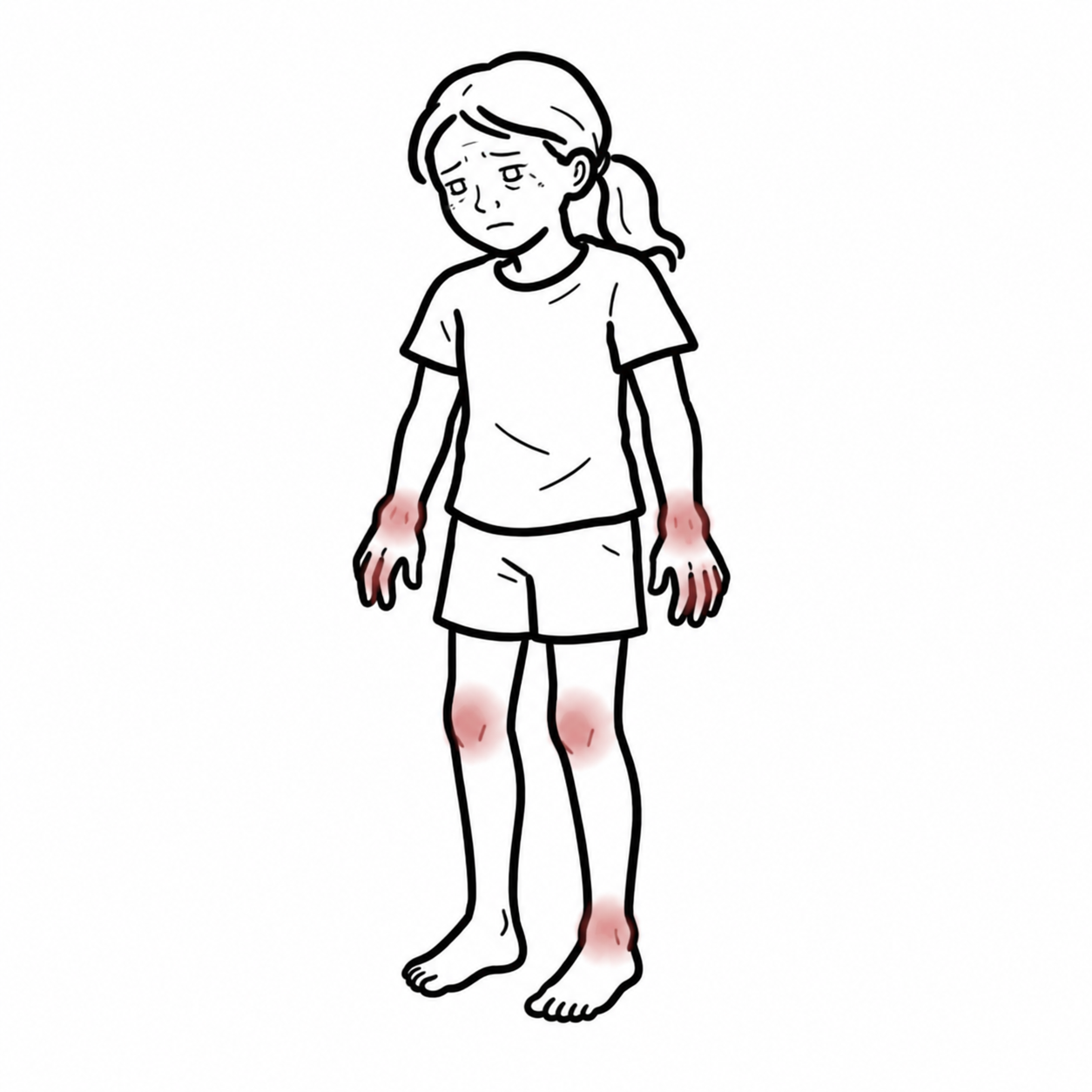
- Arthralgias
- Myalgias
- Headache.
- Ocular Manifestations
- Conjunctivitis
- Episcleritis
- Iridocyclitis
- Limbal ulceration in up to 30% of patients.
Cutaneous Lesions
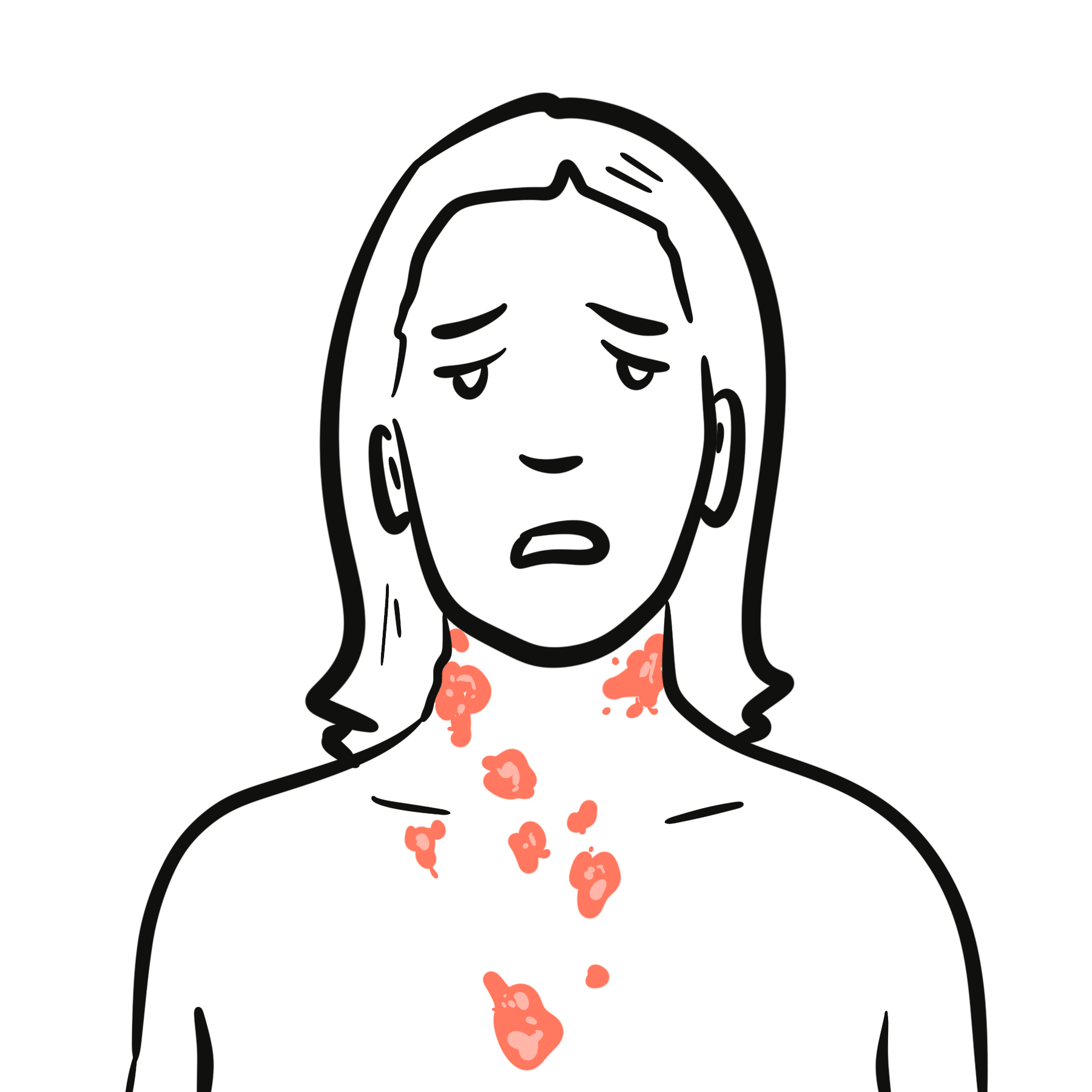
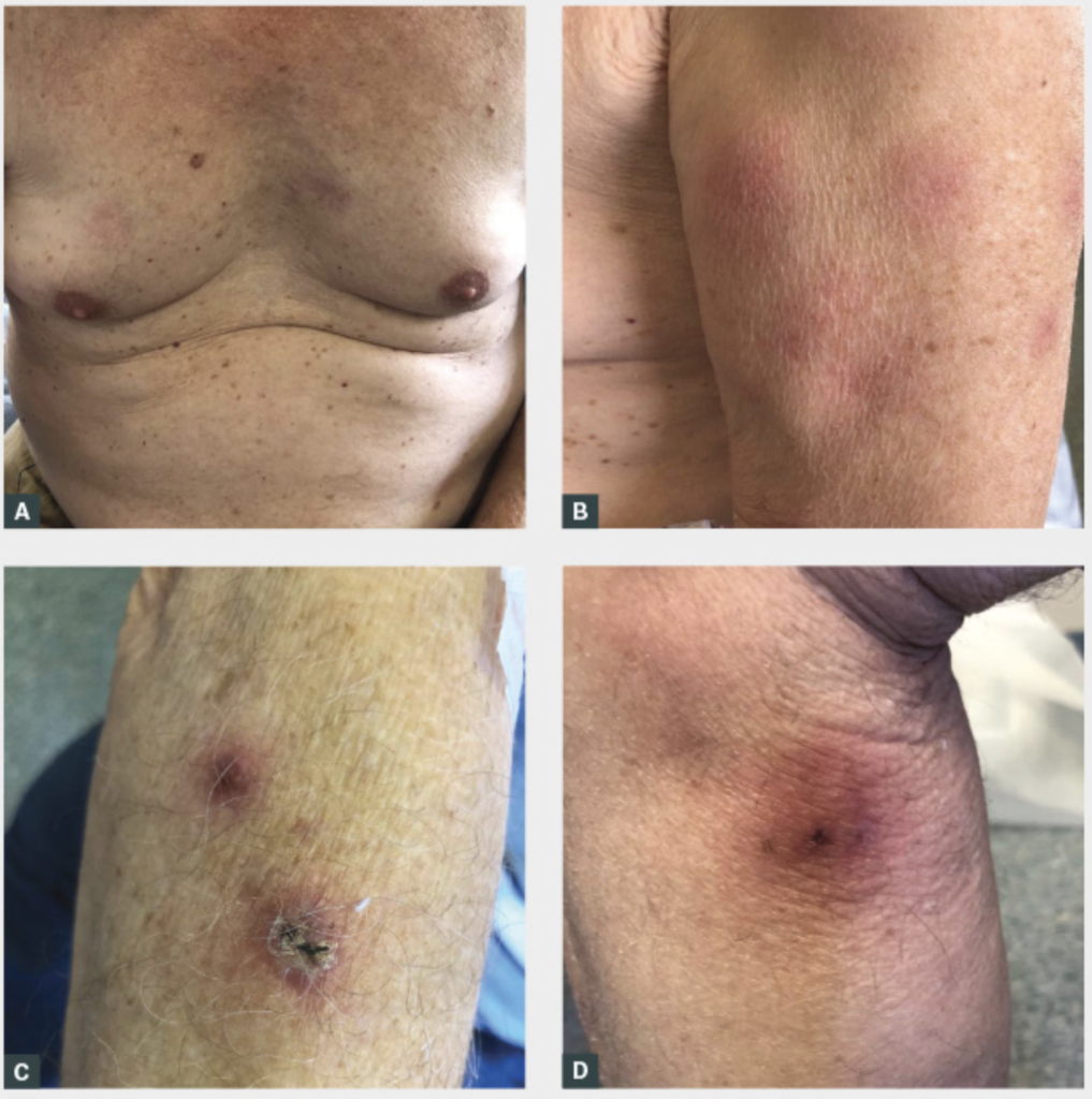
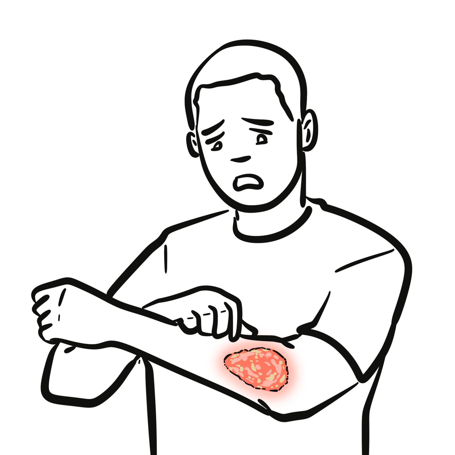
- Morphology: Tender, sharply demarcated, edematous, bright red to violaceous papules, nodules, or elevated plaques.
- “Pseudovesicular” Appearance: Marked papillary edema gives the lesions a transparent, fluid-filled appearance on inspection, though palpation reveals solid tissue.
- Distribution: Asymmetric presentation predominantly affecting the upper extremities, face, neck, and upper trunk.
- Pathergy: Lesions may erupt at sites of minor trauma, IV cannulation, or biopsy.
Extracutaneous Organ Involvement
- Musculoskeletal: Sterile mono- or oligoarthritis, myositis.
- Pulmonary: Neutrophilic alveolitis presenting with dyspnea, cough, and diffuse pulmonary infiltrates.
- Central Nervous System (Neuro-Sweet): Encephalitis, aseptic meningitis, or cranial nerve palsies.
Unlike Pyoderma Gangrenosum, which presents as deep, expanding necrotic ulcers with violaceous undermined edges, classic Sweet’s syndrome presents as raised, edematous plaques and nodules that rarely ulcerate.
Diagnosis
Diagnostic Criteria (Su and Liu / von den Driesch Criteria)
Confirmation requires meeting BOTH Major Criteria and at least TWO Minor Criteria:
| Category | Criteria Requirements |
| Major Criteria (Both Required) | 1. Abrupt onset of painful, erythematous, or violaceous plaques/nodules. 2. Histopathologic evidence of a dense neutrophilic infiltrate in the dermis without primary leukocytoclastic vasculitis. |
| Minor Criteria (At Least 2 Required) | 1. Fever (> 38.0°C). 2. Association with an underlying malignancy, inflammatory disease, pregnancy, or preceded by an infection/drug exposure. 3. Excellent clinical response to systemic corticosteroids or potassium iodide. 4. Abnormal laboratory values at presentation (3 of 4): ESR > 20 mm/hr, elevated CRP, Leukocytes > 8,000/μL, or Neutrophils > 70%. |
- Skin Biopsy (Gold Standard)
- Demonstrates a dense, band-like infiltration of mature neutrophils in the upper papillary dermis
- Marked papillary edema
- Fragmenting neutrophilic nuclei (leukocytoclasia)
- Preserved blood vessel walls (no fibrinoid necrosis or primary vasculitis).
- Full blood count & blood film
- Shows leukocytosis with marked neutrophilia
- Crucial: Inspect the smear for immature blast cells or cytopenias indicating underlying acute leukemia or myelodysplasia.
- Elevated CRP/ESR
| Feature | Sweet’s Syndrome | Pyoderma Gangrenosum | Erythema Nodosum | Cellulitis |
| Primary Lesion | “Juicy” red-purple plaques/nodules | Deep necrotic ulcer with violaceous border | Tender red nodules on anterior shins | Spreading poorly demarcated erythema |
| Primary Site | Face, neck, upper limbs | Lower extremities (shins), peristomal | Pretibial lower leg | Unilateral limb |
| Ulceration | Rare | Universal | Absent | Rare (unless bullous) |
| Histology | Upper dermal neutrophils | Full-thickness dermal/subcutis neutrophils | Septal panniculitis | Dermal edema + infectious neutrophils |
| Cultures | Sterile | Sterile | N/A | Often positive |
Histology in Sweet’s syndrome shows abundant leukocytoclasia (fragmented neutrophil nuclei), but lacks primary leukocytoclastic vasculitis (there is no fibrinoid necrosis of vessel walls).
A rapid clinical improvement following systemic corticosteroid administration—where fever and skin pain resolve within 24 to 48 hours—is so characteristic that it forms one of the official minor diagnostic criteria for Sweet’s syndrome.
Treatment
First-Line Systemic Therapy
- Systemic Corticosteroids (Gold Standard): Oral Prednisone (0.5–1.0 mg/kg/day). Produces dramatic clinical improvement—fever and pain resolve within 24 to 48 hours, with complete skin lesion clearance within 1 to 2 weeks. Taper slowly over 4 to 6 weeks to prevent relapse.
- Parenteral Corticosteroids: IV Methylprednisolone (up to 1000 mg/day for 3 days) for severe visceral organ involvement or Neuro-Sweet syndrome.
Localized First-Line Therapy
- Topical or Intralesional Corticosteroids: High-potency topical steroids (e.g., Clobetasol propionate 0.05%) or intralesional Triamcinolone acetonide injections for localized, solitary lesions.
Second-Line & Steroid-Sparing Agents
Indicated when systemic corticosteroids are contraindicated (e.g., severe uncontrolled diabetes, active systemic infection) or for frequent relapses:
- Colchicine: 0.5 mg orally 2 to 3 times daily (inhibits neutrophil chemotaxis).
- Dapsone: 100 to 150 mg orally daily (check G6PD level prior to initiation).
- Potassium Iodide: Oral solution (100 to 300 mg 3 times daily).
- Biologic Agents (Refractory Cases): Interleukin-1 receptor antagonists (Anakinra) or TNF-alpha inhibitors (Infliximab, Adalimumab).
Before initiating Dapsone as a steroid-sparing agent, always check the patient’s G6PD activity level to prevent severe hemolytic anemia.
Complications & Prognosis
- Disease Recurrence: Occurs in up to 30–50% of patients, particularly if steroid therapy is tapered too quickly or if an underlying malignancy remains untreated.
- Progressive Hematological Malignancy: Unmasking or transformation into Acute Myeloid Leukemia.
- Severe Extracutaneous Damage: Permanent neurological deficits from Neuro-Sweet syndrome, respiratory failure from neutrophilic alveolitis, or renal impairment.
- Uncomplicated / Classic Cases: Excellent prognosis; lesions heal completely without scarring (unlike Pyoderma Gangrenosum, which leaves behind cribriform scarring).
- Malignancy-Associated Cases: Prognosis is dictated entirely by the course and therapeutic response of the underlying hematological or solid organ malignancy.
Sweet’s syndrome heals without scarring because the inflammatory process is centered in the upper papillary dermis without tissue necrosis. Conversely, Pyoderma Gangrenosum causes extensive, deep liquefactive tissue necrosis extending into the subcutis, resulting in permanent cribriform (“cigarette paper”) scarring.
In rare instances, cutaneous lesions of Sweet’s syndrome can undergo localized bullous changes or ulceration. When this occurs in patients with myeloid leukemia, it is termed “bullous Sweet’s syndrome” and can clinically overlap with atypical Pyoderma Gangrenosum.
References
- Cohen PR. Sweet’s syndrome (acute febrile neutrophilic dermatosis). Orphanet J Rare Dis. 2007;2:34. doi:10.1186/1750-1172-2-34
- Nelson CA, Stephen S, Ashchyan HJ, James WD, Micheletti RG, Rosenbach M. Neutrophilic dermatoses: Disorders implied systemically, Part I. Sweet syndrome and pyoderma gangrenosum. J Am Acad Dermatol. 2018;79(6):987-1000. doi:10.1016/j.jaad.2017.11.063
- Heath MS, Ortega-Loayza AG. Insights Into the Pathogenesis of Sweet Syndrome. Front Immunol. 2019;10:414. doi:10.3389/fimmu.2019.00414














Members only discussions coming soon…