

De Quervain’s Tendinopathy
Overview
De Quervain’s tendinopathy is a painful stenosing disorder affecting the tendons within the first dorsal extensor compartment of the wrist. It involves the abductor pollicis longus (APL) and extensor pollicis brevis (EPB) tendons as they pass beneath the extensor retinaculum near the radial styloid.
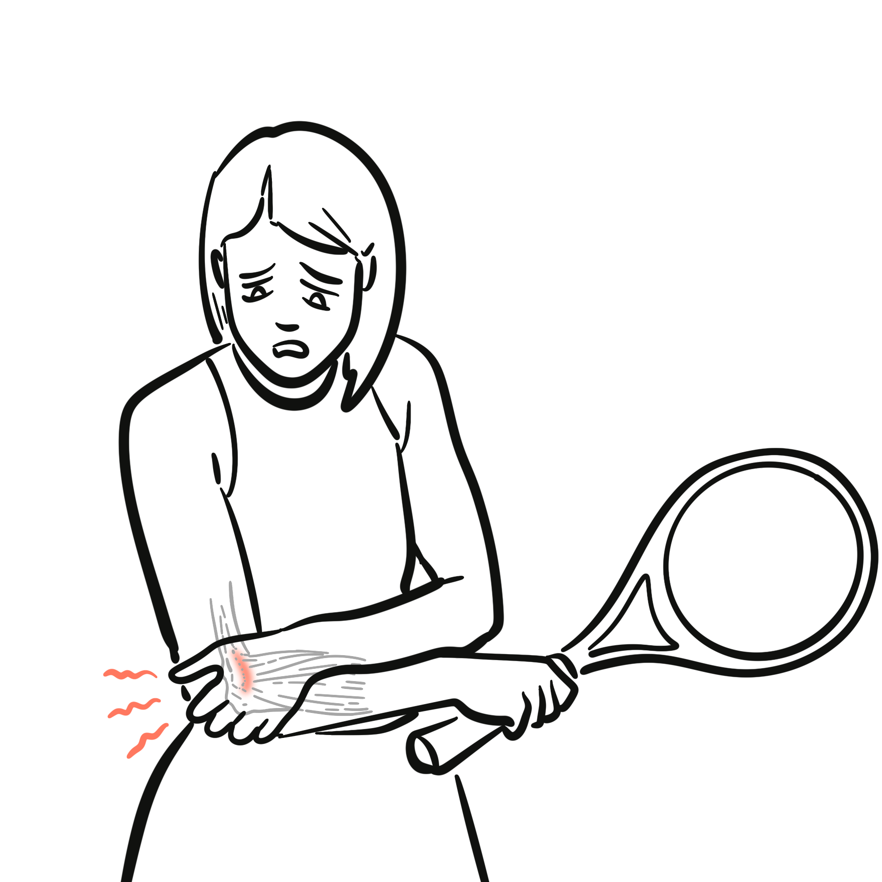
Patients typically present with radial-sided wrist pain that is aggravated by thumb movement, gripping, lifting or ulnar deviation of the wrist. Although the condition is often called de Quervain’s tenosynovitis, histological studies suggest that degenerative thickening of the tendon sheath is more prominent than acute inflammation.
Definition
- De Quervain’s tendinopathy
- A painful stenosing tendinopathy affecting the APL and EPB tendons within the first dorsal extensor compartment of the wrist.
- First dorsal extensor compartment
- A fibro-osseous tunnel located over the radial styloid that normally contains the APL and EPB tendons.
- Abductor pollicis longus (APL)
- A tendon that abducts and assists with extension of the thumb at the carpometacarpal joint.
- Extensor pollicis brevis (EPB)
- A tendon that extends the thumb primarily at the metacarpophalangeal joint.
- Finkelstein test
- A provocative examination manoeuvre in which passive ulnar deviation of the wrist tensions the first dorsal compartment tendons and reproduces characteristic radial styloid pain.
Anatomy & Physiology
The APL and EPB originate from the posterior surfaces of the radius, ulna and interosseous membrane. Their tendons travel distally around the lateral aspect of the radius before passing through the first dorsal extensor compartment at the radial styloid.
The compartment is bounded by the radius and the overlying extensor retinaculum. Its confined structure maintains the tendons close to the wrist while allowing them to glide during thumb and wrist movement.
The APL abducts the thumb away from the palm and assists with extension at the carpometacarpal joint. The EPB primarily extends the thumb at the metacarpophalangeal joint. Consequently, gripping, pinching and lifting commonly require repeated movement of both tendons through the compartment.
Anatomical variation is common. The APL may have multiple tendon slips, while a fibrous septum may separate the EPB into an independent subcompartment. These variations are clinically important because an unrecognised subcompartment may contribute to incomplete corticosteroid delivery or incomplete surgical decompression.
Aetiology & Risk Factors
Aetiology
The precise cause is not always identifiable. Repeated loading and friction involving the APL and EPB tendons are thought to promote thickening of the tendon sheath and narrowing of the available fibro-osseous tunnel.
Symptoms may follow:
- A recent increase in repetitive thumb or wrist activity
- Repeated gripping, pinching, lifting or wringing movements
- Direct trauma to the radial aspect of the wrist
- Repeated lifting of an infant with the wrist deviated and the thumb abducted
- Less commonly, inflammatory disease affecting the tendon sheath
Repetitive activity may provoke symptoms, but the condition should not automatically be attributed to a single occupational or recreational activity without considering other factors.
Risk Factors
Recognised associations include:
- Female sex
- Pregnancy and the postpartum period
- Middle age, particularly between 30 and 50 years
- Repetitive or forceful thumb and wrist use
- Frequent infant lifting or childcare activities
- Anatomical septation of the first dorsal compartment
- Multiple APL tendon slips
- Diabetes mellitus and other metabolic disorders
- Inflammatory arthropathies
Pregnancy-related fluid retention, hormonal influences and the repetitive mechanical demands of caring for an infant may all contribute to symptoms during pregnancy or after childbirth.
Pathophysiology
Repetitive mechanical loading produces microtrauma and remodelling within the tendon sheath. The extensor retinaculum and sheath become thickened, reducing the space available for the APL and EPB tendons.
Histologically, the condition is characterised predominantly by myxoid degeneration, accumulation of mucopolysaccharides and thickening of the tendon sheath rather than marked acute inflammatory cell infiltration.1 The term tendinopathy or stenosing tenosynovitis therefore describes the underlying process more accurately than a purely inflammatory “tendinitis”.
The narrowed compartment restricts normal tendon gliding. Thumb movement and wrist deviation increase friction and mechanical compression, producing pain, tenderness and occasionally crepitus or triggering.
A patient receives a corticosteroid injection but remains symptomatic despite apparently correct needle placement.
A fibrous septum may divide the first dorsal compartment into separate APL and EPB subcompartments. If medication enters only one subcompartment, the other may remain untreated. Ultrasound can identify this variation and assist accurate injection or surgical planning.
Clinical Manifestations
Patients commonly report:
- Gradual or occasionally acute onset of pain over the radial styloid
- Pain radiating into the thumb or proximally along the radial forearm
- Pain aggravated by thumb abduction or extension
- Pain during gripping, pinching, lifting or twisting activities
- Difficulty opening jars, wringing cloths or lifting an infant
- Reduced grip or pinch strength because of pain
- Local swelling or a sensation of tendon catching
- Symptoms affecting one or both wrists, particularly during the postpartum period
Clinical Examination
Typical findings include:
- Focal tenderness over the first dorsal compartment at the radial styloid
- Localised swelling or thickening
- Pain with resisted thumb abduction
- Pain with resisted thumb extension
- Pain during ulnar deviation of the wrist
- Reduced thumb or wrist movement secondary to pain
- Occasionally palpable crepitus during tendon movement
- Reproduction of characteristic pain with provocative testing
Neurological examination is usually normal. Sensory disturbance should prompt consideration of superficial radial nerve irritation or another neurological diagnosis.
Diagnosis
The diagnosis is primarily clinical and is based on the characteristic history, focal radial styloid tenderness and reproduction of symptoms during examination.
Provocative Tests
Finkelstein test
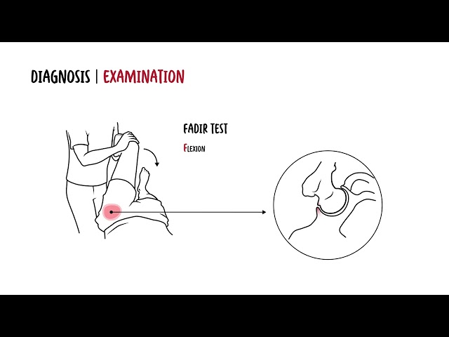
The examiner stabilises the patient’s forearm, holds the thumb and gently deviates the wrist towards the ulna. Reproduction of the patient’s characteristic pain over the first dorsal compartment supports the diagnosis.
Eichhoff test
The patient places the thumb within a closed fist and then actively deviates the wrist towards the ulna. This manoeuvre is frequently called the Finkelstein test, but it is a different and generally more provocative test. It may produce discomfort in asymptomatic individuals and should not be interpreted in isolation.
Wrist hyperflexion and abduction of the thumb test
The wrist is placed in flexion while the patient actively abducts the thumb against resistance. Pain over the first dorsal compartment constitutes a positive result. One prospective study found the WHAT test to be highly sensitive, although its specificity remained limited.3
The Eichhoff and Finkelstein manoeuvres are commonly confused.
In the Eichhoff test, the patient encloses the thumb within the fist and actively ulnar-deviates the wrist. In the Finkelstein test, the examiner holds the thumb and produces controlled passive ulnar deviation. The more forceful Eichhoff manoeuvre may cause false-positive discomfort.
A provocative test is supportive only when it reproduces the patient’s characteristic pain at the radial styloid.
Pain located several centimetres proximal to the wrist suggests intersection syndrome rather than de Quervain’s tendinopathy.
Imaging
Imaging is not routinely required when the clinical presentation is typical.
Ultrasound
Ultrasound may demonstrate:
- Thickening of the APL or EPB tendons
- Thickening of the tendon sheath or extensor retinaculum
- Fluid surrounding the tendons
- Hypervascularity on Doppler imaging
- Restricted tendon movement
- A septum dividing the first dorsal compartment
Ultrasound is particularly useful when the diagnosis is uncertain, when anatomical variation is suspected or when image-guided injection is planned.
Plain radiography
Radiographs are usually normal in de Quervain’s tendinopathy. They may be appropriate when fracture, thumb carpometacarpal osteoarthritis or another bony disorder is suspected.
Magnetic resonance imaging
MRI is rarely necessary. It may demonstrate tendon and retinacular thickening but is generally reserved for atypical or diagnostically uncertain presentations.
Treatment
Treatment aims to reduce pain, restore tendon gliding and allow a gradual return to normal activity.
Conservative Management
Initial measures may include:
- Temporarily modifying activities that repeatedly provoke symptoms
- Avoiding forceful thumb abduction, repetitive pinching and sustained wrist deviation
- Using ergonomic adaptations for work, sport or childcare
- Applying ice for short-term symptom relief
- Simple analgesia where clinically appropriate
- A short course of a non-steroidal anti-inflammatory drug when not contraindicated
- A thumb-spica orthosis that supports the wrist and thumb
- Hand therapy focusing on education, activity modification and progressive rehabilitation
Prolonged complete immobilisation is usually unnecessary. Once pain is controlled, gradual range-of-motion and strengthening exercises may be introduced according to symptoms.2,5
Corticosteroid Injection
A corticosteroid injection into the first dorsal compartment is an effective non-operative treatment. Evidence indicates that combining injection with thumb-spica immobilisation for approximately three to four weeks provides better pain and functional outcomes than either strategy alone.4
Surgery
Surgical decompression may be considered when symptoms remain functionally limiting despite appropriate non-operative treatment.
The procedure involves releasing the first dorsal compartment to restore unrestricted gliding of the APL and EPB tendons. The surgeon should identify and release any separate EPB subcompartment while protecting branches of the superficial radial nerve.
Complications & Prognosis
Complications
Untreated or persistent disease may result in:
- Chronic radial-sided wrist pain
- Reduced grip and pinch strength
- Restriction of thumb or wrist movement
- Difficulty performing work, sport or childcare activities
- Recurrent symptoms
Prognosis
The prognosis is generally favourable. Many patients improve with activity modification, a thumb-spica orthosis and corticosteroid injection. Surgery is usually effective when conservative treatment has failed, provided all relevant tendon subcompartments are adequately released.
Pregnancy-associated and postpartum symptoms may improve as hormonal and mechanical factors resolve. Persistent symptoms should nevertheless be assessed and treated according to their severity and functional effect.
Recurrence may occur when provocative loading resumes too quickly, an anatomical subcompartment remains untreated or another source of radial-sided wrist pain has been overlooked.
References
- Clarke MT, Lyall HA, Grant JW, Matthewson MH. The histopathology of de Quervain’s disease. J Hand Surg Br. 1998;23(6):732–734. doi:10.1016/S0266-7681(98)80085-5
- Huisstede BMA, Coert JH, Fridén J, Hoogvliet P; European HANDGUIDE Group. Consensus on a multidisciplinary treatment guideline for de Quervain disease: results from the European HANDGUIDE study. Phys Ther. 2014;94(8):1095–1110. doi:10.2522/ptj.20130069
- Goubau JF, Goubau L, Van Tongel A, Van Hoonacker P, Kerckhove D, Berghs B. The wrist hyperflexion and abduction of the thumb test: a more specific and sensitive test to diagnose de Quervain tenosynovitis than the Eichhoff’s test. J Hand Surg Eur Vol. 2014;39(3):286–292. doi:10.1177/1753193412475043
- Challoumas D, Ramasubbu R, Rooney E, et al. Management of de Quervain tenosynovitis: a systematic review and network meta-analysis. JAMA Netw Open. 2023;6(10):e2337001. doi:10.1001/jamanetworkopen.2023.37001
- Goel R, Abzug JM. De Quervain’s tenosynovitis: a review of the rehabilitative options. Hand (N Y). 2015;10(1):1–5. doi:10.1007/s11552-014-9649-3










Members only discussions coming soon…