

Bunion
Overview
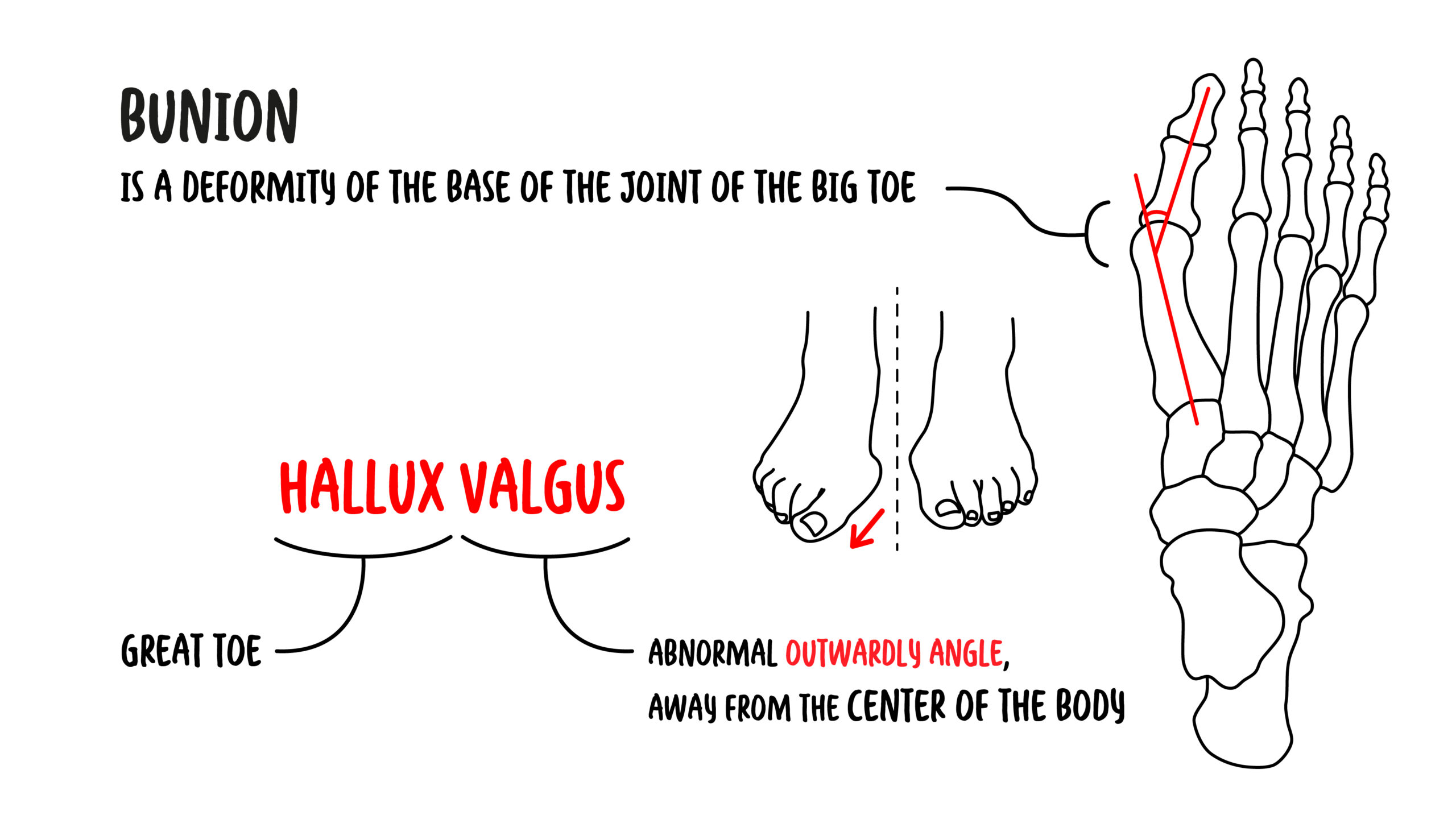
A bunion is the visible medial prominence that develops at the first metatarsophalangeal joint, most commonly as part of hallux valgus. The great toe drifts laterally towards the lesser toes while the first metatarsal drifts medially, widening the forefoot and making the metatarsal head more prominent.
Bunions are common and may be painless. When symptomatic, they can cause pain over the medial prominence, difficulty with footwear, inflammation, altered gait, lesser-toe deformity and transfer metatarsalgia. Severity on radiographs does not always match symptom severity.
Management is guided by symptoms and functional limitation rather than appearance alone. Footwear modification, padding, analgesia and selected orthoses may reduce symptoms, but they do not reliably correct the established deformity. Surgery is considered when persistent pain or functional impairment remains despite appropriate nonoperative care.
Definition
- Bunion
- A prominent bump at the medial aspect of the first metatarsophalangeal joint, usually produced by hallux valgus rather than by new bone growth alone.
- Hallux valgus
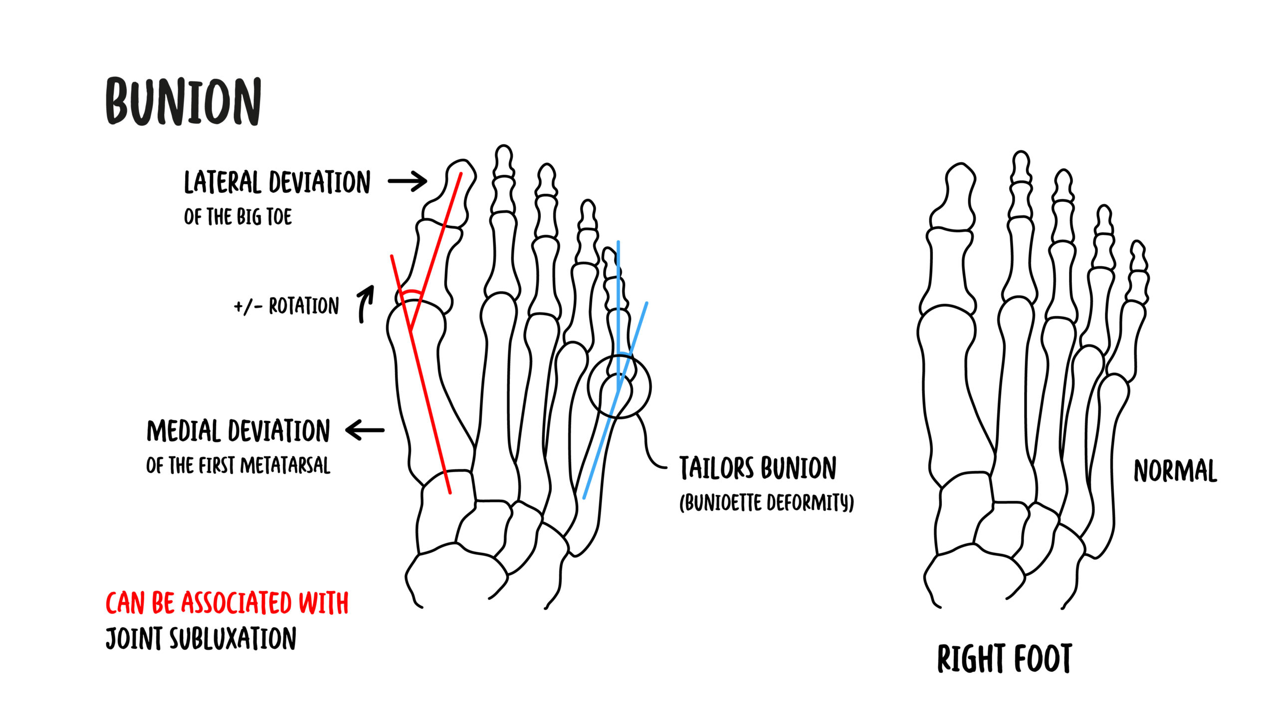
- Lateral deviation of the great toe at the first metatarsophalangeal joint, usually accompanied by medial deviation of the first metatarsal.
- Hallux valgus angle
- The angle between the longitudinal axes of the first metatarsal and proximal phalanx on a weight-bearing radiograph.
- Intermetatarsal angle
- The angle between the longitudinal axes of the first and second metatarsals.
- Bunionette
- A similar prominence at the lateral aspect of the fifth metatarsophalangeal joint.
- Transfer metatarsalgia
- Pain beneath the lesser metatarsal heads caused by redistribution of forefoot load away from the first ray.
Bunion and Hallux Valgus
The terms are often used interchangeably, but they are not identical. Hallux valgus describes the three-dimensional deformity of the great toe and first ray. A bunion is the clinically visible prominence over the medial first metatarsal head that usually accompanies this deformity.
Anatomy & Physiology
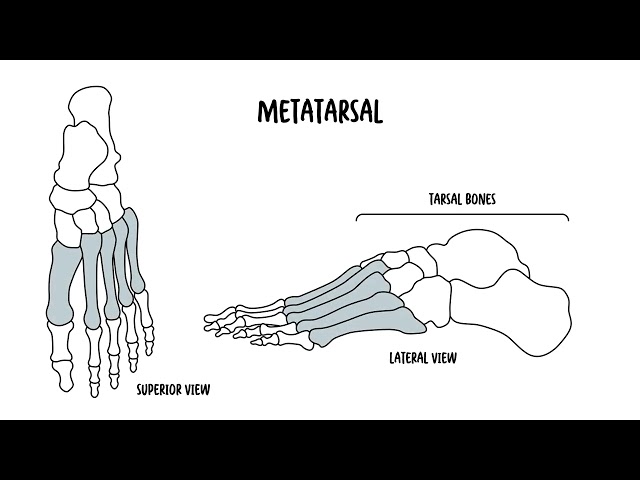
The first metatarsophalangeal joint is formed by the first metatarsal head and the base of the proximal phalanx. It is stabilised by the joint capsule, collateral ligaments, plantar plate, sesamoid apparatus and the tendons acting on the great toe.
Two sesamoid bones lie within the flexor hallucis brevis tendons beneath the first metatarsal head. They protect the flexor hallucis longus tendon, increase the mechanical advantage of the flexor muscles and help the first ray bear load during push-off.
The abductor hallucis, adductor hallucis, flexor hallucis brevis, flexor hallucis longus and extensor hallucis longus help control the great toe. Normal alignment allows the first ray to accept load and provides a stable lever during late stance and propulsion.
In hallux valgus, the first metatarsal shifts medially and the proximal phalanx deviates laterally. The metatarsal head moves medially relative to the sesamoid complex, while the tendons and soft tissues increasingly act as deforming forces.
Aetiology & Risk Factors
Bunion formation is multifactorial. Genetic predisposition and foot structure are important, while footwear and loading patterns may influence symptoms and progression.
- Family history and inherited foot morphology
- Female sex
- Increasing age
- Pes planus and excessive pronation
- First-ray hypermobility
- A long first metatarsal or other forefoot structural variation
- Generalised ligamentous laxity
- Inflammatory arthritis, particularly rheumatoid arthritis
- Neuromuscular disorders
- Previous trauma or surgery affecting the first ray
- Narrow toe-box or high-heeled footwear, which may aggravate pressure and symptoms
A bunion is not caused by tight shoes alone.
Footwear can increase pressure over the medial prominence and may contribute to progression in a susceptible foot, but intrinsic anatomy, heredity, ligamentous laxity and biomechanical factors are often central.
Pathophysiology
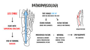
Hallux valgus begins with loss of normal alignment and stability of the first ray. The first metatarsal progressively deviates medially while the proximal phalanx moves laterally. The medial capsule stretches, the lateral soft tissues tighten and the metatarsal head becomes prominent.
As the deformity progresses, the sesamoids appear laterally displaced relative to the first metatarsal head. The abductor hallucis migrates plantarward, and the long flexor and extensor tendons may act like bowstrings that reinforce lateral deviation of the toe.
Repeated friction between footwear and the medial prominence can cause erythema, callus formation, bursitis and pain. Abnormal joint loading may damage articular cartilage and lead to stiffness or osteoarthritis.
Reduced loading through the first ray transfers pressure to the lesser metatarsal heads. This may produce plantar callosities and metatarsalgia, while crowding can contribute to hammer toes, crossover toes or lesser metatarsophalangeal instability.
Why can a bunion cause pain beneath the lesser metatarsal heads?
An inefficient or painful first ray accepts less load during stance and push-off. Load is transferred laterally to the lesser metatarsal heads, producing plantar pressure, callus and transfer metatarsalgia.
Radiographic Severity Does Not Equal Symptom Severity
A marked deformity may cause few symptoms, whereas a smaller deformity may be very painful in footwear. Treatment decisions should integrate pain, function, examination findings and patient goals.
Clinical Manifestations
Symptoms
- Pain or tenderness over the medial first metatarsal head
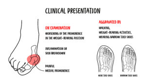
- Pain worsened by narrow footwear, prolonged standing or walking
- Difficulty finding comfortable shoes
- Intermittent redness, swelling or bursitis over the prominence
- Great-toe stiffness or pain during push-off
- Plantar forefoot pain due to transfer metatarsalgia
- Symptoms from associated calluses, hammer toes or overlapping toes
Bunion is commonly associated with tight footwear, female sex, family history, pes planus, and ligamentous laxity.
Clinical Examination
- Medial prominence at the first metatarsophalangeal joint
- Lateral deviation and possible pronation of the great toe
- Widened forefoot and crowding of the lesser toes
- Erythema, callus or inflamed bursa over the prominence
- Reduced or painful first metatarsophalangeal movement
- Plantar callosities beneath the lesser metatarsal heads
- Pes planus, hindfoot valgus or first-ray hypermobility
- Associated hammer toe, crossover toe or lesser metatarsophalangeal instability
The examination should assess whether the deformity is flexible or rigid, whether the first metatarsophalangeal joint is congruent, and whether pain arises from the medial prominence, the joint itself, the sesamoids or the lesser metatarsals. Neurovascular status, gait and footwear should also be assessed.
A painless bunion does not require treatment.
Appearance alone is not an indication for surgery. Management is appropriate when symptoms, footwear difficulty or functional impairment justify intervention.
Diagnosis
Diagnosis is usually clinical.
Imaging
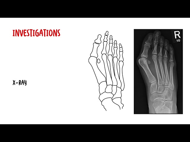
Weight-bearing anteroposterior, lateral and oblique foot radiographs are used when symptoms are significant, surgery is being considered or another diagnosis is suspected. Weight-bearing views demonstrate functional alignment and permit measurement of the hallux valgus and intermetatarsal angles.
Hallux valgus or hallux rigidus?
Hallux valgus is primarily an alignment deformity with lateral deviation of the great toe and a medial prominence. Hallux rigidus is degenerative arthritis characterised by pain and restricted dorsiflexion, often with a dorsal osteophyte. Both may coexist.
Bunions are a clinical diagnosis. Weight-bearing foot X-rays are used to assess deformity severity and guide surgical planning.
Treatment
Nonoperative Management
Initial treatment is appropriate for most symptomatic bunions and aims to reduce pressure and improve comfort. It does not reliably reverse the structural deformity.
- Wide, deep shoes with a low heel and adequate toe-box space
- Avoidance of footwear that compresses the forefoot
- Protective pads or sleeves over the medial prominence
- Activity modification during painful flares
- Simple analgesia or nonsteroidal anti-inflammatory medicines when appropriate
- Ice for short-term relief of inflammation
- Orthoses when pes planus, pronation or transfer loading contributes to symptoms
- Toe spacers or splints for comfort in selected patients
- Management of calluses and associated lesser-toe problems
Evidence for splints, toe spacers and orthoses is variable. They may improve comfort but should not be presented as a permanent correction of an established deformity.
Limits of Conservative Treatment
Nonoperative care can reduce pressure and pain, but it does not realign the first metatarsophalangeal joint. Its success is measured by symptom control and function, not by radiographic correction.
Surgical Management
Surgical referral is considered when persistent pain, recurrent inflammation, footwear difficulty or functional limitation continues despite adequate nonoperative care. Cosmetic concern alone is not an indication.
Procedures may include:
- Distal metatarsal osteotomy: commonly used for selected mild-to-moderate deformity
- Diaphyseal or proximal osteotomy: used when greater correction of the intermetatarsal angle is required
- First tarsometatarsal fusion (Lapidus procedure): considered when first-ray instability or a substantial deformity is present
- First metatarsophalangeal arthrodesis: useful for severe deformity, significant arthritis or selected revision cases
Bunion surgery corrects alignment; it is not merely removal of a bump.
Durable correction usually requires restoration of first-ray and great-toe alignment, with the procedure matched to the underlying deformity.
Surgery is considered for persistent pain or functional limitation despite conservative treatment.
Complications & Prognosis
Complications
- Progressive deformity and footwear intolerance
- Recurrent bursitis or skin irritation
- First metatarsophalangeal osteoarthritis and stiffness
- Transfer metatarsalgia and plantar callosity
- Hammer toe, crossover toe or lesser metatarsophalangeal instability
- Occasional skin ulceration in patients with neuropathy or impaired circulation
Prognosis
Many patients obtain adequate symptom relief with footwear modification and other conservative measures. Symptoms may fluctuate, and structural progression is variable.
Surgery generally improves pain and alignment in appropriately selected patients, but swelling may persist for months and recovery is not immediate. Outcome depends on accurate procedure selection, correction of the underlying deformity, adherence to postoperative restrictions and management of associated pathology.
References
- American Academy of Orthopaedic Surgeons. Bunions. OrthoInfo. Available from: https://orthoinfo.aaos.org/en/diseases–conditions/bunions/
- King CM, Richey J, Patel S, Collman DR. American College of Foot and Ankle Surgeons Clinical Consensus Statement: Hallux Valgus. J Foot Ankle Surg. 2022;61(2):369–383. doi:10.1053/j.jfas.2021.08.011
- National Health Service. Bunions. Available from: https://www.nhs.uk/conditions/bunions/
- Royal National Orthopaedic Hospital. A Patient’s Guide to Bunions (Hallux Valgus). Stanmore: RNOH; 2026.
- Ettinger S, et al. Hallux valgus: prevalence and treatment options. Dtsch Arztebl Int. 2025;122:501–509.
- Dias CR, et al. Surgical interventions for treating hallux valgus and bunions. Cochrane Database Syst Rev. 2024;7:CD013726.










Members only discussions coming soon…