

Aortic Stenosis
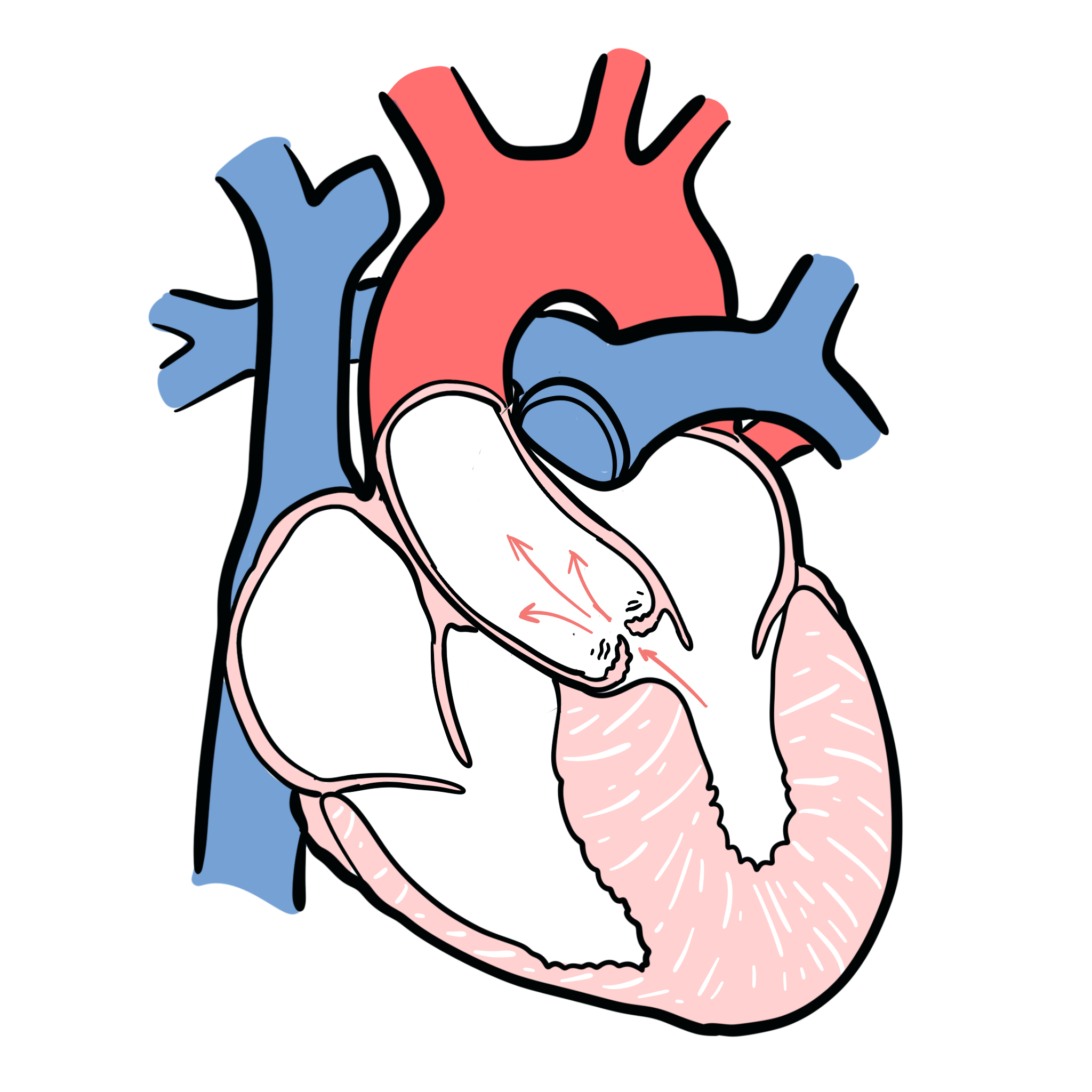
Overview
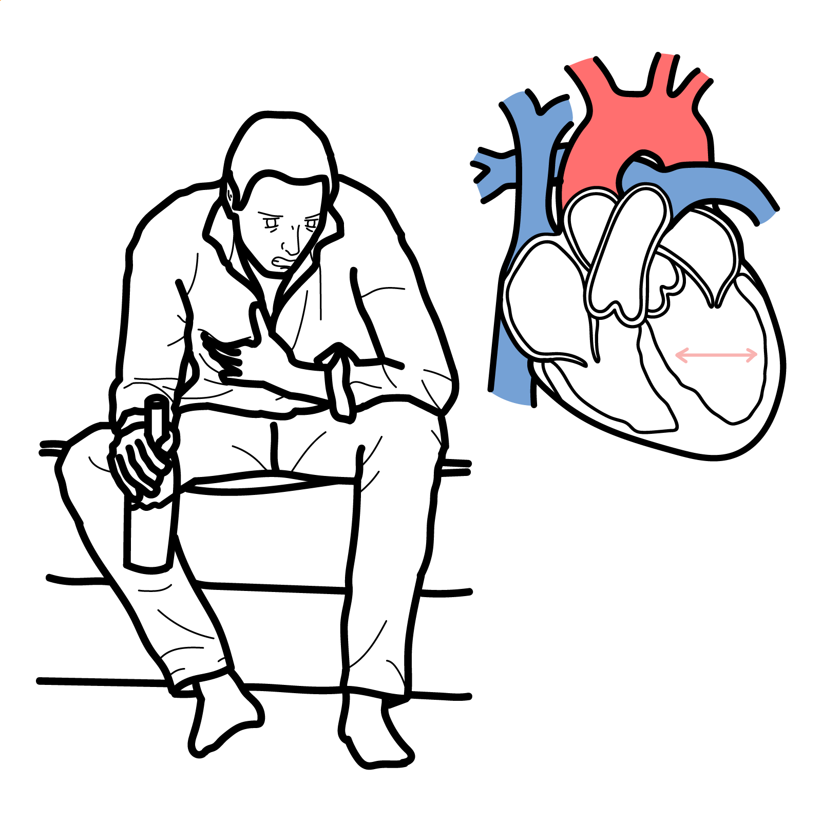
- Aortic valve stenosis is characterised by obstruction of left ventricular outflow, resulting in inadequate cardiac output, decreased exercise capacity, heart failure, and death from cardiovascular causes.
- Mild to moderate aortic stenosis is usually a symptomatic, but occasionally can be found on routine examination
- Locations of stenosis – Valvular (common), supravalvular and subvalvular
- Aortic Stenosis produces a “Mid Systolic Murmur”, which is a murmur heard between the first heart sound (S1 – AV valve closure) and ends before the second heart sound (S2 – Aortic and Pulmonary valve closure)
- Most common valvular heart disease in western countries and prevalence increase with increasing age
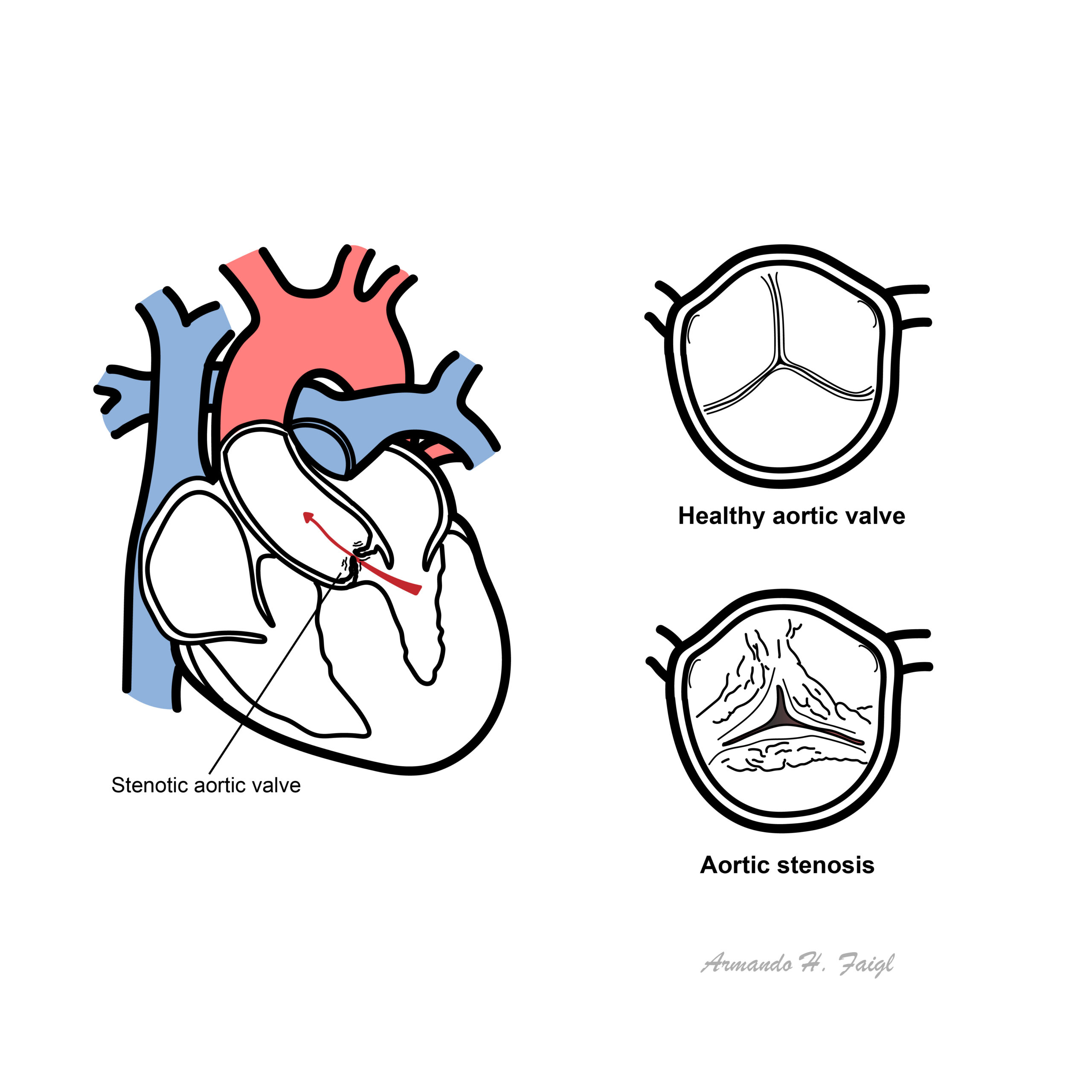
What to notice
Detailed image description
Cardiac Valves
The heart has 4 valves:
- Aortic valve
- Pulmonary valve
- Tricuspid valve
- Mitral valve (bicuspid valve)
The Mitral and tricuspid valves are atrioventricular valves meaning they allow blood to move from the atrium to the ventricles of the heart. This occurs with ventricular diastole.
The aortic and pulmonary valve are tricuspid valves which when open allow blood to move to the aorta and pulmonary system respectively. This occurs during ventricular systole when the heart contracts.
All of the heart valves except the mitral valve are usually tricuspid. However, there can be congenital bicuspid valves which can predispose one to valvular disease later on. The heart valves can be heard most prominent in the following regions of the chest
- Aortic valve – Right 2nd intercostal space parasternal
- Pulmonary valve – Left 2nd intercostal space parasternal
- Tricuspid valve – Left 4th intercostal space parasternal
- Mitral valve – Left 5 intercostal space mid-clavicular (below the nipple)
Aetiology & Risk factors
Aetiology
- A congenitally abnormal valve (bicuspid or rarely unicuspid) with superimposed calcification
- Calcific disease of a trileaflet valve
- Age related
- Typically in patients >65years
- Rheumatic valve disease
- Congenital aortic stenosis (rare)
- congenital stenosis – abnormal valve is present from birth
- acquired stenosis – abonrmal calve beomes stenotic overtime
- Age >60
- Congenitally unicuspid, bicuspid valve
- Rheumatic heart disease
- Chronic Kidney Disease
Pathophysiology
The natural history of AS begins with a prolonged asymptomatic period.
Pathology
- Calcified, stiff aortic valve leaflets
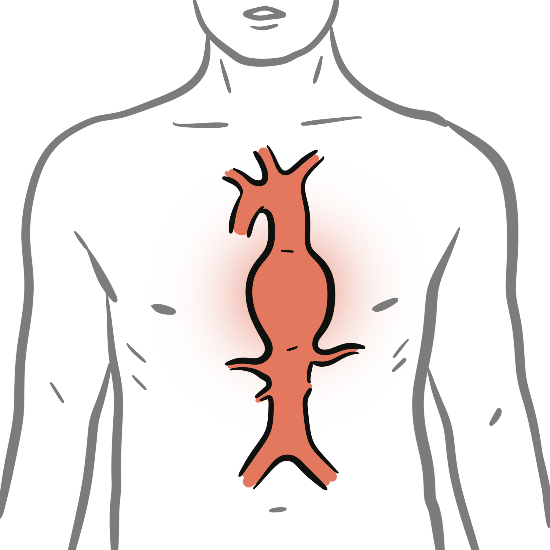
- Calcification and dilation of the ascending aorta
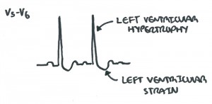
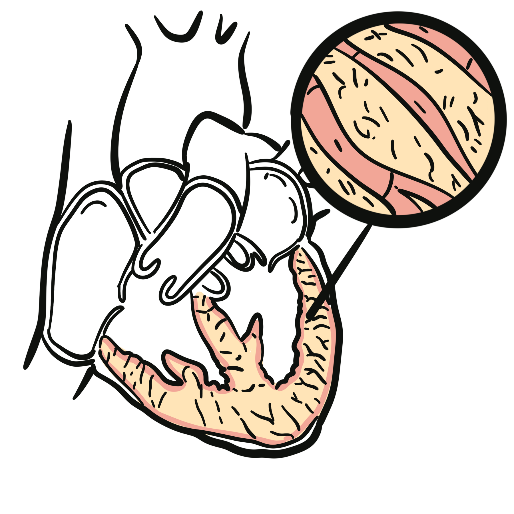
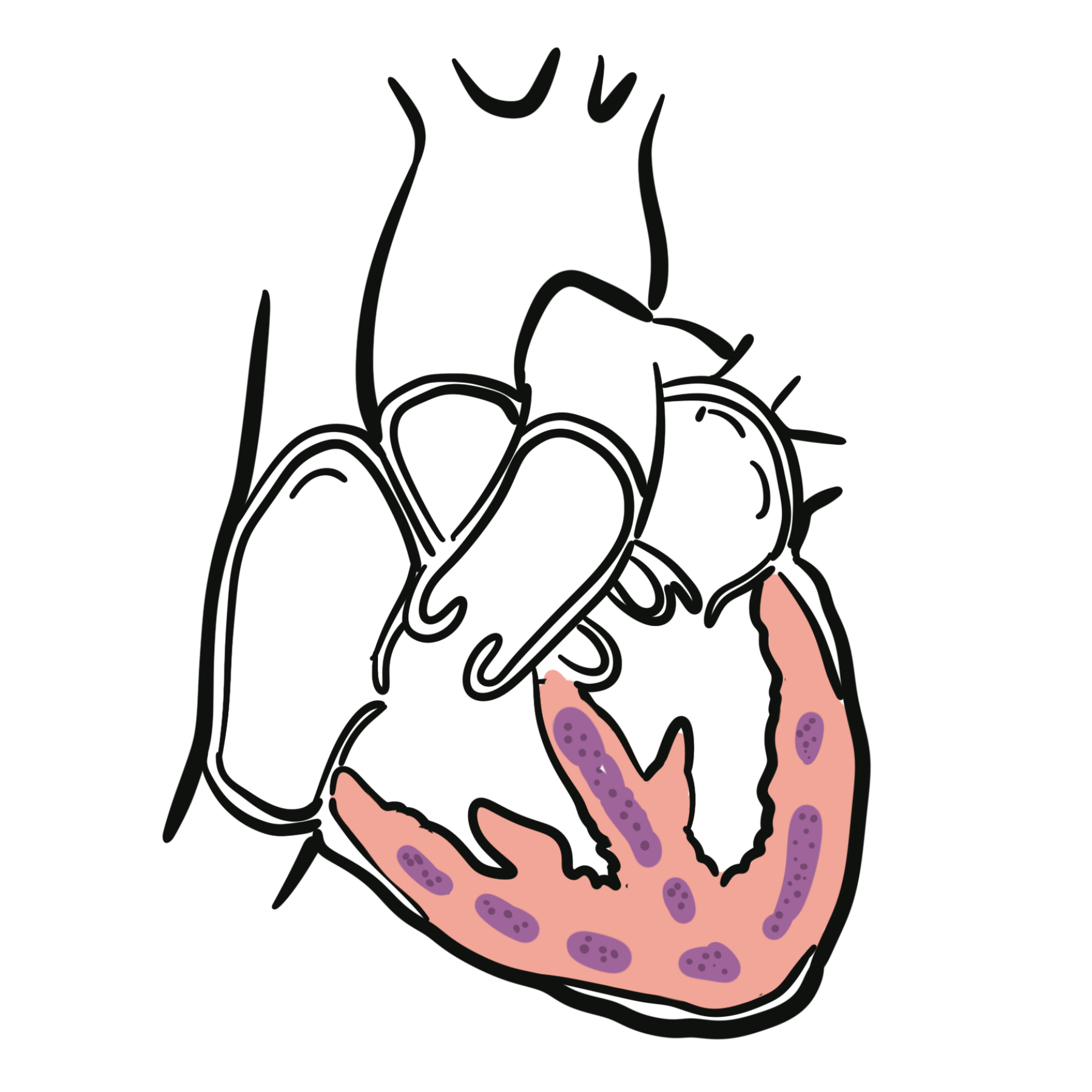
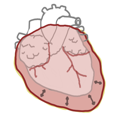
- Left ventricular hypertrophy
- Possible left atrial enlargement
- Mitral annular calcification -> mitral regurgitation
Clinical Manifestations
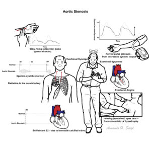
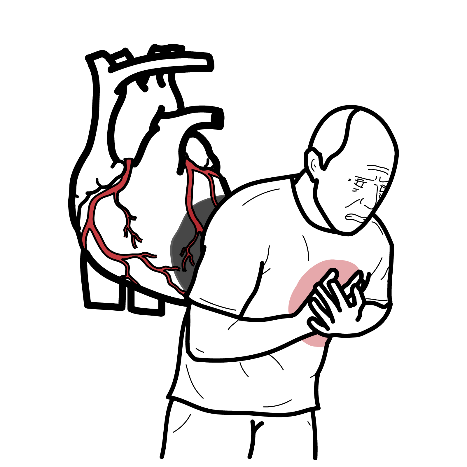
Aortic Stenosis classic triad of signs and symptoms are dizziness/syncope, dyspnoea, angina on exertion.
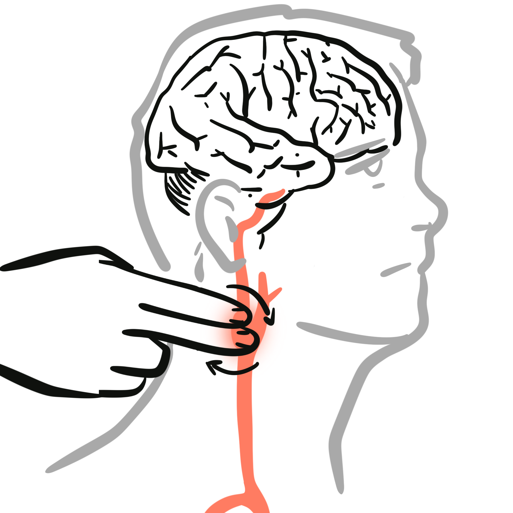
- Slow rate of rise in carotid pulse (parvus and tardus)
- Auscultation – Aortic Valve (right second intercostal space parasternal)
- Ejection systolic murmur – Mid to late peak intensity of the murmur generally begins after S1 and ends before S2 (mid systolic murmur)
- Reduced intensity of S2, closing of the aortic valve
- Atrial contraction, S4, maybe heard
- Auscultation – Apex (Left fifth intercostal space mid-clavicular)
- Murmur may radiate to apex of heart
- High frequency and louder murmur (Holosystolic murmur) can be confused with mitral regurgitation (Gallavardin phenomenon)
Cardiac Examination findings include parvus and tardus of the carotid artery and mid-systolic ejection murmur heard over the left second intercostal space parasternal. Murmur can radiate to the carotids.
Signs of severity
- Parvus et tardus
- Aortic thrill
- Length, harshness and lateness of the peak of the systolic murmur
- Atrial contraction, S4
- Parodical splitting of the second heart sound (delayed left ventricular ejection and aortic valve closure)
- Left ventricular failure
Squatting increases venous return and accentuate the murmur.
| Side note – Clinical Auscultation | ||
| Murmur | Heart Sound | |
| Mitral Stenosis | High pitched early-diastolic murmur | Loud S1 |
| Mitral Regurgitation | Pansystolic murmur radiates to the axilla | Soft S1 loud S2 |
| Aortic stenosis | Ejection systolic “crescendo decresendo” murmur radiating to the carotids | Soft S2 |
| Aortic regurgitation | early diastolic murmur | Soft S2 |
Diagnosis
- Transthoracic echocardiogram – elevated aortic pressure
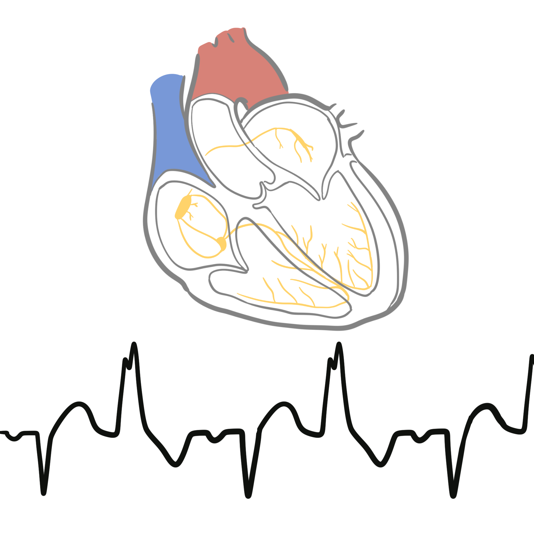
- ECG – left ventricular hypertrophy and absent Q wave
- X-ray – valve calcification
- Cardiac MRI
- Cardiac catheterisation – allows for direct measurement of the pressure gradient
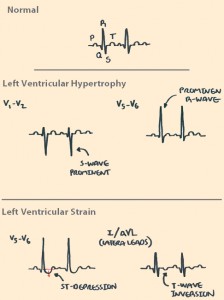
- Echocardiography is the primary test in diagnosis and evaluation of aortic stenosis
- 50% stenosis of aortic valve
Differential Diagnosis include other causes of mid-systolic murmurs
- Mitral valve dysfunction
- Hypertrophic cardiomyopathy
- Aortic sclerosis
- Atrial septal defect
- Pulmonary stenosis
Treatment
| Indications for surgery |
| Symptomatic |
| Critical obstruction based on catheterisatin |
Mild–moderate or asymptomatic disease
- Regular cardiology review and serial echocardiography.
- Educate patients to report exertional dyspnoea, chest pain or syncope promptly.
- Treat hypertension carefully and manage cardiovascular risk factors.
- Diuretics may relieve congestion but should be used cautiously because severe AS is preload-dependent.
- No medication reverses or reliably slows aortic-valve narrowing.
- Statins are used for standard lipid indications but do not treat calcific AS itself.
- Avoid excessive vasodilation, hypotension and dehydration in severe disease.
Aortic valve replacement
Definitive treatment is aortic valve replacement.
Indications include:
- Symptomatic severe AS:
- Exertional dyspnoea or heart failure
- Angina
- Exertional syncope or presyncope
- Severe AS with LVEF <50%, even without symptoms.
- Severe AS when undergoing other cardiac surgery.
- Consider intervention in selected asymptomatic patients with:
- Symptoms or hypotension during exercise testing
- Very severe stenosis
- Rapid haemodynamic progression
- Markedly elevated BNP
- Progressive ventricular dysfunction.
- There are two types of aortic valve replacements: TAVI and SAVR
TAVI versus SAVR
- TAVI/TAVR: valve inserted by catheter, usually through the femoral artery.
- SAVR: surgical removal and replacement of the valve.
- Choice requires a multidisciplinary Heart Team, considering age, frailty, life expectancy, anatomy, surgical risk, coronary disease and future valve-management needs.
- Under the 2025 European guideline:
- TAVI is generally favoured in anatomically suitable patients aged ≥70 years with tricuspid AS.
- SAVR is generally favoured in patients <70 years with low surgical risk.
- SAVR may be preferred with bicuspid anatomy, aortic-root disease, unsuitable vascular access, need for CABG or another valve operation.
- TAVI may be preferred in older, frail patients or those at increased surgical risk.
Balloon aortic valvuloplasty
- Temporarily widens the valve but restenosis is common.
- Mainly used:
- As a bridge to TAVI or SAVR
- In haemodynamically unstable patients
- Occasionally for palliation when definitive replacement is unsuitable
Complication & Prognosis
Disease-related complications
- Left-ventricular hypertrophy and diastolic dysfunction
- Left-ventricular systolic failure
- Pulmonary oedema and congestive heart failure
- Angina due to increased myocardial oxygen demand
- Exertional syncope
- Atrial fibrillation and ventricular arrhythmias
- Pulmonary hypertension
- Functional mitral regurgitation
- Sudden cardiac death
- Heyde syndrome: AS associated with acquired von Willebrand-factor dysfunction and gastrointestinal angiodysplasia bleeding
- Bicuspid AS may be associated with ascending-aortic dilatation or aneurysm.
Valve-replacement complications
- Bleeding
- Stroke or systemic embolism
- Acute kidney injury
- Infection or prosthetic-valve endocarditis
- Prosthetic-valve thrombosis or degeneration
- Paravalvular aortic regurgitation, particularly following TAVI
- Conduction block requiring permanent pacemaker, particularly following TAVI
- Vascular-access injury following TAVI
- Postoperative atrial fibrillation following SAVR
- Lifelong warfarin if a mechanical surgical valve is used.
- Mild or moderate AS may remain stable for years, but progression is variable and requires ongoing surveillance.
- Prognosis becomes poor once symptoms or left-ventricular dysfunction develop.
- Untreated symptomatic severe AS may have mortality approaching 50% within one year.
- Aortic valve replacement substantially improves symptoms, functional capacity and survival.
- Long-term prognosis after replacement depends on age, comorbidities, ventricular recovery and prosthetic-valve durability.
References
- Praz F, Borger MA, Lanz J, et al. 2025 ESC/EACTS Guidelines for the management of valvular heart disease. Eur Heart J. 2025;46(44):4635–4736. doi:10.1093/eurheartj/ehaf194.
- Otto CM, Nishimura RA, Bonow RO, et al. 2020 ACC/AHA guideline for the management of patients with valvular heart disease. Circulation. 2021;143(5):e72–e227. doi:10.1161/CIR.0000000000000923.
- Jneid H, Chikwe J, Arnold SV, et al. 2024 ACC/AHA clinical performance and quality measures for adults with valvular and structural heart disease. J Am Coll Cardiol. 2024;83(18):1803–1841. doi:10.1016/j.jacc.2023.12.006.
- Lindman BR, Clavel MA, Mathieu P, et al. Calcific aortic stenosis. Nat Rev Dis Primers. 2016;2:16006. doi:10.1038/nrdp.2016.6.


Members only discussions coming soon…