

Angina Pectoris

Overview
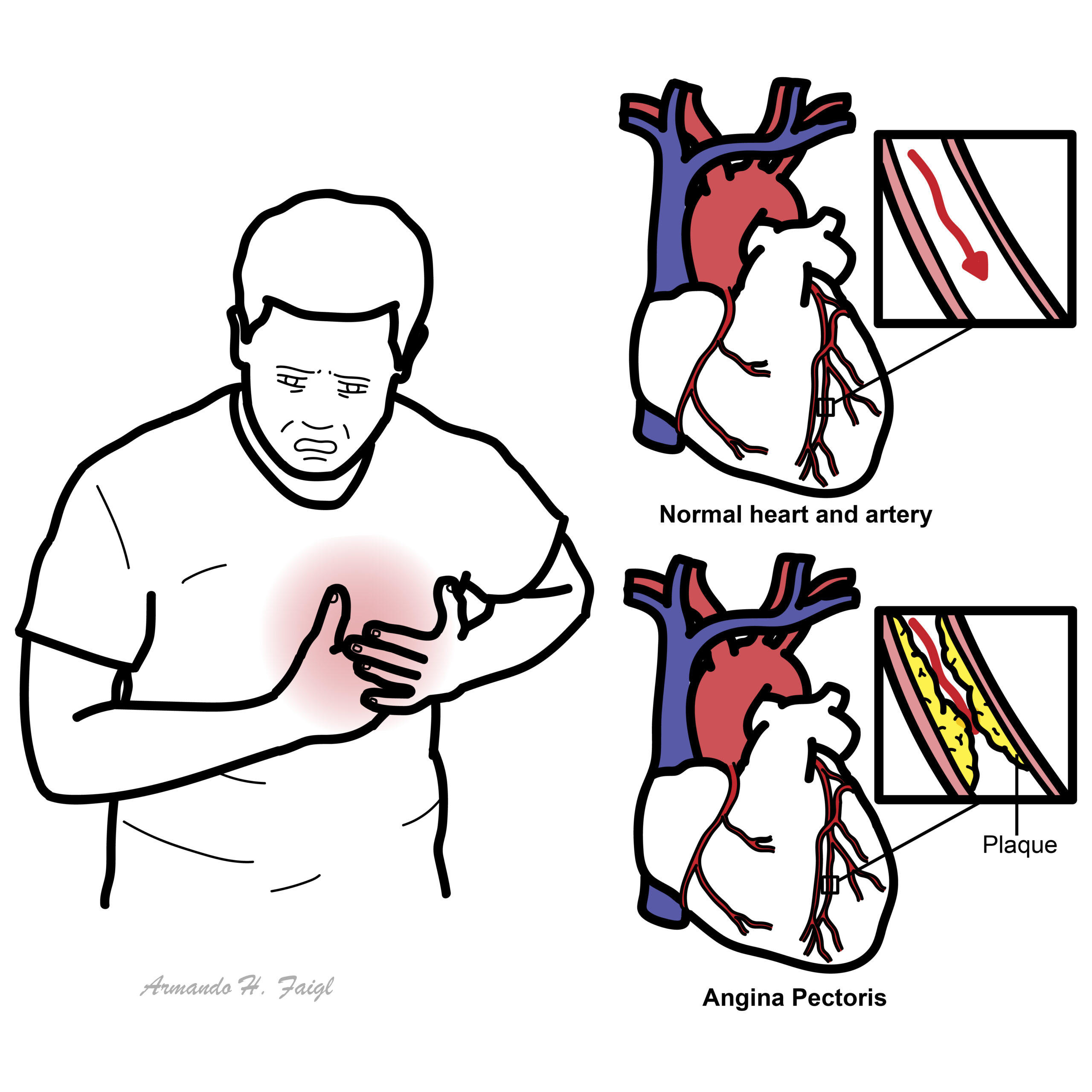
Angina pectoris is discomfort caused by transient myocardial ischaemia, usually reflecting an imbalance between oxygen supply and demand. Obstructive coronary disease, vasospasm and microvascular dysfunction can produce angina.
Clinical Goal: Identify an acute coronary syndrome (ACS) or another emergency first; then establish the cause of stable symptoms and reduce future cardiovascular risk.
A predictable exertional pattern supports stable angina. New, worsening or rest symptoms require urgent reassessment.
Relief with glyceryl trinitrate (GTN) does not establish a cardiac diagnosis.2
Approach
Step 1: Emergency Triage & Red Flags
- Assess airway, breathing, circulation, vital signs and current pain.
- Persistent pain, haemodynamic instability, syncope or severe breathlessness: arrange emergency assessment. Obtain and interpret a 12-lead ECG within 10 minutes of first clinical contact when ACS is suspected.
- Rest pain, crescendo symptoms or new severe episodes: follow the suspected-ACS pathway rather than routine outpatient testing.
Step 2: Characterise the Presentation
- Anginal pattern: Constricting chest, neck, jaw, shoulder or arm discomfort; provoked by exertion; relieved by rest or GTN within about five minutes. NICE describes all three features as typical angina and two as atypical angina.
- History: Record onset, duration, frequency, radiation, exertional threshold and change from baseline. Ask about associated breathlessness, sweating, nausea and palpitations.
- Context: Review known cardiovascular disease, smoking, diabetes, hypertension, lipids, family history, medications and stimulant use.
Step 3: Focused Examination & Branching
- Examine heart, lungs and circulation; look for heart failure, murmurs and alternative causes.
- Stable pattern: Arrange coronary assessment based on clinical likelihood.
- Persistent symptoms without obstructive disease: Consider angina/ischaemia with non-obstructive coronary arteries (ANOCA/INOCA), including vasospasm and microvascular dysfunction.
A normal resting ECG does not exclude angina or ACS.2
Differential Diagnosis
Coronary Ischaemia
- Stable angina: Distinguishing features: reproducible exertional discomfort with a consistent threshold and relief at rest.
- ACS: Distinguishing features: new or worsening symptoms, prolonged/rest pain, autonomic symptoms or instability. Unstable angina lacks acute infarction; NSTEMI involves myocardial infarction.
- Vasospastic / microvascular angina: Distinguishing features: recurrent angina despite non-obstructive arteries; rest episodes with transient ECG changes suggest vasospasm. Symptoms alone cannot establish the endotype.
Other Urgent Causes
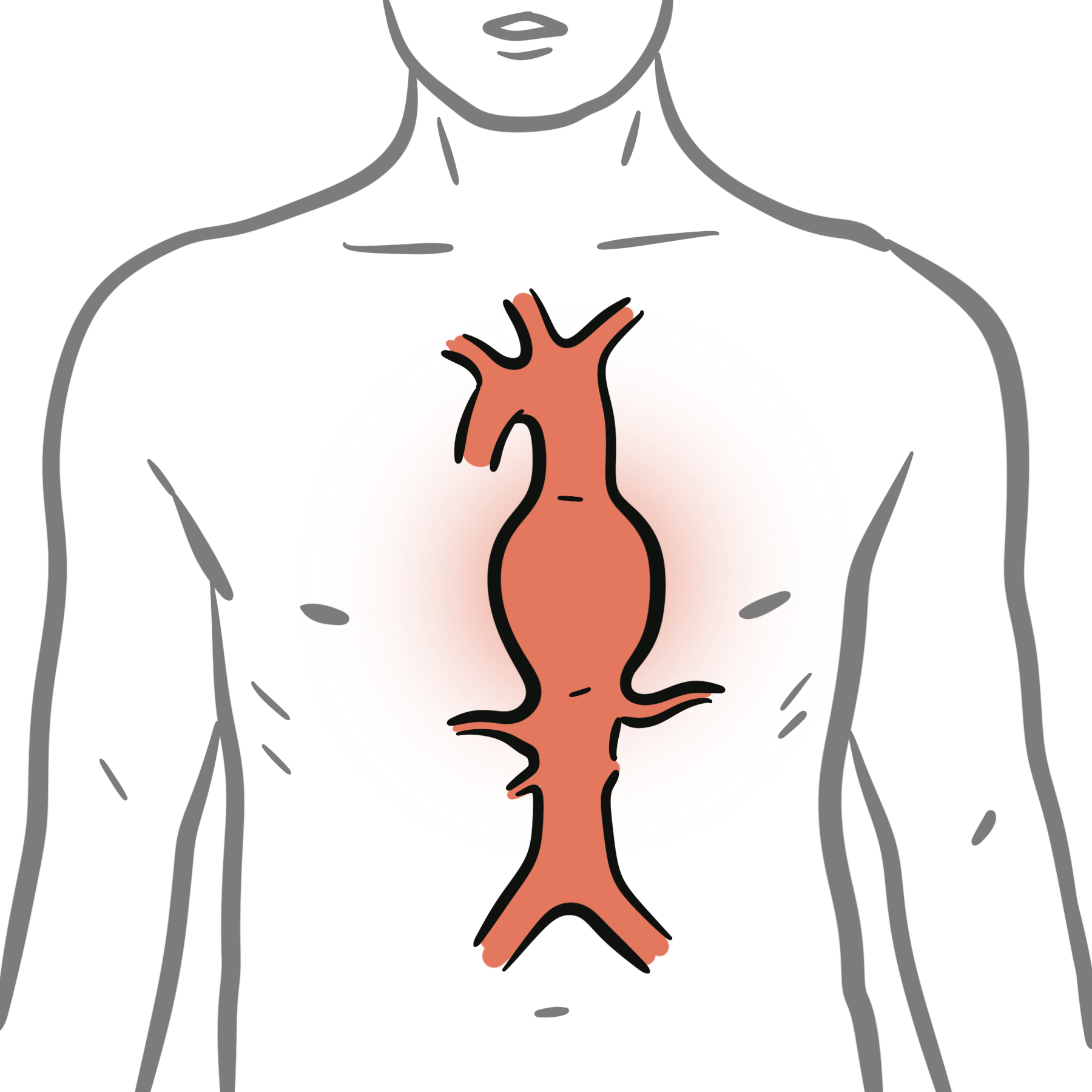
- Aortic dissection: Distinguishing features: abrupt severe chest/back pain, pulse asymmetry or neurological deficit.
- Pulmonary embolism: Distinguishing features: dyspnoea, pleuritic pain, hypoxaemia or venous thromboembolism risk.
- Pneumothorax / myopericarditis: Distinguishing features: unilateral respiratory findings or pleuritic/positional pain. Use the whole clinical picture and targeted testing.
Common Mimics
- Reflux / oesophageal disease: Distinguishing features: meal-related burning, regurgitation or swallowing symptoms.
- Musculoskeletal pain: Distinguishing features: local tenderness or movement-related pain.
- Anxiety: Distinguishing features: panic symptoms or hyperventilation; consider only after appropriate assessment of dangerous causes.
Tenderness, indigestion-like symptoms or an unusual pain description cannot independently rule out coronary ischaemia.2
Investigations
First-Line / Initial Workup
- ECG: Repeat during recurrent symptoms or diagnostic uncertainty. For suspected ACS, use high-sensitivity troponin within an assay-specific validated pathway; do not delay urgent reperfusion awaiting results.
- Stable presentation: Check full blood count, renal function, lipids and HbA1c/fasting glucose. Echocardiography assesses ventricular function and structural disease.
Targeted / Specialised Diagnostic Strategy
- CCTA: Preferred by ESC for low-to-moderate likelihood of obstructive coronary disease.
- Stress imaging: Assesses inducible ischaemia; useful when CCTA is inconclusive or stenosis significance is uncertain.
- Invasive angiography: Consider for high-risk findings, low-exertion angina or refractory symptoms. Persistent ANOCA/INOCA may require specialist coronary function testing.
Choose the test for the clinical question, patient characteristics and local expertise; exclude an acute presentation before elective testing.1,3
Critical Management
Suspected ACS
- Activate emergency transfer and cardiac monitoring as indicated.
- Australian 2025 guidance: Give aspirin 300 mg orally, chewed or dissolved, unless contraindicated. GTN may relieve pain, but avoid with hypotension, right ventricular infarction or recent PDE5 inhibitor use. Routine oxygen is unnecessary at SpO₂ ≥90%.
Confirmed Stable Angina
- Symptom relief: Provide a short-acting nitrate and clear instructions. Start a beta blocker or calcium channel blocker, considering contraindications and comorbidities; reassess response and adverse effects after treatment changes.
- Prevention: Address smoking, activity, diet, blood pressure, diabetes and lipids. Use statin therapy and consider antiplatelet treatment according to established coronary disease, bleeding risk and other antithrombotics.
- Escalation: Consider revascularisation when symptoms remain uncontrolled despite optimal treatment. Vasospastic angina requires mechanism-specific treatment, commonly a calcium channel blocker.
Give a written action plan: new, severe, worsening or persistent symptoms need emergency help. Do not delay emergency assessment while repeatedly trying GTN.3,4
References
- European Society of Cardiology. 2024 ESC Guidelines for the management of chronic coronary syndromes [Internet]. 2024 [cited 2026 Oct 5]. Available from: Guideline PDF.
- National Institute for Health and Care Excellence. Recent-onset chest pain of suspected cardiac origin: assessment and diagnosis. CG95 [Internet]. 2010 [updated 2016 Nov 30; cited 2026 Oct 5]. Available from: NICE CG95.
- Brieger D, Cullen L, Briffa T, et al. National Heart Foundation of Australia & Cardiac Society of Australia and New Zealand: Comprehensive Australian clinical guideline for diagnosing and managing acute coronary syndromes 2025 [Internet]. 2025 [cited 2026 Oct 5]. Available from: ACS guideline.
- National Institute for Health and Care Excellence. Stable angina: management. CG126 [Internet]. 2011 [cited 2026 Oct 5]. Available from: NICE CG126.








Members only discussions coming soon…