

Calf Pain
OVERVIEW
Calf pain is discomfort localized to the posterior lower leg between the popliteal fossa and the calcaneus, arising from muscular, tendinous, vascular, osseous, or neurogenic structures.
The primary clinical priority is rapidly ruling out life-threatening thromboembolism (deep vein thrombosis) and limb-threatening emergencies (acute compartment syndrome or acute limb ischemia) before treating benign musculoskeletal conditions.
Common Pathological Categories
- Vascular: Deep vein thrombosis, superficial thrombophlebitis, popliteal artery entrapment syndrome, peripheral artery disease (intermittent claudication).
- Musculoskeletal: Gastrocnemius tear (“tennis leg”), Achilles tendon rupture, plantaris tendon rupture, ruptured Baker cyst (pseudothrombophlebitis).
- Neurological / Other: Lumbar radiculopathy (first sacral nerve root compression), compartment syndrome, cellulitis.
Do not rely on Homans sign (calf pain on forced ankle dorsiflexion) to diagnose or exclude deep vein thrombosis. It has a sensitivity of < 30% and an unacceptably low specificity (< 50%). Vigorous forced dorsiflexion can also theoretically risk dislodging a non-occlusive thrombus.
TRIAGE & ALGORITHM
Step 1: Emergency Triage & Red Flag Screening
- Critical Limb-Threatening Ischemia (The “6 Ps”):
- Pain, Pallor, Pulselessness, Paresthesia, Paralysis, Perishingly cold -> Emergency vascular surgery consultation for catheter-directed embolectomy or surgical revascularization.
- Acute Compartment Syndrome:
- Severe pain out of proportion to injury, tense calf firmness, severe pain aggravated by passive toe extension, progressive sensory deficit in the deep peroneal or tibial nerve distribution -> Emergency orthopedic or vascular consult for urgent four-compartment fasciotomy.
- Massive Thromboembolism / Phlegmasia Cerulea Dolens:
- Massive calf and thigh swelling, intense cyanosis, severe pain, loss of peripheral pulses due to venous gangrene -> Emergent catheter-directed thrombolysis or surgical thrombectomy.
Step 2: Suspected Deep Vein Thrombosis Stratification (Wells Score)
- Clinical Criteria (+1 point each): Active cancer (treatment within 6 months), paralysis or plaster immobilization of lower limb, bedridden > 3 days or major surgery within 12 weeks, localized tenderness along deep venous system, entire leg swollen, calf swelling > 3 cm compared to asymptomatic side (measured 10 cm below tibial tuberosity), pitting edema confined to symptomatic leg, collateral superficial non-varicose veins; Minus 2 points: Alternative diagnosis at least as likely as deep vein thrombosis.
- Wells Score <= 1 (Deep Vein Thrombosis Unlikely):
- -> Obtain high-sensitivity D-dimer.
- D-dimer negative (below age-adjusted cutoff): -> Deep vein thrombosis excluded safely; evaluate for musculoskeletal causes.
- D-dimer positive: -> Proceed to comprehensive compression ultrasound.
- Wells Score >= 2 (Deep Vein Thrombosis Likely):
- -> Proceed directly to comprehensive compression ultrasound.
- If compression ultrasound is delayed > 4 hours, initiate empiric therapeutic anticoagulation while awaiting imaging (if bleeding risk is low).
Step 3: Musculoskeletal & Soft Tissue Branching
- Sudden “Pop” / Sensation of Being Kicked in Back of Leg:
- Inability to plantarflex against resistance + palpable tendon defect -> Perform Simmonds-Thompson test (Achilles tendon rupture).
- Mid-belly gastrocnemius tenderness with ecchymosis -> Medial gastrocnemius tear (“tennis leg”).
- Pre-existing Knee Swelling with Sudden Severe Calf Pain + Crescent Sign:
- Rapid crescent-shaped ecchymosis below the lateral malleolus -> Ruptured Baker (popliteal) cyst.
Use the age-adjusted D-dimer threshold for patients aged >50 years (Age × 10 µg/L for assays with a standard cutoff of 500 µg/L). Applying the standard 500 µg/L cutoff in elderly patients causes an unacceptable number of false-positive tests, whereas age-adjusting safely maintains >97% sensitivity while significantly increasing specificity.
DIFFERENTIAL DIAGNOSIS
Vascular Emergencies:
- Deep Vein Thrombosis (Proximal vs Distal Calf Vein):
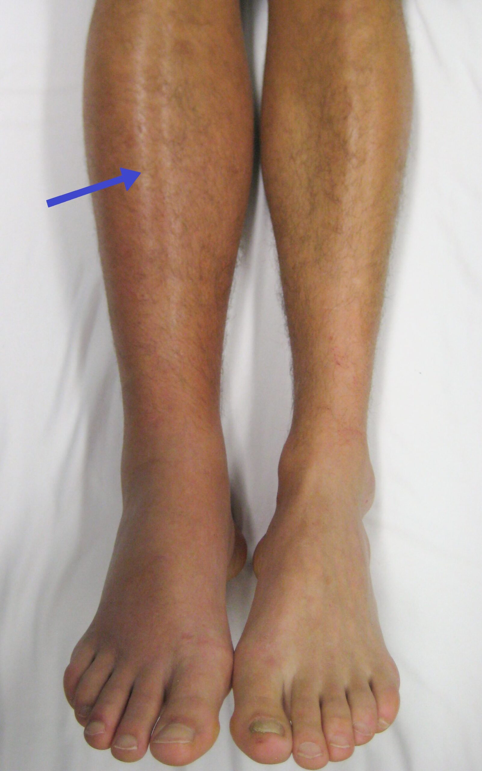
- Distinguishing features: Unilateral, pitting calf edema; progressive ache; localized tenderness along the posterior tibial, peroneal, or popliteal veins; calf circumference asymmetry > 3 cm measured 10 cm below the tibial tuberosity.
- Superficial Thrombophlebitis:
- Distinguishing features: Palpable, tender, indurated, erythematous cord along the course of the great or small saphenous vein; no deep calf swelling (duplex ultrasound required if within 3 cm of saphenofemoral or saphenopopliteal junction to exclude extension into deep system).
Acute Musculoskeletal Disorders:
- Achilles Tendon Rupture:
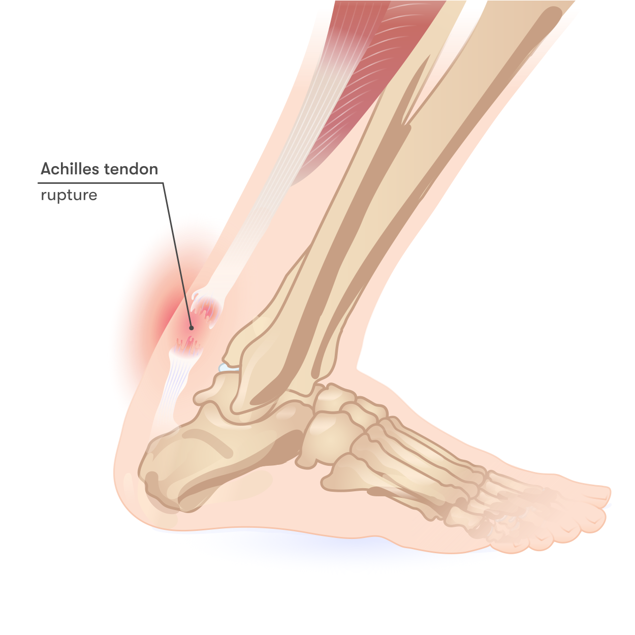
- Distinguishing features: Middle-aged adult playing racquet sports; sudden audible pop with sharp pain at posterior heel or ankle; palpable defect 2–6 cm proximal to calcaneal insertion; positive Simmonds-Thompson test (squeezing calf muscle belly fails to produce passive ankle plantarflexion).
- Medial Gastrocnemius Tear (“Tennis Leg”):
- Distinguishing features: Sudden, acute tearing pain at the musculotendinous junction (medial aspect of middle third of calf) during forceful push-off; focal point tenderness and swelling; Simmonds-Thompson test is normal (Achilles intact).
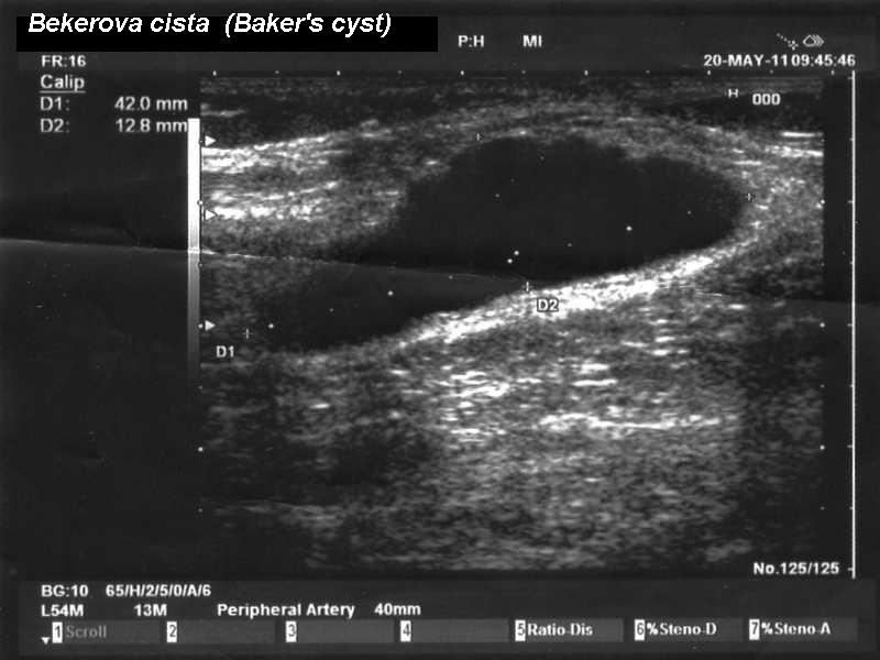
- Ruptured Baker (Popliteal) Cyst (Pseudothrombophlebitis):
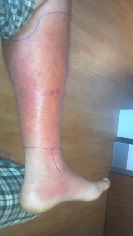
- Distinguishing features: History of knee osteoarthritis, inflammatory arthritis, or meniscal pathology; sudden leakage of synovial fluid into calf tissues causing acute pain, swelling, and an erythematous flare mimicking deep vein thrombosis; pathognomonic “crescent sign” (ecchymosis tracking beneath the lateral or medial malleolus).
- Chronic Exertional Compartment Syndrome:
- Distinguishing features: Young athletes or runners; predictable, progressive, aching calf tightness developing after a specific duration or intensity of exercise, completely resolving with rest; normal resting physical examination.
Infectious & Neurological Causes:
- Cellulitis:
- Distinguishing features: Spreading, warm, erythematous skin plaque with poorly demarcated borders; systemic fever; unilateral lymphadenopathy; entry point (tinea pedis, macerated web spaces, ulcer).
- Lumbar Radiculopathy (First Sacral Nerve Root Impingement):
- Distinguishing features: Sharp, lancinating pain radiating from lower back down posterior thigh and calf to lateral foot; diminished or absent Achilles tendon reflex; positive straight leg raise test.
A ruptured Baker cyst is the great clinical mimic of deep vein thrombosis. It causes acute calf pain, warmth, and marked swelling. Look carefully at the ankle for the crescent sign (a crescent of dependent bruising inferior to the malleoli), which indicates extravasated synovial fluid tracking along tissue planes.
INVESTIGATIONS
- First-Line / Bedside & Laboratory Testing:
- Wells Clinical Score: Mandatory pre-test probability stratification.
- High-Sensitivity D-dimer Assay: High negative predictive value (> 99%) when combined with an “unlikely” Wells score; not indicated if Wells score indicates high probability (proceed directly to ultrasound).
- Simmonds-Thompson Test & Palpation: Bedside physical confirmation of Achilles tendon integrity.
- Calf Circumference Measurement: Measured with a tape measure precisely 10 cm below the tibial tuberosity bilaterally (difference > 3 cm is clinically significant).
- Targeted / Specialized Diagnostic Imaging:
- Venous Duplex Ultrasound (Compression Ultrasound):
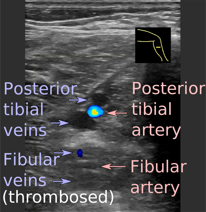
- Gold standard for deep vein thrombosis: Inability to fully compress the venous lumen under direct probe pressure is the definitive diagnostic sign of intraluminal thrombus; spectral Doppler evaluates absence of flow or blunted respiratory phasicity.
- Musculoskeletal Ultrasound of the Calf / Ankle:
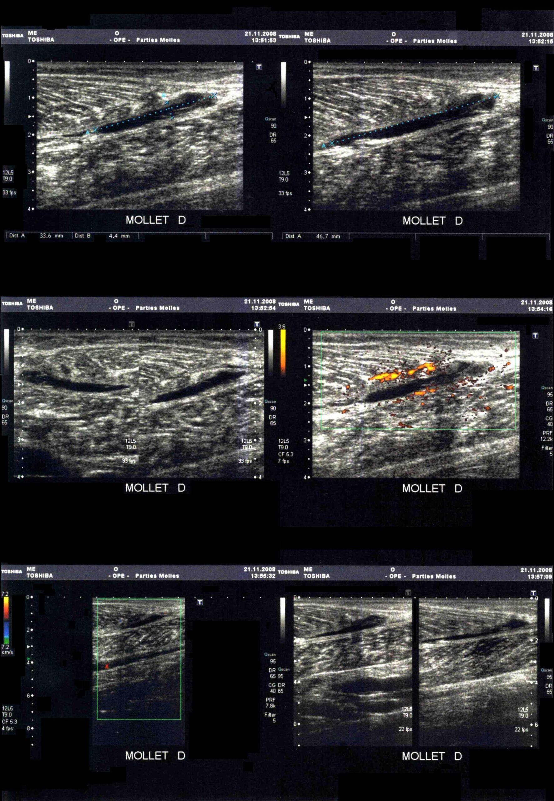
- Accurately distinguishes gastrocnemius tears (hypoechoic fluid collection at the myotendinous junction), plantaris tendon rupture, and detects gaps in Achilles tendon fibers.
- Intracompartmental Pressure Monitoring:
- Indicated for suspected acute compartment syndrome in obtunded or uncooperative patients (resting compartment pressure > 30 mmHg or a delta pressure [diastolic blood pressure minus compartment pressure] <= 30 mmHg confirms compartment syndrome).
- Magnetic Resonance Imaging of the Lumbar Spine: Modality of choice if calf pain is accompanied by dermatomal sensory loss, motor weakness, or refractory sciatica to identify first sacral nerve root disc herniation.
- Venous Duplex Ultrasound (Compression Ultrasound):
If a proximal deep vein thrombosis is clinically suspected (high pre-test probability) but initial whole-leg or proximal venous ultrasound is negative, repeat compression ultrasound in 5 to 7 days. This rules out a propagating distal isolated calf vein thrombosis that was radiographically undetectable during the initial scan.
CRITICAL MANAGEMENT
- Deep Vein Thrombosis Anticoagulation Protocol:
- Direct Oral Anticoagulants (First-Line):
- Apixaban: 10 mg orally twice daily for 7 days, then 5 mg orally twice daily maintenance OR
- Rivaroxaban: 15 mg orally twice daily with food for 21 days, then 20 mg orally once daily maintenance.
- Low Molecular Weight Heparin (Enoxaparin 1.5 mg/kg subcutaneously once daily): Preferred in active malignancy, severe renal impairment (dose-adjusted), antiphospholipid syndrome, or pregnancy.
- Duration of Anticoagulation:
- Provoked deep vein thrombosis (transient major risk factor, such as surgery or plaster cast):3 months.
- Unprovoked deep vein thrombosis / persistent risk factor (active cancer): Extended or indefinite therapy, provided bleeding risk is acceptable.
- Direct Oral Anticoagulants (First-Line):
- Acute Compartment Syndrome (Surgical Emergency):
- Immediately remove all constrictive casts, dressings, or splints down to the skin (bivalving a cast reduces compartment pressure by > 50%).
- Maintain leg at heart level (do NOT elevate above heart level, as this reduces arterial perfusion pressure).
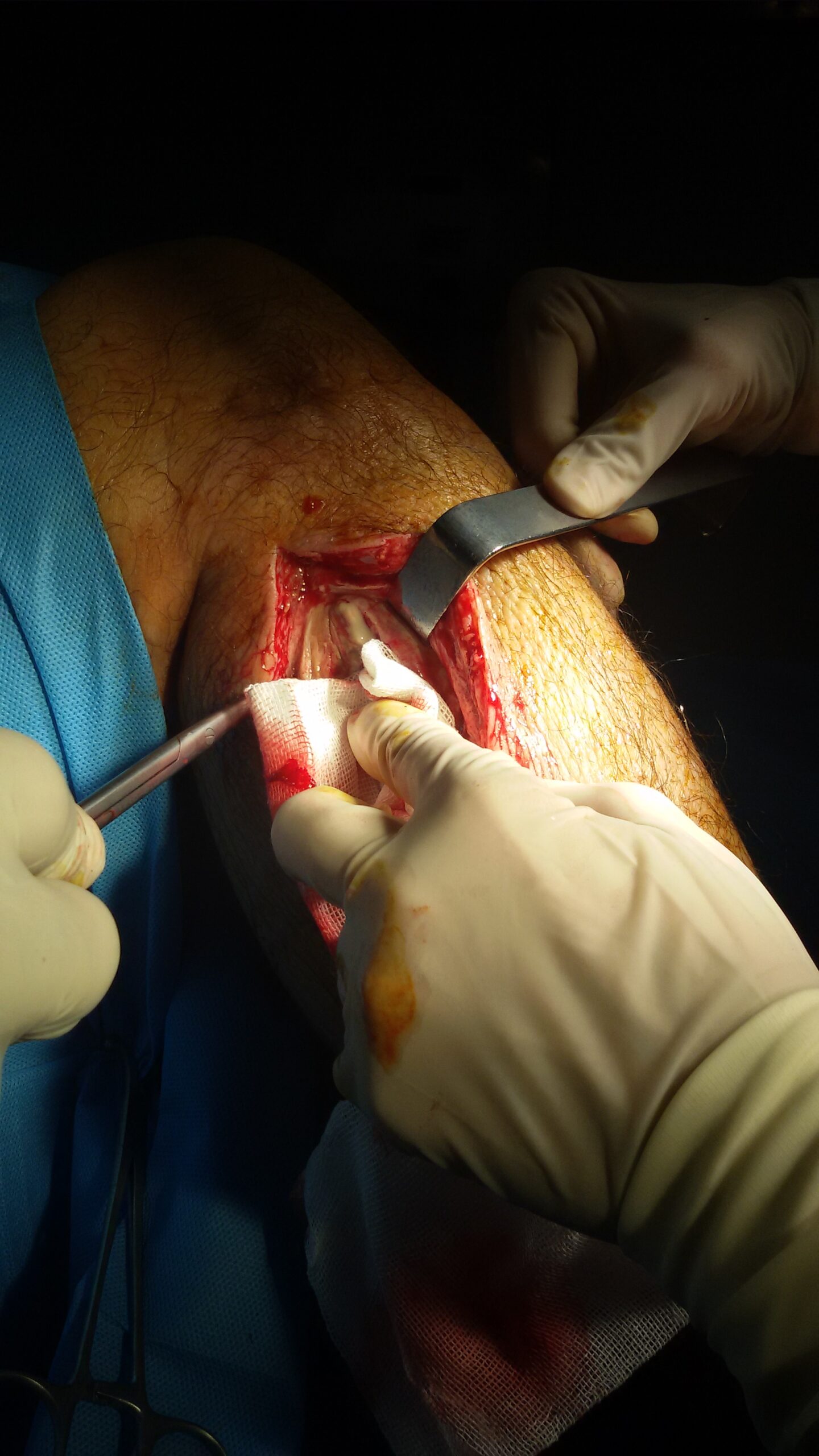
- Emergency four-compartment fasciotomy (anterolateral and posteromedial incisions releasing the anterior, lateral, superficial posterior, and deep posterior compartments).
- Achilles Tendon Rupture Management:
- Immediate Immobilization: Rigid splint or specialized functional walking boot in equinus position (maximal passive plantarflexion, approximately 20–30 degrees) to appose the torn tendon ends and avoid lengthening.
- Definitive Pathway: Fast-track orthopedic referral for accelerated functional rehabilitation protocol (non-operative management with progressive wedge removal) or surgical percutaneous or open repair (favored in high-level athletes or delayed presentations).
- Medial Gastrocnemius Tear (“Tennis Leg”) Protocol:
- Conservative management: Relative rest, ice, compression bandage, heel lift in shoe for 2–4 weeks to reduce tension on healing muscle fibers, followed by progressive isometric and eccentric calf loading.
When managing an acute Achilles tendon rupture, immediately immobilize the ankle in equinus (plantarflexion). Splinting or casting the foot in neutral (90 degrees) separates the ruptured tendon ends, resulting in tendon elongation, chronic calf weakness, and high rates of non-union.













Members only discussions coming soon…