

Bone Lump (Bone Tumour)

DEFINITION & OVERVIEW
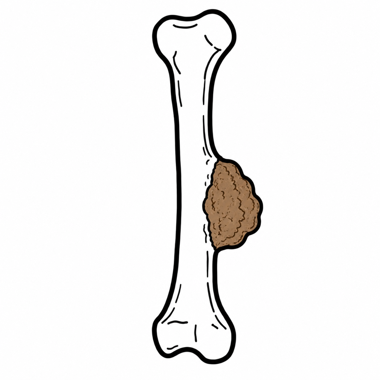
- Definition: A bone lump is an osseous or subperiosteal mass arising from the cortex, medulla, or surface of a bone, which may represent benign neoplasms, primary bone sarcomas, metastatic disease, infection (osteomyelitis), or non-neoplastic reactive processes.
- Epidemiology & Demographics:
- In patients age > 40 years, bone lesions are overwhelmingly Metastatic Carcinoma or Multiple Myeloma.
- In children and young adults (age < 30 years), benign bone lesions (e.g., osteochondroma, non-ossifying fibroma) predominate, but primary bone sarcomas (Osteosarcoma, Ewing Sarcoma) must be excluded.
- Anatomical Subregions: Epiphyseal (e.g., Giant Cell Tumor, Chondroblastoma), Metaphyseal (e.g., Osteosarcoma, Osteochondroma), or Diaphyseal (e.g., Ewing Sarcoma, Osteoid Osteoma, Fibrous Dysplasia).
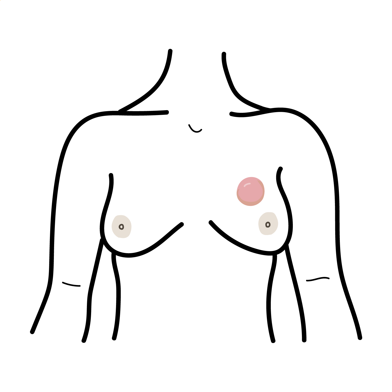
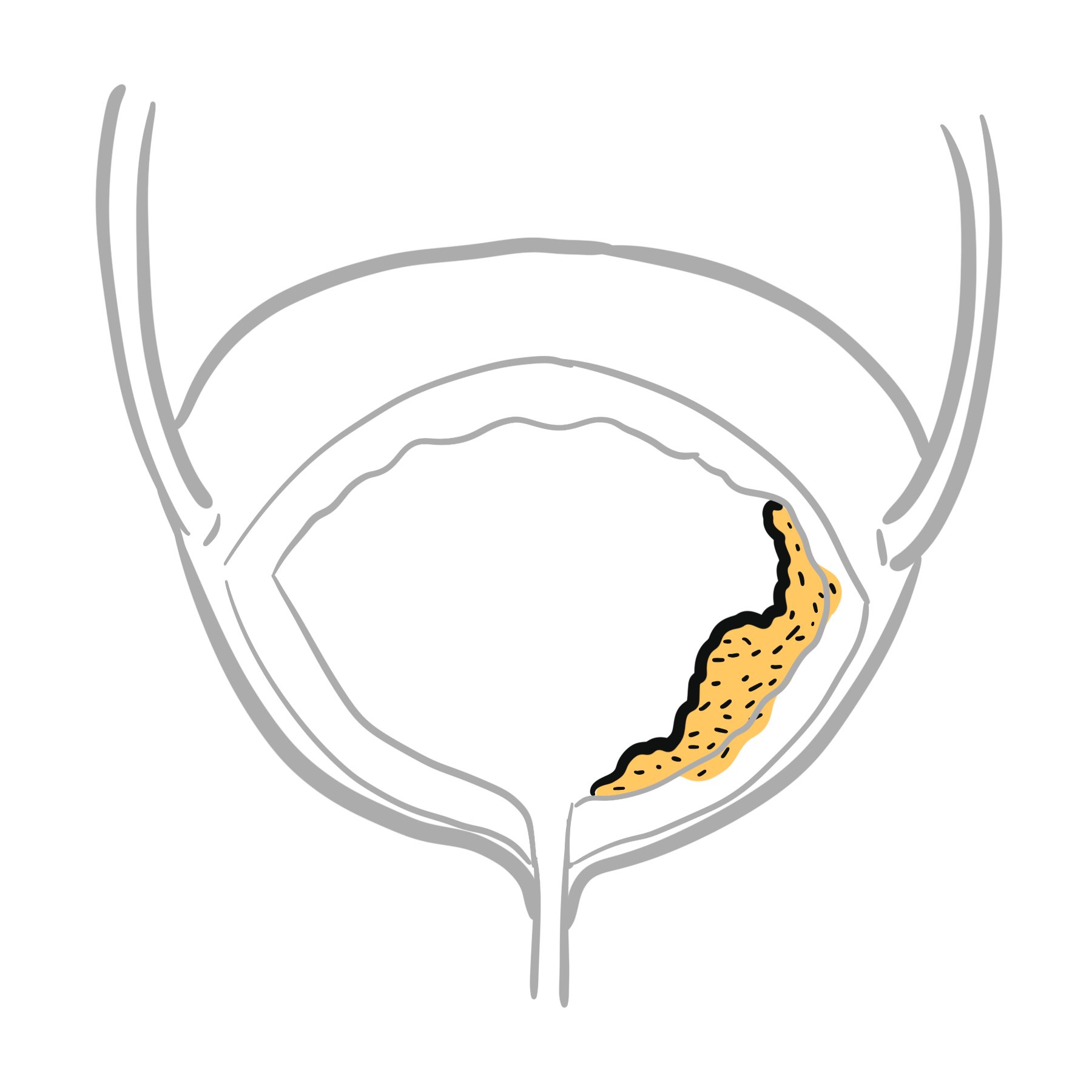
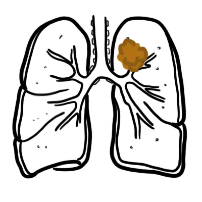
In any patient aged > 40 years presenting with a new, destructive, or painful bone lesion, think “BLT with a Kosher Pickle” for the common osteolytic/osteoblastic metastases: Breast, Lung, Thyroid, Kidney, and Prostate (plus Multiple Myeloma).
APPROACH
- Step 1: Clinical Red Flag Screening
- Pain Pattern: Constant, progressive, deep, aching bone pain, especially night pain waking the patient from sleep or unprovoked resting pain (malignancy red flag).
- Systemic Features: Unexplained weight loss, night sweats, fevers, malaise, hypercalcaemia.
- Mass Characteristics: Rapidly enlarging lesion, hard fixed mass > 5 cm, warm/erythematous skin over bone, or associated soft tissue mass extension.
- Structural Instability: Impending Pathological Fracture (Mirels’ score >= 9 -> urgent prophylactic orthopedic stabilization).
- Step 2: Plain Radiograph Assessment (The “Lodwick” Criteria)
- Obtain orthogonal views (AP and Lateral) of the entire affected bone.
- Assess Radiographic Aggressiveness:
- Benign / Non-Aggressive: Well-defined margin, narrow zone of transition, sclerotic border, intact cortex, no periosteal reaction, no soft-tissue mass.
- Malignant / Aggressive: Ill-defined / permeative / “moth-eaten” margin, wide zone of transition, cortical destruction, aggressive periosteal reaction (Codman’s triangle, “sunburst”, or “onion-skin” multilamellated pattern), soft tissue mass.
- Step 3: Diagnostic Branching Logic
- Aggressive Radiographic Features / Red Flags Present: -> Do NOT Biopsy in Primary Care. Obtain urgent staging MRI and refer directly to a specialized Sarcoma / Orthopaedic Oncology Multidisciplinary Team (MDT).
- Classic Incidental Benign Lesion (e.g., Non-Ossifying Fibroma, Osteochondroma): -> Conservative follow-up or reassurance.
- Lytic Bone Lesion in Age > 40: -> Staging CT chest/abdomen/pelvis + Myeloma screen (Serum Free Light Chains, SPEP/UPEP) + Bone Scan.
Never perform an unplanned excisional biopsy or puncture of a suspected primary bone tumor outside of a specialist sarcoma center. A poorly placed biopsy tract can contaminate tissue planes, necessitating limb amputation rather than limb-sparing surgery.
DIFFERENTIAL DIAGNOSIS
- Malignant Bone Lesions:
- Osteosarcoma (Most Common Primary Bone Malignancy in Youth):
- Distinguishing features: Bimodal age peak (10–25 years and > 60 years); metaphyseal (distal femur > proximal tibia > proximal humerus); mixed blastic/lytic lesion, Codman’s triangle, and “sunburst” spiculation of periosteal bone formation.
- Ewing Sarcoma:
- Distinguishing features: Children/adolescents (age 5–20); diaphyseal/metadiaphyseal region of long bones and pelvis; permeative bone destruction, “onion-skin” lamellated periosteal reaction, large soft tissue component; fever/leukocytosis mimicking osteomyelitis ($t(11;22)$ translocation).
- Chondrosarcoma:
- Distinguishing features: Adults age 40–70; axial skeleton/pelvis/proximal femur; lobulated matrix with “rings and arcs” / “popcorn” calcifications on plain radiograph; resistant to radiotherapy/chemotherapy.
- Multiple Myeloma / Bone Metastases:
- Distinguishing features: Age > 50; multiple discrete “punched-out” lytic lesions without sclerotic rims on skull/spine X-rays; elevated serum protein, Bence-Jones proteinuria, hypercalcaemia, and renal impairment (CRAB criteria).
- Osteosarcoma (Most Common Primary Bone Malignancy in Youth):
- Benign Bone Tumors & Pseudotumors:
- Osteochondroma (Exostosis – Most Common Benign Tumor):
- Distinguishing features: Bony outgrowth on bone surface pointing away from adjacent joint, with continuous marrow and cortex cavity; cartilage cap < 1.5–2 cm (cartilage cap > 2 cm indicates malignant transformation to secondary chondrosarcoma).
- Osteoid Osteoma:
- Giant Cell Tumor of Bone (GCTB):
- Distinguishing features: Young adults age 20–40 (skeletally mature with closed epiphyses); strictly epiphyseal extending to subarticular surface (around the knee); purely lytic, eccentric, expansile “soap-bubble” appearance without peripheral sclerosis.
- Non-Ossifying Fibroma (NOF) / Fibrous Cortical Defect:
- Distinguishing features: Incidental finding in pediatric/adolescent patients; eccentric, bubbly lytic lesion with well-defined scalloped sclerotic margins in the metaphysis; spontaneously ossifies and disappears with age.
- Osteomyelitis / Brodie’s Abscess:
- Osteochondroma (Exostosis – Most Common Benign Tumor):
The classic hallmark of an Osteoid Osteoma is severe, localized bone pain that worsens at night and is dramatically and rapidly relieved by low-dose Aspirin or NSAIDs (due to high prostaglandin production by the vascular nidus).
INVESTIGATIONS
- First-Line / Mandatory Baseline Imaging & Labs:
- Orthogonal Plain Radiographs (AP & Lateral): Mandatory first test. Evaluates lesion location, margins, cortical breach, matrix type (osteoid vs chondroid), and periosteal reaction.
- Baseline Blood Panel: Full Blood Count (cytopenias, infection), ESR & CRP, Serum Corrected Calcium, Phosphate, Alkaline Phosphatase (ALP – elevated in osteosarcoma and Paget’s disease), Serum Creatinine.
- Myeloma Screen (If age > 40): Serum Protein Electrophoresis (SPEP), Urine Protein Electrophoresis (UPEP), Serum Free Light Chains (sFLC).
- Targeted / Specialized Diagnostic & Staging Workup:
- MRI of the Entire Affected Bone (with IV Gadolinium): Gold standard to delineate intraosseous tumor extent, marrow replacement, joint space involvement, skip lesions, and neurovascular proximity.
- Staging CT Chest / Abdomen / Pelvis: Detects pulmonary metastases (lungs are the most common site of metastasis for primary bone sarcomas) and identifies occult primary carcinoma in metastatic disease.
- Technetium-99m Whole-Body Bone Scintigraphy / 18F-FDG PET-CT: Assesses multifocal skeletal lesions, metabolic activity, and occult distant metastases.
- Image-Guided Core Needle Biopsy (Specialist Center Only): Mandatory for definitive histopathological diagnosis prior to neoadjuvant therapy or operative intervention.
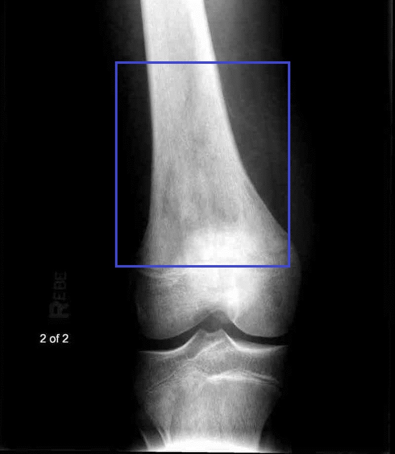
Radiograph showing osteosarcoma of the distal femur. Credit: Scott Dulebohn, MD and S. Bhimji, MD, via Wikimedia Commons, CC BY 4.0.
What to notice
Detailed image description
Source: https://commons.wikimedia.org/wiki/File:Osteosarcoma1.jpg Licence: Creative Commons Attribution 4.0 International (CC BY 4.0): https://creativecommons.org/licenses/by/4.0/ Modifications: None.
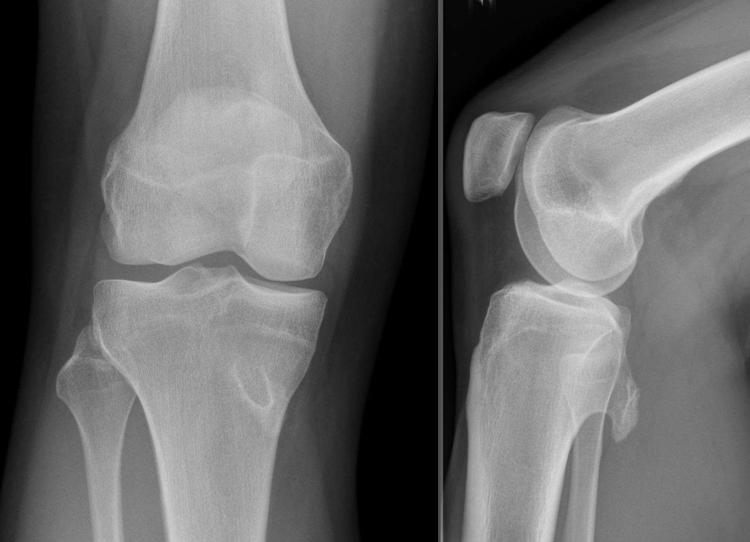
Radiograph showing an osteochondroma arising from the proximal tibia. Credit: Hellerhoff, via Wikimedia Commons, CC BY-SA 3.0.
What to notice
Detailed image description
Source: https://commons.wikimedia.org/wiki/File:Kartilaginäre_Exostose.png Licence: Creative Commons Attribution-ShareAlike 3.0 Unported (CC BY-SA 3.0): https://creativecommons.org/licenses/by-sa/3.0/ Modifications: None.
The skull X-ray in Multiple Myeloma demonstrates classic “punched-out” lytic lesions that lack reactive sclerotic borders because myeloma cells release DKK-1 and other cytokines that completely suppress osteoblast bone repair.

Skull radiograph showing multiple punched-out osteolytic lesions, a classic but not pathognomonic appearance of multiple myeloma. Credit: Medicalpal, via Wikimedia Commons, CC BY-SA 4.0.
What to notice
Detailed image description
Source: https://commons.wikimedia.org/wiki/File:Osteolytic_Lesions.jpg Licence: Creative Commons Attribution-ShareAlike 4.0 International (CC BY-SA 4.0): https://creativecommons.org/licenses/by-sa/4.0/ Modifications: None.
CRITICAL MANAGEMENT
- Primary Bone Sarcoma Pathway (Osteosarcoma, Ewing Sarcoma):
- Immediate MDT Referral: Refer to a dedicated tertiary orthopedic oncology and sarcoma center before any surgical intervention.
- Neoadjuvant Chemotherapy: Multimodal systemic chemotherapy (e.g., MAP: Methotrexate, Doxorubicin [Adriamycin], Cisplatin) prior to definitive surgery to shrink tumor and treat micrometastatic disease.
- Limb-Salving Resection & Reconstruction: Wide local surgical excision with clear oncological margins + modular endoprosthetic replacement or biological reconstruction.
- Pathological Fracture Risk Assessment & Management (Mirels’ Score):
- Mirels’ Criteria (Score 1–3 for each: Site, Pain, Lesion Type, Size):
- Score >= 9: High fracture risk (> 33%) -> Prophylactic Orthopedic Internal Fixation (Intramedullary nailing / cemented arthroplasty) prior to radiation therapy or mobilization.
- Score <= 7: Low risk (< 5%) -> Conservative management / radiation therapy.
- Mirels’ Criteria (Score 1–3 for each: Site, Pain, Lesion Type, Size):
- Osteoid Osteoma Definitive Treatment:
- First-Line Medical Therapy: Daily NSAIDs (if pain is controlled and patient tolerates).
- Minimally Invasive Interventional Procedure: CT-guided Percutaneous Radiofrequency Ablation (RFA) of the nidus (cure rate > 90%).
Always calculate Mirels’ Score for metastatic or lytic long-bone lesions. A score of 9 or greater is a surgical emergency requiring immediate prophylactic internal fixation before a catastrophic pathological fracture occurs.
REFERENCES
- Gerrand C, Amary F, Anwar HA, et al. UK guidelines for the management of bone sarcomas. Br J Cancer. 2025;132(1):32–48. doi:10.1038/s41416-024-02868-4
- Ahlawat S, Lenchik L, Baker JC, et al. ACR Appropriateness Criteria® Suspected Primary Bone Tumors: 2024 Update. J Am Coll Radiol. 2025;22(5 Suppl):S440–S454. doi:10.1016/j.jacr.2025.02.020
- National Institute for Health and Care Excellence. Suspected cancer: recognition and referral [Internet]. London: NICE; 2015 [updated 2026; cited 2026 Aug 31]. Available from: https://www.nice.org.uk/guidance/ng12
- National Cancer Institute. Osteosarcoma and undifferentiated pleomorphic sarcoma of bone treatment (PDQ®)–health professional version [Internet]. Bethesda (MD): National Cancer Institute; [updated 2024 Dec 2; cited 2026 Aug 31]. Available from: https://www.cancer.gov/types/bone/hp/osteosarcoma-treatment-pdq
- National Cancer Institute. Ewing sarcoma treatment (PDQ®)–health professional version [Internet]. Bethesda (MD): National Cancer Institute; [updated 2024 Nov 27; cited 2026 Aug 31]. Available from: https://www.cancer.gov/types/bone/hp/ewing-treatment-pdq
- Mirels H. Metastatic disease in long bones: a proposed scoring system for diagnosing impending pathologic fractures. Clin Orthop Relat Res. 1989;(249):256–264. PMID:2684463.
- Tepelenis K, Skandalakis GP, Papathanakos G, et al. Osteoid osteoma: an updated review of epidemiology, pathogenesis, clinical presentation, radiological features, and treatment option. In Vivo. 2021;35(4):1929–1938. doi:10.21873/invivo.12459














Members only discussions coming soon…