

Hypoglycaemia
Overview
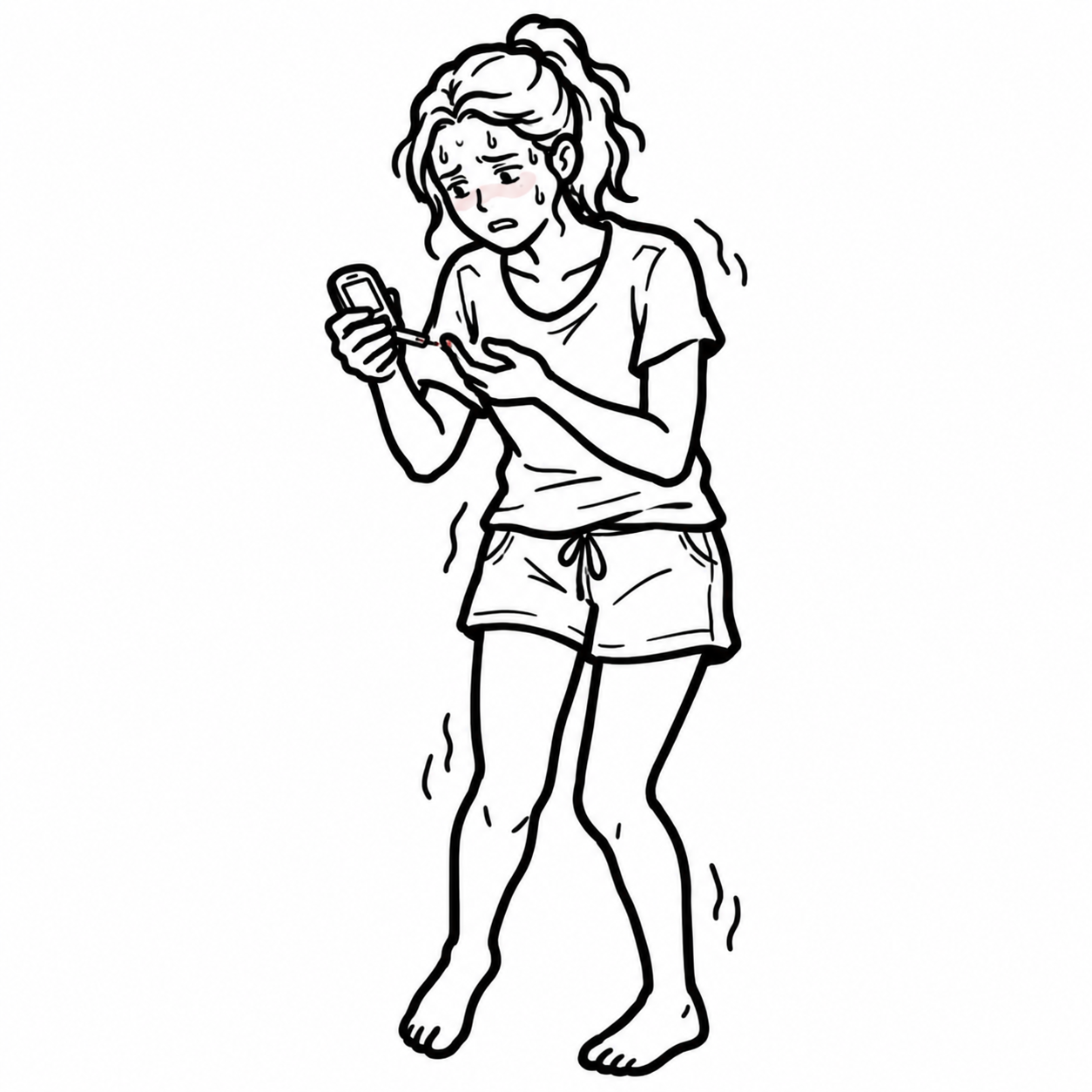
Hypoglycaemia is a plasma glucose concentration low enough to cause autonomic or neuroglycopenic symptoms. Severe or prolonged hypoglycaemia can cause seizures, permanent neurological injury, cardiac arrhythmia, coma and death.
In people with diabetes, a blood glucose level below 4.0 mmol/L should be treated promptly, even if symptoms are absent.1
In adults without diabetes, investigate a hypoglycaemic disorder when Whipple’s triad is documented:
- Symptoms or signs consistent with hypoglycaemia
- A low laboratory-measured plasma glucose concentration
- Resolution of symptoms after the plasma glucose concentration is corrected.2
Symptoms are divided into:
- Autonomic or neurogenic: tremor, sweating, palpitations, anxiety, hunger and perioral tingling
- Neuroglycopenic: behavioural change, confusion, visual disturbance, weakness, dysarthria, seizure, focal neurological deficit and coma.2
Hypoglycaemia is a medical emergency. Treat immediately when severe symptoms are present—do not delay glucose administration while waiting for laboratory confirmation or diagnostic specimens.
Triage & Approach
Step 1: Immediate Assessment and Stabilisation
- Apply an ABCDE approach.
- Check bedside BGL immediately in any patient with altered consciousness, seizure, behavioural change, diaphoresis, weakness or an unexplained neurological presentation.
- Assess airway protection and ability to swallow.
- Obtain vital signs, cardiac monitoring and intravenous access.
- Treat immediately if the patient is unconscious, seizing, unable to swallow or clinically unstable.
- Recheck BGL approximately 10–15 minutes after treatment and continue monitoring for recurrence.
Step 2: Determine Whether Diabetes Treatment Is Involved
Ask about:
- Insulin type, dose and timing
- Sulfonylureas, particularly long-acting preparations
- Recent dose changes or medication errors
- Missed or delayed meals
- Vomiting or reduced carbohydrate intake
- Unplanned or prolonged exercise
- Alcohol consumption
- Renal or hepatic impairment
- Recent weight loss
- Pregnancy
- Continuous glucose monitor or insulin-pump data
Hypoglycaemia in a person taking insulin or an insulin secretagogue is usually medication-related until proven otherwise. However, infection, organ failure, adrenal insufficiency and other acute illnesses may lower the glucose threshold.
Step 3: If Diabetes Medication Is Not the Obvious Cause
Look for:
- Critical illness or sepsis
- Liver, renal or cardiac failure
- Malnutrition, starvation or prolonged fasting
- Alcohol-related hypoglycaemia
- Adrenal insufficiency or hypopituitarism
- Insulinoma or another cause of endogenous hyperinsulinism
- Non-islet-cell tumour
- Accidental, surreptitious or malicious exposure to insulin or a sulfonylurea
- Post-bariatric or other postprandial hypoglycaemia
Confirm Whipple’s triad before beginning an extensive investigation in an otherwise well adult without diabetes.2
Step 4: Classify the Pattern
- Fasting hypoglycaemia: medication, alcohol, critical illness, hormone deficiency, insulinoma, organ failure or metabolic disease
- Postprandial hypoglycaemia: post-bariatric hypoglycaemia, endogenous hyperinsulinism or a functional postprandial syndrome
- Ketotic hypoglycaemia: prolonged fasting, reduced intake, illness, cortisol or growth hormone deficiency and some metabolic disorders
- Hypoketotic hypoglycaemia: hyperinsulinism or impaired fatty-acid oxidation
In an otherwise well adult without diabetes, symptoms such as tremor, sweating or anxiety with a normal plasma glucose do not establish hypoglycaemia. Document Whipple’s triad before undertaking an extensive endocrine work-up.
Differential Diagnosis
Medication and Toxin-Related
- Exogenous insulin: Most common in insulin-treated diabetes. During hypoglycaemia, insulin is inappropriately detectable or elevated, with suppressed C-peptide and proinsulin.
- Sulfonylurea or meglitinide: Endogenous insulin and C-peptide are both elevated. Confirm using a plasma sulfonylurea/meglitinide screen. Long-acting agents may cause recurrent or prolonged hypoglycaemia.
- Alcohol: Inhibits hepatic gluconeogenesis, particularly after fasting or poor nutritional intake. May coexist with liver disease, hypothermia or trauma.
- Other medications: Consider quinine, pentamidine and medication interactions, particularly in patients with renal or hepatic impairment.
Critical Illness and Organ Failure
- Sepsis: Increased glucose consumption, reduced intake and impaired hepatic glucose production. Look for fever or hypothermia, hypotension, altered mental state and elevated lactate.
- Hepatic failure: Impaired glycogenolysis and gluconeogenesis. Associated jaundice, coagulopathy and elevated transaminases.
- Renal failure: Reduced insulin clearance and impaired gluconeogenesis. Insulin or sulfonylurea doses may become excessive as renal function declines.
- Cardiac failure: Severe disease may impair hepatic perfusion and glucose production.
- Malnutrition or starvation: Depleted glycogen stores and limited gluconeogenic substrate. Consider eating disorders, frailty, malignancy and gastrointestinal disease.
Hormonal Deficiency
- Adrenal insufficiency: Hypotension, weight loss, nausea, abdominal pain, hyponatraemia and hyperkalaemia. Primary adrenal insufficiency may cause hyperpigmentation.
- Hypopituitarism: Cortisol and growth hormone deficiency. May coexist with hypothyroidism, amenorrhoea or other pituitary deficits.
Endogenous Hyperinsulinism
- Insulinoma: Recurrent fasting or exercise-related hypoglycaemia. Symptoms may occur in the morning or after missed meals and improve with eating. Demonstrated biochemically before localisation imaging.
- Non-insulinoma pancreatogenous hypoglycaemia: Rare endogenous hyperinsulinaemic disorder. May be predominantly postprandial.
- Insulin autoimmune syndrome: Hypoglycaemia with very high insulin levels and insulin antibodies without exogenous insulin use.
Postprandial Causes
- Post-bariatric hypoglycaemia: Typically occurs 1–3 hours after eating, especially after high-glycaemic-index carbohydrate. Associated with an exaggerated incretin and insulin response.
- Functional postprandial syndrome: Adrenergic symptoms following meals without documented biochemical hypoglycaemia. Does not fulfil Whipple’s triad.
Non-Islet-Cell Tumour Hypoglycaemia
- Usually associated with a large mesenchymal, hepatic or epithelial tumour.
- Often mediated by incompletely processed insulin-like growth factor 2.
- Insulin, C-peptide and ketones are suppressed during hypoglycaemia.
Investigations
Immediate Bedside and Laboratory Tests
- Bedside capillary BGL
- Confirmatory venous plasma glucose when feasible
- Blood gas: glucose, pH, bicarbonate and lactate
- Electrolytes, urea, creatinine and liver function tests
- Full blood count and inflammatory markers if infection is suspected
- Blood ketones, particularly beta-hydroxybutyrate
- ECG and cardiac monitoring in severe or prolonged hypoglycaemia
- Toxicology or medication screen when exposure is possible
Point-of-care glucometers and continuous glucose monitors are less accurate in the hypoglycaemic range. Confirm unexplained low readings using a laboratory or blood-gas glucose measurement, but do not delay emergency treatment.
Critical Sample
In unexplained hypoglycaemia, collect a critical blood and urine sample during the episode and before glucose is given, if this can be done without delaying treatment:
- Laboratory plasma glucose
- Insulin
- C-peptide
- Proinsulin
- Beta-hydroxybutyrate
- Cortisol
- Growth hormone when hypopituitarism is clinically suspected
- Sulfonylurea and meglitinide screen
- Insulin antibodies
- Electrolytes, liver function and lactate
- Free fatty acids
Interpret insulin concentrations relative to the low glucose. During genuine hypoglycaemia, insulin should normally be nearly completely suppressed.
Interpreting the Critical Sample
- High insulin + low C-peptide: Exogenous insulin administration
- High insulin + high C-peptide + positive sulfonylurea screen: Sulfonylurea or meglitinide exposure
- High insulin + high C-peptide + negative medication screen: Endogenous hyperinsulinism
- Low insulin + elevated ketones: Fasting, malnutrition, alcohol, adrenal insufficiency or another non-insulin-mediated cause
- Low insulin + suppressed ketones: Consider insulin-like growth factor 2-mediated tumour or a fatty-acid oxidation disorder
Plasma glucose below approximately 3.0 mmol/L, insulin at least 18 pmol/L, C-peptide at least 0.2 nmol/L and proinsulin at least 5 pmol/L during symptoms support endogenous hyperinsulinism.2
Provocative Testing
- Supervised fast: Used for suspected fasting hypoglycaemia when a spontaneous episode cannot be captured. It must be conducted under specialist supervision, with critical samples obtained when biochemical hypoglycaemia occurs.
- Mixed-meal test: Used for suspected postprandial hypoglycaemia. An oral glucose tolerance test is not recommended for diagnosing postprandial hypoglycaemia.
Imaging
Only perform pancreatic localisation imaging after biochemical confirmation of endogenous hyperinsulinism:
- Pancreatic protocol CT or MRI
- Endoscopic ultrasound
- Functional imaging or selective arterial calcium stimulation when conventional imaging is non-diagnostic
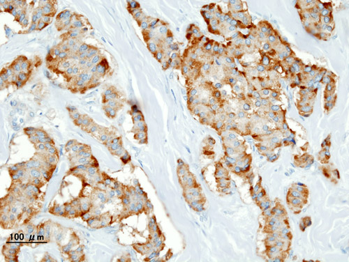
Chromogranin A immunostaining highlights tumour cells in this pancreatic insulinoma.
What to notice
Detailed image description
Histopathology of a pancreatic neuroendocrine tumour (insulinoma) stained for chromogranin A, with brown positive immunostaining.
Do not order pancreatic imaging merely because insulin is “within the normal range.” Insulin should be suppressed during hypoglycaemia; endogenous hyperinsulinism must first be demonstrated using a properly collected critical sample.
Critical Management
Conscious and Able to Swallow
- Give approximately 15 g of fast-acting carbohydrate, such as glucose tablets or gel containing 15 g carbohydrate, 6–7 regular jellybeans, 150 mL regular soft drink, 125 mL fruit juice, or 3 teaspoons of sugar or honey.
- Recheck BGL after 15 minutes.
- If BGL remains below 4.0 mmol/L, repeat the fast-acting carbohydrate.
- Once BGL is above 4.0 mmol/L and symptoms have resolved, provide a meal or longer-acting carbohydrate if the next meal is not imminent.1,3
Altered Consciousness, Seizure or Unsafe Swallow
- Call for emergency assistance.
- Place the patient in the lateral position and protect the airway.
- Do not give food, drink or glucose gel orally.
- Administer intravenous glucose according to the local emergency protocol.
- If intravenous access is unavailable, administer glucagon IM or subcutaneously if available.
- Recheck BGL after approximately 10 minutes.
- Repeat treatment or commence a glucose infusion if hypoglycaemia persists.
- Investigate and treat the underlying cause.
Glucagon may be less effective when hepatic glycogen stores are depleted, including prolonged fasting, malnutrition, severe liver disease and alcohol-related hypoglycaemia.
Sulfonylurea-Associated Hypoglycaemia
- Expect recurrent hypoglycaemia after initial glucose correction.
- Admit for prolonged glucose monitoring.
- Use intravenous glucose for immediate correction.
- Discuss octreotide with toxicology or endocrinology to suppress recurrent insulin secretion.
- Do not discharge after a single normal glucose reading.
Prevention of Recurrence
Before discharge or transfer:
- Identify and correct the precipitating factor.
- Review insulin and glucose-lowering medication doses.
- Consider renal function, hepatic function, food intake, exercise and alcohol use.
- Ensure access to fast-acting carbohydrate and glucagon where appropriate.
- Provide education to the patient, family and carers.
- Review hypoglycaemia awareness and driving safety.
- Consider continuous glucose monitoring for people at high risk of recurrent hypoglycaemia.4
- Arrange diabetes or endocrinology follow-up where indicated.
After correcting the glucose, always determine why the episode occurred. Long-acting insulin and sulfonylureas can cause recurrent hypoglycaemia many hours after the initial apparent recovery.
References
- Diabetes Australia. Hypoglycaemia (hypo) and hyperglycaemia [Internet]. Canberra (AU): Diabetes Australia; [cited 2026 Aug 13]. Available from: https://www.diabetesaustralia.com.au/managing-diabetes/hypo-hyperglycaemia/
- Cryer PE, Axelrod L, Grossman AB, Heller SR, Montori VM, Seaquist ER, et al. Evaluation and management of adult hypoglycemic disorders: an Endocrine Society clinical practice guideline. J Clin Endocrinol Metab. 2009;94(3):709-728. doi:10.1210/jc.2008-1410
- National Diabetes Services Scheme. Low blood glucose levels (hypoglycaemia) [Internet]. Canberra (AU): Diabetes Australia; [cited 2026 Aug 13]. Available from: https://www.ndss.com.au/managing-diabetes/blood-glucose-monitoring/hypoglycaemia/
- McCall AL, Lieb DC, Gianchandani R, MacMaster HW, Maynard GA, Murad MH, et al. Management of individuals with diabetes at high risk for hypoglycemia: an Endocrine Society clinical practice guideline. J Clin Endocrinol Metab. 2023;108(3):529-562. doi:10.1210/clinem/dgac596


Members only discussions coming soon…