

Fungal Pneumonia
Overview
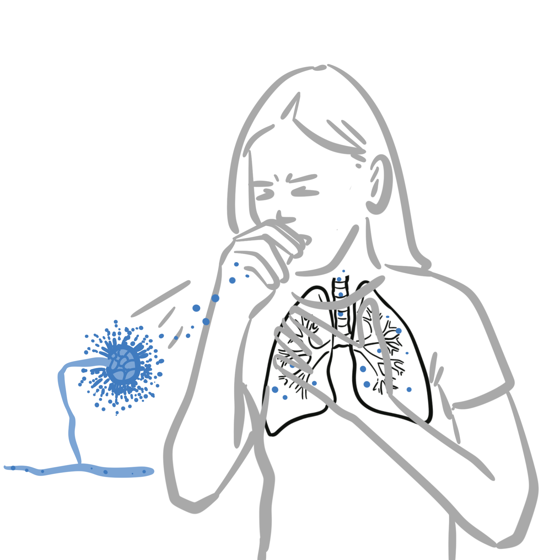
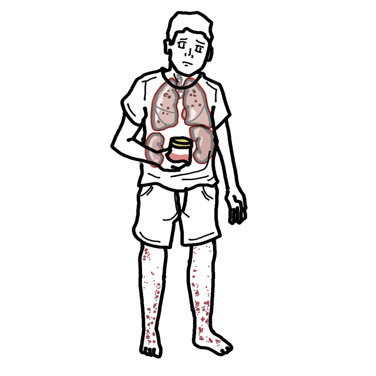
Fungal pneumonia refers to infection of the lung parenchyma caused by pathogenic fungi. It is not a single disease: presentation, diagnostic testing and treatment depend on the organism, the patient’s immune status, underlying lung disease, and environmental or geographic exposure. Endemic fungal pneumonias such as histoplasmosis, coccidioidomycosis and blastomycosis may occur in otherwise healthy people after inhalational exposure, whereas invasive aspergillosis, cryptococcosis and Pneumocystis jirovecii pneumonia (PCP) occur predominantly in susceptible hosts.
Fungal infection should be considered when a presumed community-acquired pneumonia is unusually prolonged, recurrent or unresponsive to antibacterial therapy, when imaging is atypical, or when the host is immunocompromised. Delay in recognising endemic fungal pneumonia is common because symptoms overlap substantially with bacterial and viral pneumonia.
Definition
- Fungal pneumonia
- Infection of the lung parenchyma caused by a pathogenic fungus.
- Endemic mycosis
- A fungal infection linked to environmental exposure in a defined geographic region; important pulmonary examples include histoplasmosis, coccidioidomycosis and blastomycosis.
- Opportunistic mycosis
- A fungal infection that occurs mainly when host immune defences or normal lung architecture are impaired.
Host factors first. Invasive fungal pneumonia is most important in patients with neutropenia, haematological malignancy, transplantation, prolonged corticosteroid use, advanced immunosuppression, or other major defects in host defence.
Classification
| Pattern | Important organisms | Typical setting | Useful clue |
|---|---|---|---|
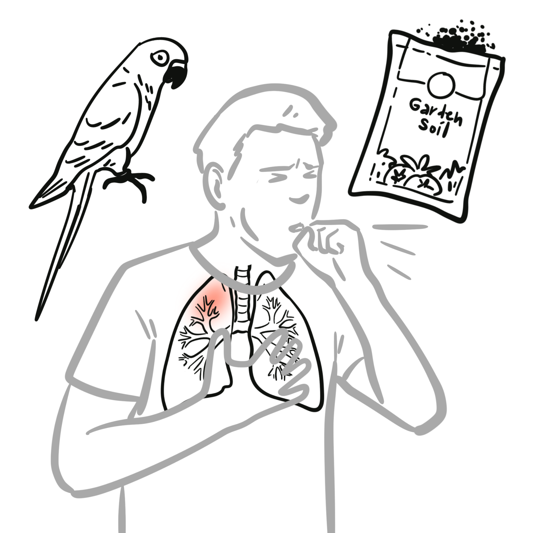
| Endemic fungal pneumonia | Histoplasma, Coccidioides, Blastomyces | Exposure in an endemic area; may affect immunocompetent or immunocompromised hosts | Travel, residence, soil disruption, cave/bird/bat exposure or outdoor activities |
| Invasive mould infection | Aspergillus | Prolonged neutropenia, haematological malignancy, stem-cell or solid-organ transplantation, high-dose corticosteroids or other major immunosuppression | Persistent fever, pleuritic pain, haemoptysis, nodules or infarct-like lesions on CT |
| Cryptococcal pneumonia | Cryptococcus neoformans, C. gattii | Especially advanced HIV or other impaired cell-mediated immunity; C. gattii can also affect immunocompetent people | Pulmonary nodules or pneumonia with possible CNS dissemination |
| Pneumocystis pneumonia | Pneumocystis jirovecii | Advanced HIV and other significant T-cell immunosuppression | Subacute dyspnoea, dry cough, hypoxaemia and diffuse bilateral ground-glass change |
Aetiology & Risk Factors
Aetiology
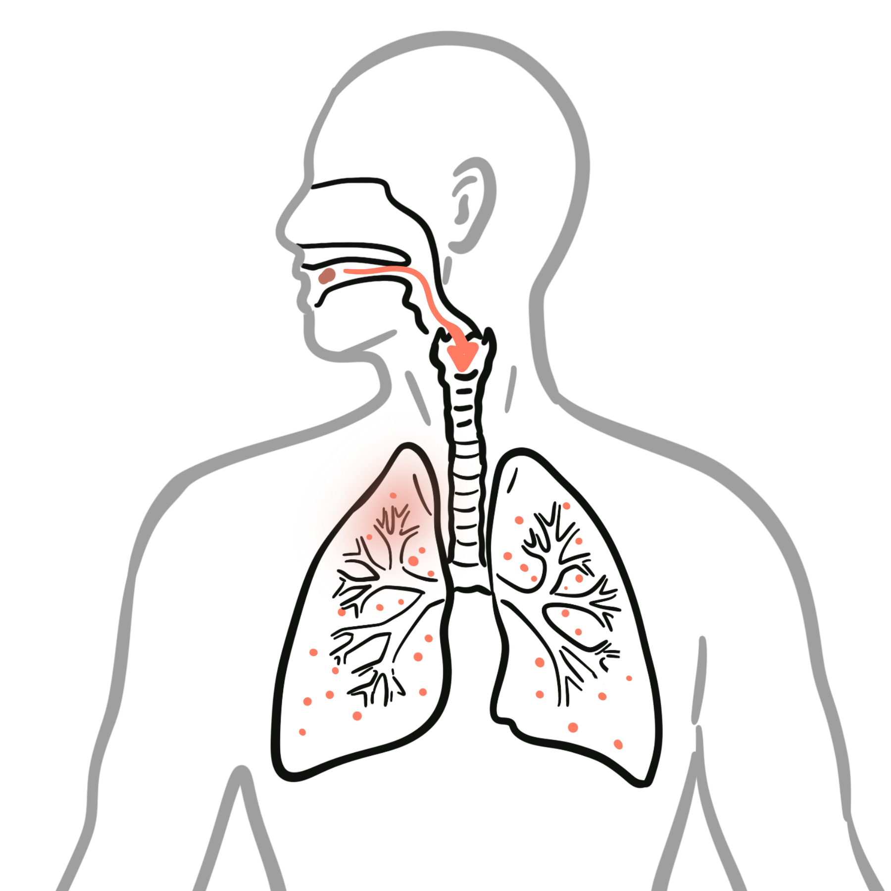
Most pulmonary fungal infections are acquired by inhalation of environmental spores or other infectious propagules. Endemic dimorphic fungi establish primary pulmonary infection after environmental exposure, while Aspergillus spores are inhaled routinely but cause invasive disease mainly when host defences are impaired. Cryptococcus is also acquired by inhalation and may later disseminate, particularly to the central nervous system. PCP reflects infection with P. jirovecii in a susceptible host.1,5,7,8
True Candida pneumonia is very uncommon. Growth of Candida from sputum, tracheal aspirate or bronchoalveolar lavage usually represents airway colonisation rather than invasive lung infection, and antifungal therapy should not be started on the basis of a respiratory culture alone. See Candida Infections.
Risk Factors
- Residence in or travel to an endemic fungal region, particularly with soil-disturbing activities or heavy environmental exposure.
- Advanced HIV infection, especially for PCP and cryptococcosis.
- Prolonged neutropenia, haematological malignancy, haematopoietic stem-cell transplantation or solid-organ transplantation.
- Systemic corticosteroids, biologic immunosuppression, cytotoxic therapy or other impairment of cell-mediated immunity.
- Structural lung disease, particularly for chronic pulmonary aspergillosis.
Persistent or recurrent fever despite appropriate antibacterial therapy in a high-risk immunocompromised patient should raise suspicion for an invasive fungal infection.
Pathophysiology
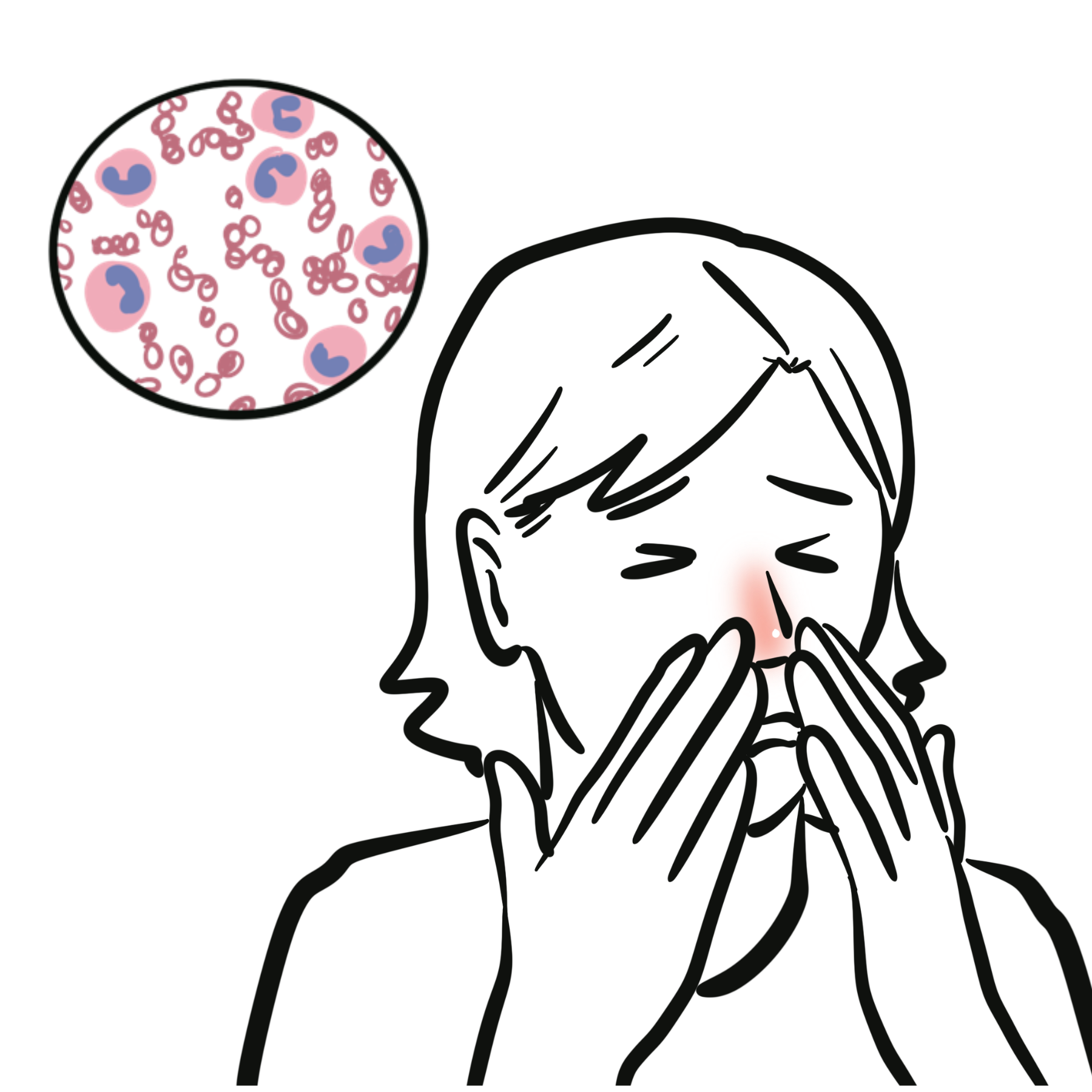
After inhalation, fungal organisms interact with alveolar macrophages, neutrophils and cell-mediated immunity. In immunocompetent hosts, many exposures are asymptomatic or contained within granulomatous inflammation. When inoculum is large, local pulmonary defences are abnormal, or cellular and neutrophil function is impaired, organisms can proliferate and produce necrosis, diffuse alveolar injury or angioinvasion. Angioinvasive aspergillosis can cause vascular thrombosis, pulmonary infarction and haemorrhage, while cryptococcosis and endemic mycoses may disseminate haematogenously beyond the lung.
Clinical Manifestations
Symptoms are often non-specific and may resemble bacterial pneumonia: fever, cough, dyspnoea, malaise, chest discomfort and constitutional symptoms. A subacute course over days to weeks, weight loss, night sweats, haemoptysis, persistent symptoms despite antibiotics, unusual exposure history or marked immunosuppression should increase suspicion for a fungal cause.
Specific features based on fungal cause
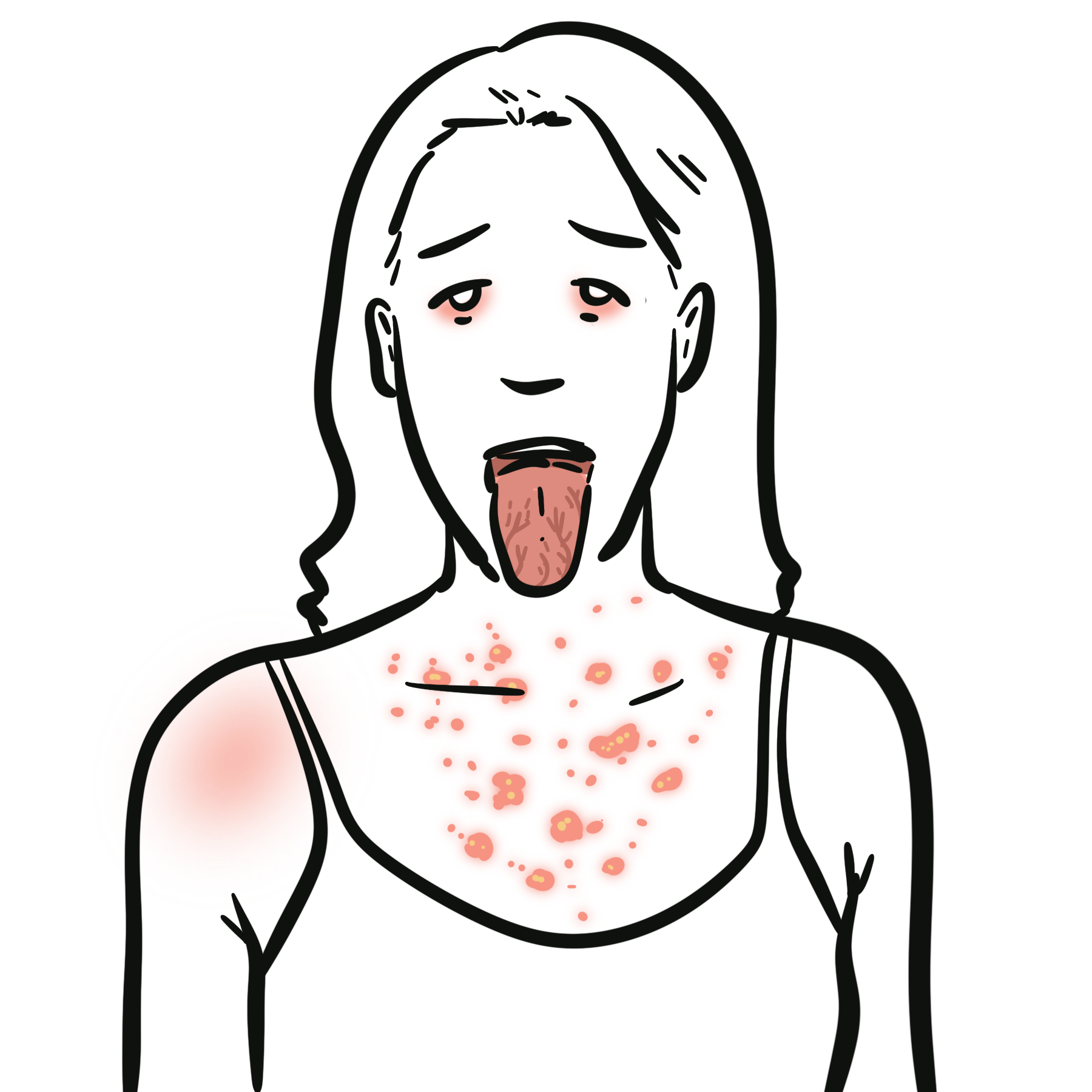
- Histoplasmosis: may be asymptomatic or cause an acute or subacute febrile pneumonia; hilar or mediastinal lymphadenopathy may occur. Severe pulmonary or disseminated disease is more likely with major immunosuppression.
- Coccidioidomycosis: commonly causes a community-acquired pneumonia syndrome after residence or travel in an endemic region; fatigue can be prolonged, and dissemination is more likely in selected high-risk hosts.
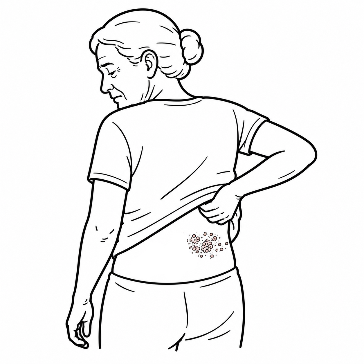
- Blastomycosis: may present with acute or chronic pneumonia and can disseminate to skin, bone, genitourinary tract or CNS.
- Invasive pulmonary aspergillosis: fever, cough, pleuritic chest pain, dyspnoea and haemoptysis in a high-risk immunocompromised host; dissemination may involve the brain, skin or other organs.
- Pulmonary cryptococcosis: cough, fever, chest pain or pulmonary nodules; headache, confusion or other neurological symptoms raise concern for CNS dissemination.
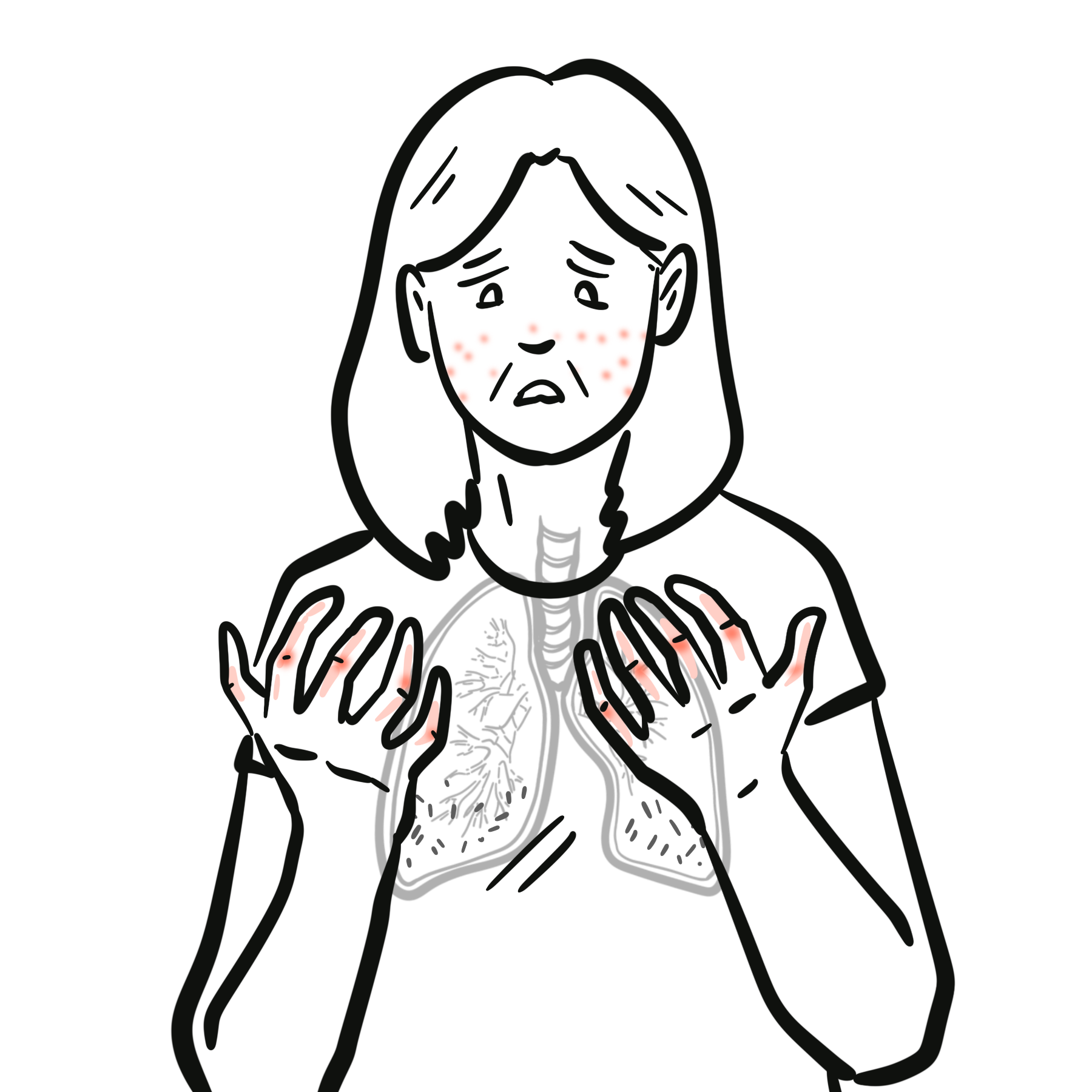
- PCP: classically causes progressive exertional dyspnoea, non-productive cough, fever and hypoxaemia, often with diffuse bilateral ground-glass opacities.
Diagnosis & Investigations
There is no single investigation that diagnoses all fungal pneumonias. The diagnostic approach should combine host risk, geography and exposure, chest imaging, targeted fungal biomarkers, respiratory sampling and—when needed—histopathology or tissue culture. Testing should be organism-directed rather than ordering every fungal assay indiscriminately.
Remember: immune status and exposure history are often as important as the chest CT. Ask where the patient has lived or travelled, what environmental exposures occurred, and which immune defects are present before selecting fungal tests.
Initial assessment
- Full exposure and travel history, including endemic regions, soil disruption, caves, bird or bat-associated environments, construction and occupational exposures.
- Immune status: HIV risk/status and CD4 count where relevant, neutrophil count, transplant history, malignancy and immunosuppressive medicines.
- Chest radiograph initially; CT chest when disease is severe, radiography is non-diagnostic, an opportunistic infection is suspected or complications need definition.
- Routine blood tests including full blood count, renal and liver function; arterial blood gas when hypoxaemia or PCP severity needs assessment.
Targeted microbiological tests
- Histoplasma: urine and/or serum antigen are useful particularly in more extensive disease; serology, respiratory culture and histopathology may contribute depending on the syndrome.
- Coccidioides: serology is central to diagnosis; culture or tissue examination may be required when serology is negative but suspicion remains high.
- Blastomyces: urine/serum antigen can provide a rapid clue, with culture and microscopy or histopathology used for confirmation; antigen cross-reactivity with histoplasmosis is important.
- Aspergillus: serum and bronchoalveolar lavage galactomannan, fungal culture, microscopy/histopathology and selected molecular assays are used according to host and syndrome.
- Cryptococcus: serum cryptococcal antigen and culture are important; suspected dissemination requires evaluation for CNS disease, often including lumbar puncture when clinically appropriate.
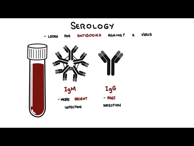
- PCP: respiratory PCR or direct organism detection on induced sputum or bronchoalveolar lavage is used for diagnosis; serum beta-D-glucan can support but is not specific for PCP.
Bronchoscopy with bronchoalveolar lavage is particularly useful in severe disease, immunocompromised patients, non-diagnostic non-invasive testing or when several opportunistic infections remain possible. Tissue biopsy may be required when malignancy, inflammatory disease and fungal infection cannot otherwise be distinguished.
Treatment
Treatment is pathogen-specific and depends on disease severity, immune status, pregnancy, renal and hepatic function, antifungal interactions and local susceptibility patterns. Severe disease or invasive fungal infection generally warrants early infectious diseases and respiratory/microbiology input. In a patient with strongly suspected invasive pulmonary aspergillosis, antifungal therapy should not be delayed while diagnostic confirmation is pursued.
| Infection | Typical treatment approach |
|---|---|
| Acute pulmonary histoplasmosis | Mild disease in an immunocompetent patient often does not require routine antifungal therapy. When treatment is indicated, itraconazole is preferred; severe acute pulmonary disease is treated initially with amphotericin B, preferably liposomal amphotericin B. |
| Pulmonary coccidioidomycosis | Mild or improving uncomplicated disease may be observed. Debilitating, extensive or higher-risk disease is generally treated with an oral azole such as fluconazole or itraconazole. |
| Blastomycosis | Itraconazole is used for mild-to-moderate disease; amphotericin B is recommended for moderate-to-severe or CNS disease, usually followed by an azole step-down course. |
| Invasive pulmonary aspergillosis | Voriconazole is recommended primary therapy; isavuconazole or liposomal amphotericin B are important alternatives in selected patients. Treatment is usually prolonged and guided by clinical, radiological and immune recovery. |
| Pulmonary cryptococcosis | Mild-to-moderate isolated pulmonary disease is generally treated with fluconazole. Severe pulmonary disease is treated similarly to CNS disease with an amphotericin B plus flucytosine induction strategy followed by fluconazole. |
| PCP | Trimethoprim–sulfamethoxazole is first-line therapy. In HIV-associated moderate-to-severe PCP, adjunctive corticosteroids are indicated when PaO2 is <70 mmHg on room air or the alveolar–arterial oxygen gradient is ≥35 mmHg. |
Antifungal pharmacology is clinically important. Azoles have major drug–drug interactions and some require therapeutic drug monitoring; amphotericin formulations can cause nephrotoxicity and electrolyte disturbance; and treatment often extends for weeks to months. Reversal or reduction of immunosuppression, where feasible and safe, is an important adjunct in invasive fungal infection.
Complications & Prognosis
Complications
- Acute hypoxaemic respiratory failure and acute respiratory distress syndrome
- Pulmonary necrosis, cavitation, infarction or haemorrhage
- Chronic pulmonary infection or residual nodules/cavities
- Dissemination to the CNS, skin, bone or other organs, depending on the organism
- Relapse or progression when immune suppression persists
- Antifungal toxicity and clinically important drug interactions
Prognosis
Prognosis varies widely. Many mild endemic fungal pneumonias in immunocompetent people resolve without complication, while invasive aspergillosis, severe cryptococcosis and PCP can be life-threatening. Outcome is strongly influenced by early recognition, severity at presentation, the underlying immune defect, ability to achieve immune recovery, dissemination, antifungal susceptibility and treatment toxicity.
References
- Centers for Disease Control and Prevention. Fungal community-acquired pneumonias [Internet]. Atlanta: CDC; 2024 [updated 2024 May 8; cited 2026 Sep 30]. Available from: https://www.cdc.gov/fungal/cap/index.html
- Infectious Diseases Society of America. Histoplasmosis in adults, children, and pregnant individuals [Internet]. Arlington (VA): IDSA; 2026 [updated 2026 Sep 16; cited 2026 Sep 30]. Available from: https://www.idsociety.org/practice-guideline/histoplasmosis
- Galgiani JN, Ampel NM, Blair JE, et al. 2016 Infectious Diseases Society of America clinical practice guideline for the treatment of coccidioidomycosis. Clin Infect Dis. 2016;63(6):e112-e146. doi:10.1093/cid/ciw360
- Centers for Disease Control and Prevention. Clinical overview of blastomycosis [Internet]. Atlanta: CDC; 2024 [updated 2024 Apr 24; cited 2026 Sep 30]. Available from: https://www.cdc.gov/blastomycosis/hcp/clinical-overview/index.html
- Centers for Disease Control and Prevention. Clinical overview of aspergillosis [Internet]. Atlanta: CDC; 2024 [updated 2024 Apr 24; cited 2026 Sep 30]. Available from: https://www.cdc.gov/aspergillosis/hcp/clinical-overview/
- Patterson TF, Thompson GR III, Denning DW, et al. Practice guidelines for the diagnosis and management of aspergillosis: 2016 update by the Infectious Diseases Society of America. Clin Infect Dis. 2016;63(4):e1-e60. doi:10.1093/cid/ciw326
- Chang CC, Harrison TS, Bicanic TA, et al. Global guideline for the diagnosis and management of cryptococcosis: an initiative of the ECMM and ISHAM in cooperation with the ASM. Lancet Infect Dis. 2024;24(8):e495-e512. doi:10.1016/S1473-3099(23)00731-4
- National Institutes of Health. Pneumocystis pneumonia: adult and adolescent opportunistic infections guidelines [Internet]. Bethesda (MD): NIH; 2026 [updated 2026 May 27; cited 2026 Sep 30]. Available from: https://clinicalinfo.hiv.gov/en/guidelines/hiv-clinical-guidelines-adult-and-adolescent-opportunistic-infections/pneumocystis
- Pappas PG, Kauffman CA, Andes DR, et al. Clinical practice guideline for the management of candidiasis: 2016 update by the Infectious Diseases Society of America. Clin Infect Dis. 2016;62(4):e1-e50. doi:10.1093/cid/civ933





Members only discussions coming soon…