

Candida Infections
Overview
Candida species are yeasts that normally colonise human skin and the gastrointestinal and genitourinary tracts. Disease occurs when local microbial balance or epithelial defences are disrupted, or when organisms enter normally sterile tissues.
Candida infections range from oropharyngeal, oesophageal, vulvovaginal and cutaneous candidiasis to invasive disease such as Candidaemia, intra-abdominal candidiasis, endocarditis, endophthalmitis, osteoarticular infection, central nervous system infection and chronic disseminated candidiasis.
Candida albicans remains important, but non-albicans Candida species increasingly influence treatment because epidemiology and antifungal susceptibility differ. Isolation of Candida does not always indicate infection; interpretation depends on the anatomical site, immune status, inflammatory response and whether the sample came from a normally sterile site.
Definition
- Candidiasis
- Infection caused by a Candida species, ranging from superficial mucosal disease to invasive infection involving the bloodstream or internal organs.
- Candidaemia
- The presence of Candida in the bloodstream. It should be treated as clinically significant and prompts evaluation for an invasive source and metastatic complications.
- Invasive candidiasis
- Candida infection involving the bloodstream, a normally sterile body site or a deep organ.
- Colonisation
- The presence of Candida without tissue invasion or clinically significant host injury.
Epidemiology
Candida species are among the most important causes of healthcare-associated fungal infection. Invasive candidiasis predominantly affects people who are critically ill, immunocompromised or exposed to invasive medical care.1
The most frequent invasive pathogens include C. albicans, C. glabrata, C. parapsilosis, C. tropicalis and C. krusei. Species distribution varies with geography, patient population, previous antifungal exposure and local infection-control conditions.
Candida auris
Candida auris—also known under the revised name Candidozyma auris—is an emerging healthcare-associated pathogen. It can cause outbreaks, persist on skin and environmental surfaces, be misidentified by some laboratory systems and show resistance to multiple antifungal classes. Infection requires specialist treatment and enhanced infection-prevention measures.
Taxonomic revisions have changed the names of several clinically important yeasts. Laboratories may report both an established clinical name and a newer taxonomic name. Treatment should be guided by accurate species identification and susceptibility rather than the name alone.
Aetiology and Risk Factors
Mucocutaneous candidiasis
- Recent broad-spectrum antibiotics
- Inhaled or systemic corticosteroids
- Diabetes mellitus, particularly when poorly controlled
- Pregnancy and increased oestrogen exposure
- HIV infection or impaired cell-mediated immunity
- Chemotherapy or other immunosuppression
- Dentures, poor oral hygiene or xerostomia
- Extremes of age
- Obesity, moist skin folds, occlusion and maceration
Recurrent, severe or persistent mucosal candidiasis should prompt assessment for an underlying predisposition rather than repeated empirical treatment alone.
Invasive candidiasis
- Critical illness and prolonged intensive care admission
- Central venous catheters
- Broad-spectrum antibacterial therapy
- Major abdominal surgery, gastrointestinal perforation or anastomotic leak
- Necrotising pancreatitis
- Total parenteral nutrition
- Haematological malignancy, neutropenia or transplantation
- Renal replacement therapy
- Severe immunosuppression
- Prematurity and very low birth weight
- Extensive burns
- Injection drug use
- Previous multifocal Candida colonisation or azole exposure
Most invasive infections arise from endogenous flora after disruption of mucosal or cutaneous barriers. Infection may also arise from a colonised intravascular device or, less commonly, healthcare-associated transmission.
A critically ill patient with persistent fever despite broad-spectrum antibacterial therapy, a central venous catheter, recent gastrointestinal surgery and total parenteral nutrition has a substantially increased pre-test probability of invasive candidiasis. Obtain appropriate cultures, assess the source and consider timely antifungal therapy.
Pathophysiology
Local disease develops when ecological or immune changes permit excessive growth and epithelial invasion. Virulence mechanisms include adhesion, yeast-to-hyphal transition, secretion of tissue-damaging enzymes, immune evasion and biofilm formation. Neutrophils are particularly important in controlling invasive infection, whereas cell-mediated immunity helps prevent chronic or recurrent mucosal disease.
Biofilm formation
Candida biofilms form on central and urinary catheters, prosthetic valves, cardiac devices, dentures and other implanted material. Biofilms protect organisms from host defences and reduce susceptibility to some antifungals, so device removal may be essential.
Bloodstream invasion and dissemination
Organisms may enter blood through disrupted gastrointestinal mucosa, an intra-abdominal infection, a colonised central line, damaged skin or injection. Haematogenous spread can seed the eyes, heart valves, brain, kidneys, liver, spleen, bones and joints.
Successful management of invasive candidiasis requires more than choosing an antifungal. Look for a removable intravascular device, undrained collection, gastrointestinal leak, infected prosthesis or another source requiring procedural control.
Clinical Manifestations
Oropharyngeal candidiasis
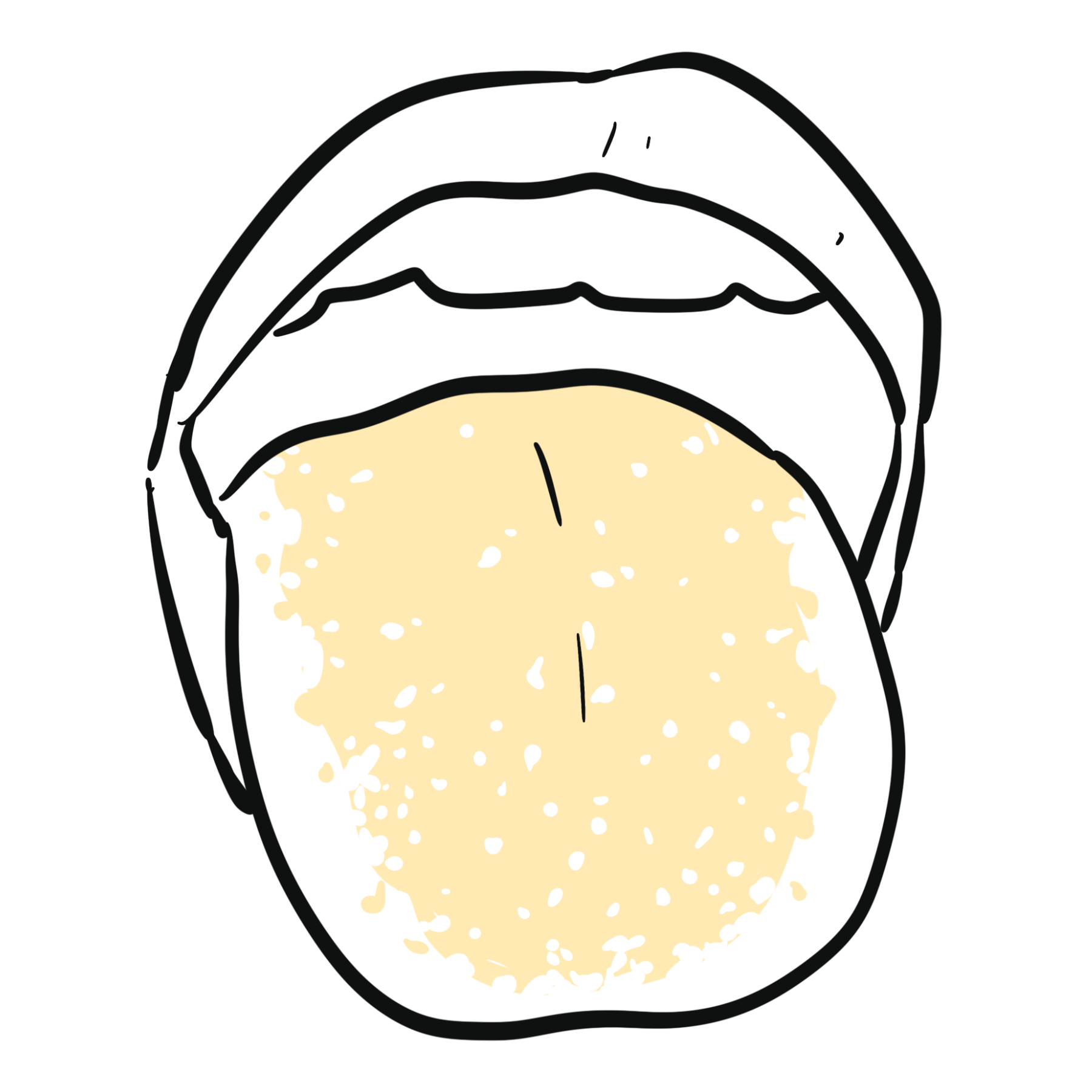
Typical findings are creamy white plaques on the tongue, palate or buccal mucosa that can often be wiped away, leaving an erythematous or friable surface. Oral burning, altered taste, angular cheilitis and feeding difficulty may occur. Erythematous candidiasis produces painful red mucosa without prominent plaques.
Oesophageal candidiasis
Oesophageal disease causes odynophagia, dysphagia, retrosternal discomfort and reduced oral intake. Oral thrush may be absent. The diagnosis should raise concern for significant immunosuppression.
Vulvovaginal candidiasis
Features include vulval pruritus, burning, soreness, external dysuria, erythema, oedema, fissures and thick white discharge. These symptoms are not specific and overlap with bacterial vaginosis, sexually transmitted infections, dermatitis, inflammatory dermatoses and vulvodynia.
Cutaneous candidiasis
Cutaneous infection affects moist opposing surfaces and causes bright erythema, maceration, peripheral scale and satellite papules or pustules. Common sites include inframammary folds, groin, axillae, abdominal folds, interdigital spaces and the perineal or napkin area.
Candidaemia and invasive candidiasis
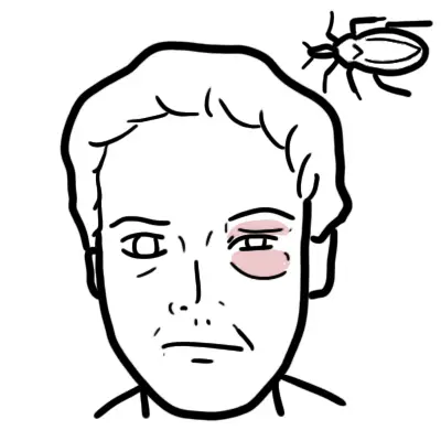
Invasive disease often presents non-specifically with persistent fever, rigors, hypotension, sepsis, organ dysfunction or deterioration despite antibacterial therapy. Endocarditis may cause embolic phenomena or heart failure; endophthalmitis may cause floaters, pain or visual loss; osteoarticular disease produces focal pain; and intra-abdominal candidiasis causes persistent postoperative or perforation-associated sepsis.
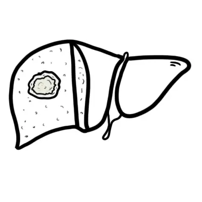
Hepatosplenic candidiasis typically causes persistent fever and hepatic or splenic lesions during neutrophil recovery.
Persistent positive blood cultures, focal symptoms or failure to improve should trigger a directed search for an uncontrolled source, antifungal resistance, endocarditis, endophthalmitis or another deep-organ focus.
Diagnosis
Investigation should determine whether Candida represents colonisation or infection, whether disease is superficial or invasive, the source and extent of infection, and the species and susceptibility profile.
Mucocutaneous candidiasis
- Typical uncomplicated oral or cutaneous disease is diagnosed clinically.
- Microscopy or culture is most useful when the appearance is atypical, treatment fails, infection recurs, a resistant or non-albicans species is suspected, or an alternative diagnosis remains plausible.
- For Vaginal candidiasis: A positive vaginal culture without compatible symptoms does not establish vulvovaginal candidiasis.
- Endoscopy is reserved for uncertain or refractory suspected oesophageal disease
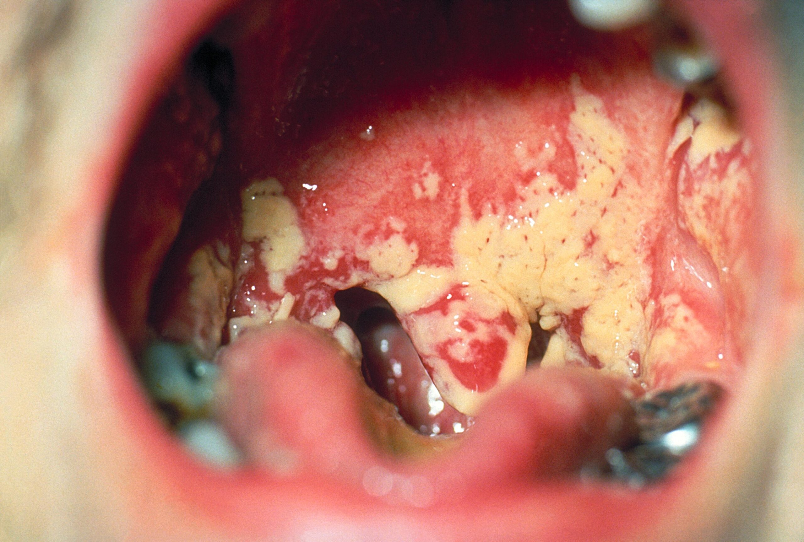
Pseudomembranous oral candidiasis showing extensive white plaques over the oral mucosa. Centers for Disease Control and Prevention, PHIL 1217; public domain.
What to notice
Detailed image description
Source: https://commons.wikimedia.org/wiki/File:Oral_thrush_Aphthae_Candida_albicans._PHIL_1217_lores.jpg | Creator/rights holder: Centers for Disease Control and Prevention, Public Health Image Library 1217 | Rights: public domain, US federal government work | Modifications: none.
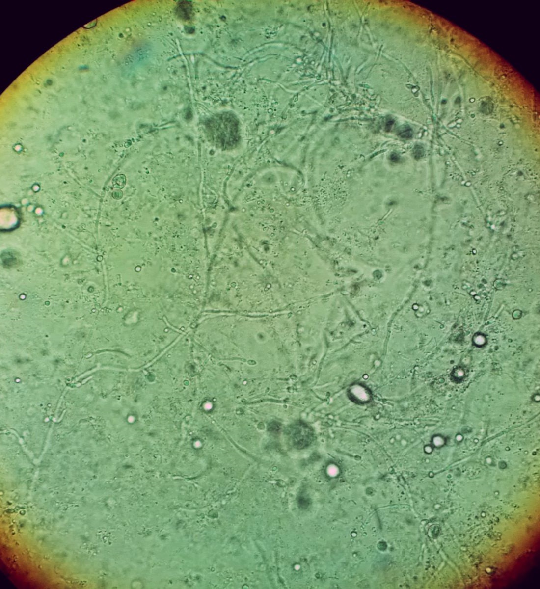
Potassium-hydroxide vaginal wet mount showing Candida pseudohyphae. The larger pale circles are preparation artefacts. Mikael Häggström, CC0 1.0.
What to notice
Detailed image description
Source: https://commons.wikimedia.org/wiki/File:Vaginal_wet_mount_in_candidal_vulvovaginitis.jpg | Creator: Mikael Häggström | Licence: CC0 1.0 Universal | Modifications: none.
Suspected Candidaemia
Obtain appropriately collected blood-culture sets before antifungal therapy when this will not delay urgent treatment. Once Candida grows from blood, identify the species, perform susceptibility testing as appropriate, repeat cultures daily or every second day until clearance, review vascular devices and assess for an intra-abdominal, urinary, cardiac or other source.
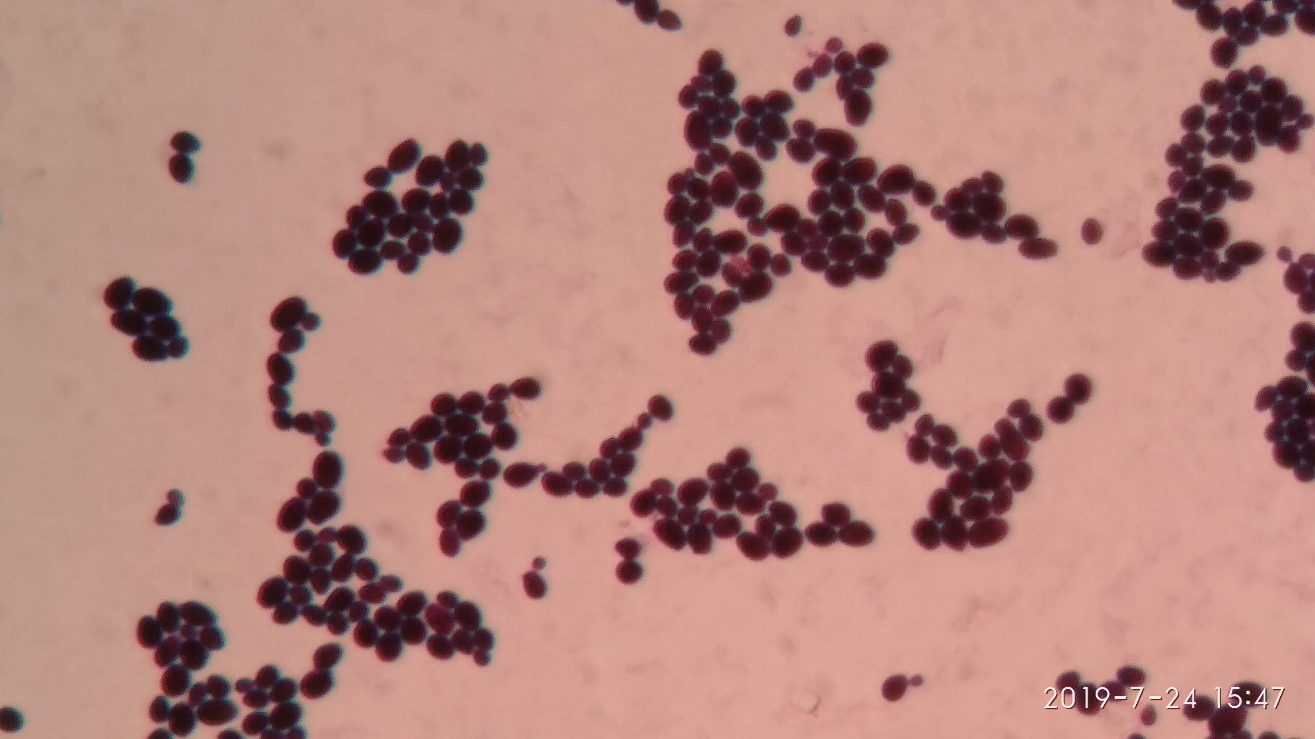
Gram-stained culture showing oval Gram-positive yeast cells of Candida albicans. Ajay Kumar Chaurasiya, CC0 1.0.
What to notice
Detailed image description
Source: https://commons.wikimedia.org/wiki/File:Yeast_cells_of_Candida_albicans_in_Gram_staining_of_culture_microscopy.jpg | Creator: Ajay Kumar Chaurasiya | Licence: CC0 1.0 Universal | Modifications: none.
Blood cultures have imperfect sensitivity and negative cultures do not exclude deep-seated candidiasis, particularly infection confined to the abdomen or another organ.1
Candida in a blood culture should not be dismissed as a contaminant. It requires treatment, repeat cultures to document clearance, source assessment and evaluation for clinically suspected metastatic complications.
Non-culture fungal biomarkers
- Serum beta-D-glucan: can support the diagnosis of invasive fungal infection but is not specific for Candida
Directed evaluation for complications
- Echocardiography for persistent Candidaemia, prosthetic valves, cardiac devices, injection drug use, embolic features or suspected endocarditis
- Ophthalmological assessment for ocular symptoms or signs, inability to report symptoms, or selected high-risk patients according to specialist protocols
- CT abdomen and pelvis for a suspected intra-abdominal source, recent surgery, anastomotic leak or persistent sepsis
- MRI for suspected vertebral osteomyelitis, central nervous system disease or another focal deep-tissue infection
- Image-guided aspiration or biopsy when a focal lesion requires microbiological and histological diagnosis
Routine indiscriminate imaging is less useful than careful source assessment and symptom-directed investigation.
Candiduria
Candida in urine frequently represents colonisation, especially with a urinary catheter. Assess urinary symptoms, catheter necessity, recent instrumentation, obstruction, neutropenia, pregnancy, planned urological procedures and evidence of bloodstream infection. Removing or replacing an unnecessary catheter may be the most important intervention. Asymptomatic candiduria usually does not require antifungal treatment except in selected high-risk situations.
Respiratory specimens
Candida commonly colonises hospitalised patients’ airways. Isolation from sputum, tracheal aspirate or bronchoalveolar lavage usually represents colonisation rather than Candida pneumonia; convincing evidence of tissue invasion is generally required.
Interpret Candida by site: blood or another sterile site indicates clinically significant infection until assessed; urine commonly reflects colonisation; respiratory secretions almost always reflect colonisation rather than Candida pneumonia.
Differential Diagnosis
Oral lesions
- Leukoplakia
- Oral lichen planus
- Aphthous ulceration
- Geographic tongue
- Oral hairy leukoplakia
- Mucositis
- Nutritional deficiency
- Oral malignancy
Unlike candidal plaques, leukoplakia is generally not easily wiped away.
Vulvovaginal symptoms
- Bacterial vaginosis
- Trichomoniasis
- Genital herpes
- Contact or irritant dermatitis
- Lichen sclerosus or lichen planus
- Desquamative inflammatory vaginitis
- Vulvodynia
Persistent fever in a high-risk patient
- Uncontrolled bacterial infection
- Invasive aspergillosis or another mould infection
- Infected intravascular catheter
- Drug fever
- Venous thromboembolism
- Transfusion reaction
- Malignancy-related fever
- Inflammatory disease
Treatment
Treatment depends on anatomical site, severity, immune status, previous antifungal exposure, species, susceptibility, organ function, drug interactions and source control.
Mucocutaneous disease
Mild oropharyngeal candidiasis can be treated with topical nystatin, miconazole oral gel or clotrimazole where available. Systemic fluconazole is used for extensive, severe or refractory disease. Address denture hygiene, inhaled corticosteroid technique, xerostomia, glycaemic control and unnecessary antibiotic exposure. Miconazole can substantially increase warfarin’s anticoagulant effect.
Oesophageal candidiasis requires systemic therapy, generally fluconazole when susceptibility is expected. Failure should prompt assessment for adherence, resistance or another cause of oesophagitis.
Uncomplicated vulvovaginal candidiasis is treated with a topical azole or oral fluconazole when appropriate. During pregnancy, topical azole therapy is preferred and oral fluconazole is generally avoided. Severe, recurrent or non-albicans infection requires confirmation and a longer or specialist-directed regimen.
Cutaneous disease is managed by keeping the area dry, reducing friction and occlusion, addressing maceration and diabetes, and using a topical azole or nystatin.
Candidaemia in non-neutropenic adults
An intravenous echinocandin is generally recommended as initial treatment for most adults.1,2 Fluconazole may be used initially in a clinically stable patient with no important recent azole exposure and a low probability of resistance. Step-down to fluconazole is appropriate when the patient is stable, the isolate is susceptible and repeat cultures are negative.
Uncomplicated Candidaemia is generally treated for at least 14 days after documented bloodstream clearance and resolution of attributable symptoms and signs. Deep-organ disease requires a longer site-specific course.
Source control
- Remove an implicated central venous catheter when feasible
- Drain intra-abdominal collections
- Repair or control gastrointestinal leaks
- Remove infected prosthetic material where possible
- Relieve urinary obstruction
- Remove or replace unnecessary urinary catheters
Neutropenic patients
An echinocandin is commonly used initially, with liposomal amphotericin B as an alternative in selected circumstances. Management must consider immune recovery, mucosal injury, previous prophylaxis and possible mould infection. Chronic disseminated candidiasis often requires prolonged therapy.
Candida auris
Management requires accurate identification, susceptibility testing, infectious diseases and microbiology involvement, an echinocandin as usual initial therapy for invasive disease, and enhanced infection-control precautions. Colonisation alone is not routinely treated with systemic antifungals.
If Candidaemia persists despite appropriate therapy, ask whether the catheter remains, an abdominal collection is undrained, the species is resistant, endocarditis or another metastatic focus is present, and antifungal dose and exposure are adequate.
Complications & Prognosis
Complications
- Septic shock and multiorgan failure
- Persistent or recurrent Candidaemia
- Endocarditis
- Endophthalmitis and visual loss
- Osteomyelitis or septic arthritis
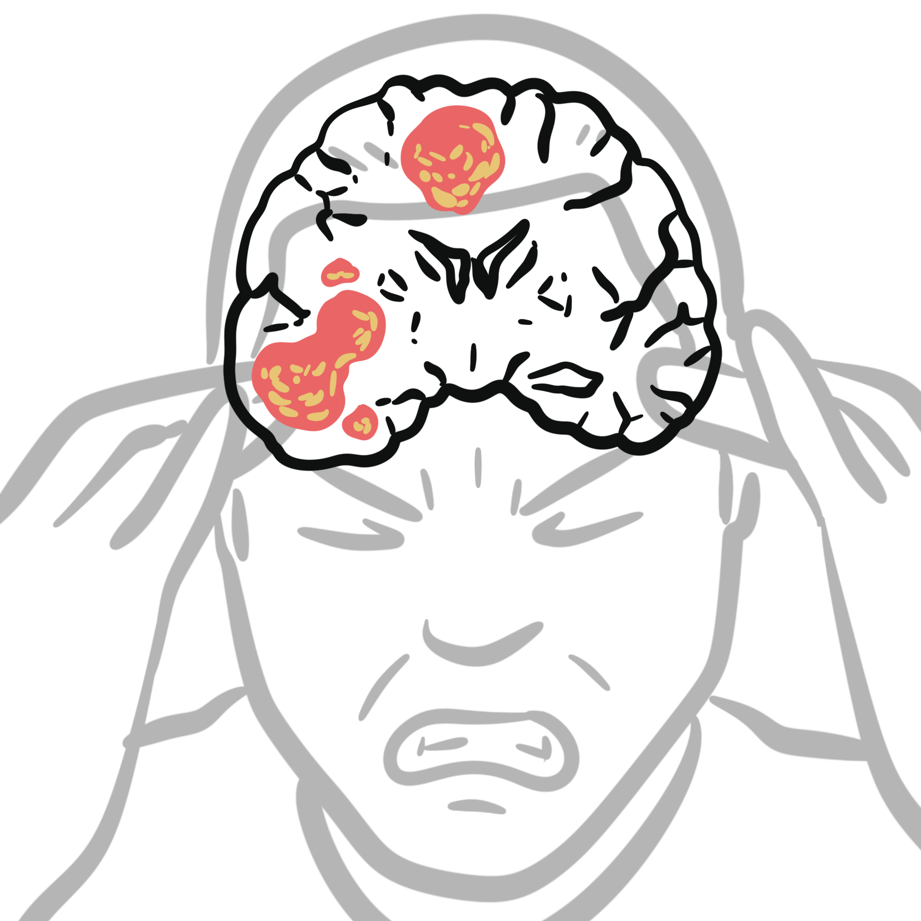
- Meningitis or brain abscess
- Renal microabscesses
- Hepatosplenic candidiasis
- Antifungal toxicity
- Emergence of resistance
- Relapse caused by inadequate source control
Prevention
- Hand hygiene and Candida auris infection-control precautions
- Evidence-based insertion and maintenance of central venous catheters
- Early removal of unnecessary invasive devices
- Antibacterial and antifungal stewardship
- Good oral and denture hygiene
- Mouth rinsing after inhaled corticosteroids
- Optimising diabetes control
- Keeping skin folds clean and dry
- Prompt management of gastrointestinal leaks
Antifungal prophylaxis is reserved for selected high-risk groups and should follow local epidemiology, resistance patterns and specialist protocols.
Prognosis
Superficial candidiasis generally responds well when the infection and its predisposing factors are addressed. Prognosis in invasive disease depends on severity at recognition, timing of active therapy, source control, species and susceptibility, immune recovery, septic shock, dissemination and comorbidity. Persistent or recurrent disease should prompt assessment for an unresolved source, infected prosthesis, inadequate duration or metastatic focus.
References
- Cornely OA, Sprute R, Bassetti M, et al. Global guideline for the diagnosis and management of candidiasis: an initiative of the ECMM in cooperation with ISHAM and ASM. Lancet Infect Dis. 2025;25(5):e280–e293. doi:10.1016/S1473-3099(24)00749-7.
- Pappas PG, Kauffman CA, Andes DR, et al. Clinical practice guideline for the management of candidiasis: 2016 update by the Infectious Diseases Society of America. Clin Infect Dis. 2016;62(4):e1–e50. doi:10.1093/cid/civ933.
- Centers for Disease Control and Prevention. Clinical overview of invasive candidiasis. Updated April 24, 2024. Accessed August 24, 2026. https://www.cdc.gov/candidiasis/hcp/clinical-overview/
- Centers for Disease Control and Prevention. Treatment of candidiasis. Updated April 24, 2024. Accessed August 24, 2026. https://www.cdc.gov/candidiasis/treatment/
- Centers for Disease Control and Prevention. Drug-resistant candidiasis. Updated December 15, 2025. Accessed August 24, 2026. https://www.cdc.gov/candidiasis/antimicrobial-resistance/








Members only discussions coming soon…