

Brain Abscess
Overview
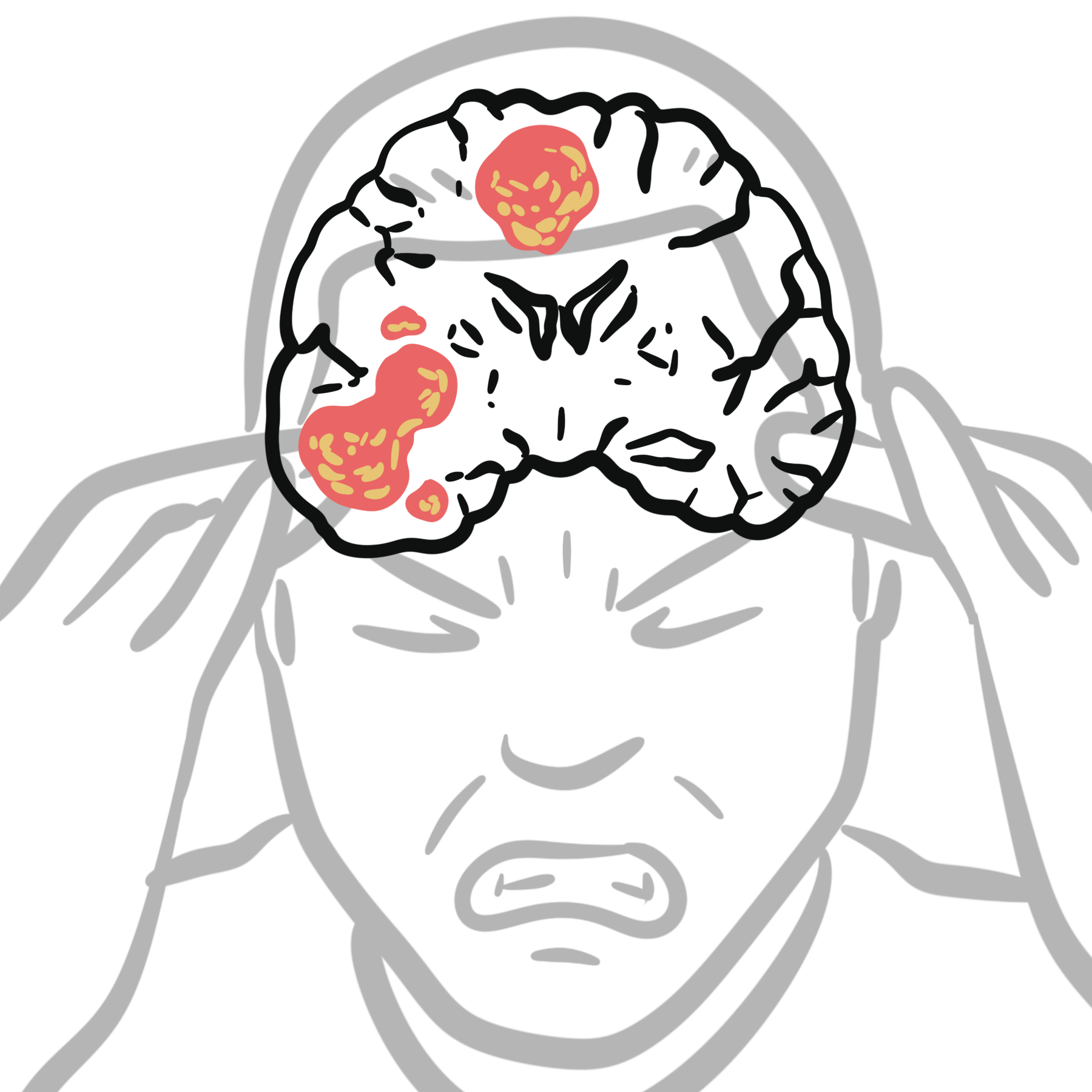
A brain abscess is a focal, encapsulated suppurative infection originating within the brain parenchyma. It begins as a localized area of unencapsulated parenchymal inflammation (cerebritis) that evolves over days to weeks into a pus-filled core surrounded by a vascularized collagen capsule.
Despite significant advances in neuroimaging, stereotactic neurosurgery, and broad-spectrum antimicrobial therapy, a brain abscess remains a severe, life-threatening neurological emergency requiring rapid medical and surgical intervention.
Definition
Cerebritis: The initial, unencapsulated stage of parenchymal brain inflammation characterized by local neutrophilic infiltration, microvascular congestion, and tissue necrosis.
Ring-Enhancing Lesion: A classic neuroimaging finding on contrast-enhanced CT or MRI demonstrating a central hypodense/hypointense non-enhancing necrotic core encircled by a brightly enhancing collagen capsule.
Contiguous Spread: Direct extension of infection into the brain parenchyma from adjacent parameningeal structures (e.g., otitis media, mastoiditis, or sinusitis).
Hematogenous (Metastatic) Dissemination: Bloodborne spread of microorganisms from a distant primary source of infection (e.g., infective endocarditis, pulmonary arteriovenous malformations) to the brain.
Aetiology & Risk Factors
Aetiology
Brain abscesses are frequently polymicrobial (~30–60% of cases).
- Streptococcus species: Most common overall (~30–50%), specifically the Streptococcus anginosus (S. milleri) group.
- Staphylococcus aureus: Common in post-traumatic, neurosurgical, or infective endocarditis cases.
- Anaerobes (Bacteroides, Peptostreptococcus, Fusobacterium): Frequent in otogenic, sinusogenic, and dental infections.
- Gram-Negative Bacilli (Enterobacteriaceae, Pseudomonas aeruginosa): Associated with otitis media, trauma, or hospital-acquired infections.
- Fungal & Parasitic (Aspergillus, Toxoplasma gondii, Nocardia, Cryptococcus): Primary causes in severely immunocompromised patients (e.g., HIV/AIDS, organ transplant recipients).
Risk Factors
- Chronic sinusitis, otitis media, or mastoiditis.
- Cyanotic congenital heart disease with right-to-left shunts (e.g., Tetralogy of Fallot).
- Pulmonary arteriovenous malformations (e.g., Hereditary Hemorrhagic Telangiectasia / Osler-Weber-Rendu syndrome).
- Immunosuppression (chemotherapy, biological agents, advanced HIV).
- Penetrating head injury or recent neurosurgery.
Pathophysiology
Mechanism of Acquisition
- Contiguous Spread (~45–50%): Direct extension from neighboring cranial infections.
- Otogenic (Middle ear / Mastoid): Spreads to the temporal lobe or cerebellum.
- Sinusogenic (Paranasal sinuses): Spreads primarily to the frontal lobe.
- Dental / Facial: Spreads to the frontal or temporal lobes.
- Hematogenous / Metastatic Spread (~25–35%): Bloodborne seeded from distant organs (e.g., bronchiectasis, lung abscess, cyanotic congenital heart disease, endocarditis). Characteristically produces multiple abscesses localized at the gray-white matter junction within the Middle Cerebral Artery (MCA) distribution.
- Post-Traumatic / Iatrogenic (~10%): Resulting from penetrating head trauma or neurosurgical procedures (e.g., open skull fractures, retained foreign bodies).
- Cryptogenic (~15–20%): Primary source of infection remains unidentified despite extensive clinical workup.
The histological evolution of a brain abscess follows four distinct stages (Britt and Enzmann criteria):
- Early Cerebritis (Days 1–3): Localized tissue destruction, acute neutrophilic infiltration, microvascular congestion, and surrounding cerebral edema.
- Late Cerebritis (Days 4–9): Central liquefactive necrosis develops; maximum surrounding vasogenic edema.
- Early Capsule Formation (Days 10–13): Fibroblastic proliferation and neovascularization around the necrotic core, creating a reticulin grid and early collagen capsule.
- Late Capsule Formation (Days 14+): Dense, mature collagenous capsule with surrounding reactive gliosis.
The medial (ventricular) wall of the abscess capsule is thinner and less vascular than the lateral (cortical) wall due to poorer deep white matter blood supply. This makes rupture into the ventricular system a critical risk, leading to fatal ventriculitis.
Clinical Manifestations
Symptom Triad
The classic triad is present in less than 50% of patients:
- Fever (often low-grade or absent in late stages)
- Headache (most common symptom; dull, constant, localized or diffuse)
- Focal Neurological Deficit (dependent on abscess location)
Systemic & Intracranial Signs
- Signs of Raised Intracranial Pressure (ICP)
- Seizures: Focal or generalized seizures occur in 25–50% of patients.
Anatomical Localizing Features
| Abscess Location | Key Localizing Features |
| Frontal Lobe | Hemiparesis, executive dysfunction, behavioral changes, expressive aphasia (Broca’s). |
| Temporal Lobe | Receptive aphasia (Wernicke’s), superior homonymous quadrantanopia (“pie in the sky” visual defect). |
| Cerebellum | Ipsilateral ataxia, dysmetria, nystagmus, intention tremor. |
| Parietal Lobe | Contralateral sensory loss, hemianopia, spatial neglect. |
Diagnosis & Investigations
Neuroimaging
- Magnetic Resonance Imaging (MRI Brain with Contrast + DWI): Gold standard diagnostic modality.
- Diffusion-Weighted Imaging (DWI): Demonstrates restricted diffusion (bright signal on DWI, dark on ADC map) within the central necrotic core—essential for differentiating an abscess from a necrotic tumor.
- T1-Weighted Contrast: Shows smooth, thin, uniform ring-enhancement.
- Computed Tomography (CT Brain with Contrast): Rapid emergency initial test; shows a hypodense central core with a smooth ring-enhancing capsule and surrounding low-density vasogenic edema.
Laboratory Workup
- Elevated Inflammatory Markers: Leukocytosis, elevated ESR and CRP.
- Blood Cultures: Positive in 25–30% of cases; obtain prior to starting antimicrobial therapy.
- Stereotactic Aspiration Sample: Gold standard for microbiological culture and Gram stain.
Differential Diagnosis of Ring-Enhancing Lesions (“MAGIC DR” Mnemonic)
- M: Metastasis
- A: Abscess
- G: Glioblastoma (GBM)
- I: Infarct (subacute / hemorrhagic)
- C: Contusion / Demyelination (tumescent MS)
- D: Radiation necrosis
- R: Resolving hematoma
Lumbar Puncture (LP) is strictly CONTRAINDICATED in suspected brain abscess due to the high risk of fatal transtentorial or tonsillar brain herniation secondary to focal mass effect.
Treatment
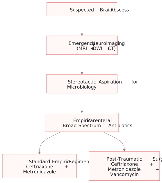
Initial brain abscess management: urgent neuroimaging, microbiological aspiration and empiric IV antibiotics, with vancomycin added for post-traumatic or neurosurgical infection.
What to notice
Detailed image description
Suspected brain abscess is followed by emergency MRI with diffusion-weighted imaging or CT, then stereotactic aspiration for microbiology before or at the time antibiotics are started. Empiric parenteral broad-spectrum antibiotics are then given. A standard regimen uses ceftriaxone plus metronidazole; post-traumatic or neurosurgical infection adds vancomycin.
1. Empiric Antimicrobial Therapy (Parenteral)
- Standard Empiric Regimen: 3rd/4th generation Cephalosporin (Ceftriaxone 2g IV q12h) + Metronidazole 500mg IV q6h (for anaerobic coverage).
- Post-Traumatic / Neurosurgical / Suspected MRSA: Add Vancomycin (15–20 mg/kg IV q8–12h).
- Immunocompromised Patients: Add coverage for Nocardia (Trimethoprim-Sulfamethoxazole), Toxoplasma (Pyrimethamine + Sulfadiazine), or Fungi (Voriconazole / Amphotericin B).
- Duration: High-dose IV antibiotics for 4 to 8 weeks, followed by oral step-down therapy based on culture results and follow-up MRI imaging.
2. Surgical Interventions
- Stereotactic Aspiration (Needle Aspiration): First-line diagnostic and therapeutic intervention. Preferred for deep-seated, eloquent area, or multiple abscesses, and for lesions > 2.5 cm.
- Surgical Excision (Craniotomy): Reserved for multiloculated abscesses, gas-forming organisms, fungal infections, retained foreign bodies, or failure of prior aspiration.
3. Adjunctive Medical Management
- Corticosteroids (Dexamethasone): Indicated ONLY when significant mass effect with impending brain herniation or severe vasogenic edema is present (corticosteroids reduce antibiotic penetration and slow capsule formation).
- Anticonvulsants: Prophylactic antiepileptics (e.g., Levetiracetam) during the acute phase.
Complications & Prognosis
Complications
- Intraventricular Rupture: Catastrophic complication caused by abscess rupture into the ventricular system, precipitating severe purulent ventriculitis and ependymitis. Mortality approaches 80%.
- Permanent Neurological Sequelae: Seizures / post-abscess epilepsy (30–50%), hemiparesis, cognitive impairment, visual field deficits.
- Hydrocephalus: Secondary to aqueductal or ventricular outflow obstruction.
Prognosis
- Overall mortality has decreased from > 50% to 5–15% with modern neuroimaging and stereotactic aspiration. Poor prognostic factors include delayed diagnosis, coma at presentation, multiple lesions, and intraventricular rupture.
References
- Brouwer MC, Tunkel AR, van de Beek D. Brain abscess. N Engl J Med. 2014;371(5):447-456. doi:10.1056/NEJMra1301635
- Cantiera M, Tattevin P, Sonneville R. Brain abscess. Curr Opin Infect Dis. 2019;32(3):240-253. doi:10.1097/QCO.0000000000000548
- Bodilsen J, Brouwer MC, Nielsen H, van de Beek D. Emergency management of brain abscess. Curr Opin Infect Dis. 2021;34(3):239-245. doi:10.1097/QCO.0000000000000727







Members only discussions coming soon…