

Atrophic Gastritis
Overview
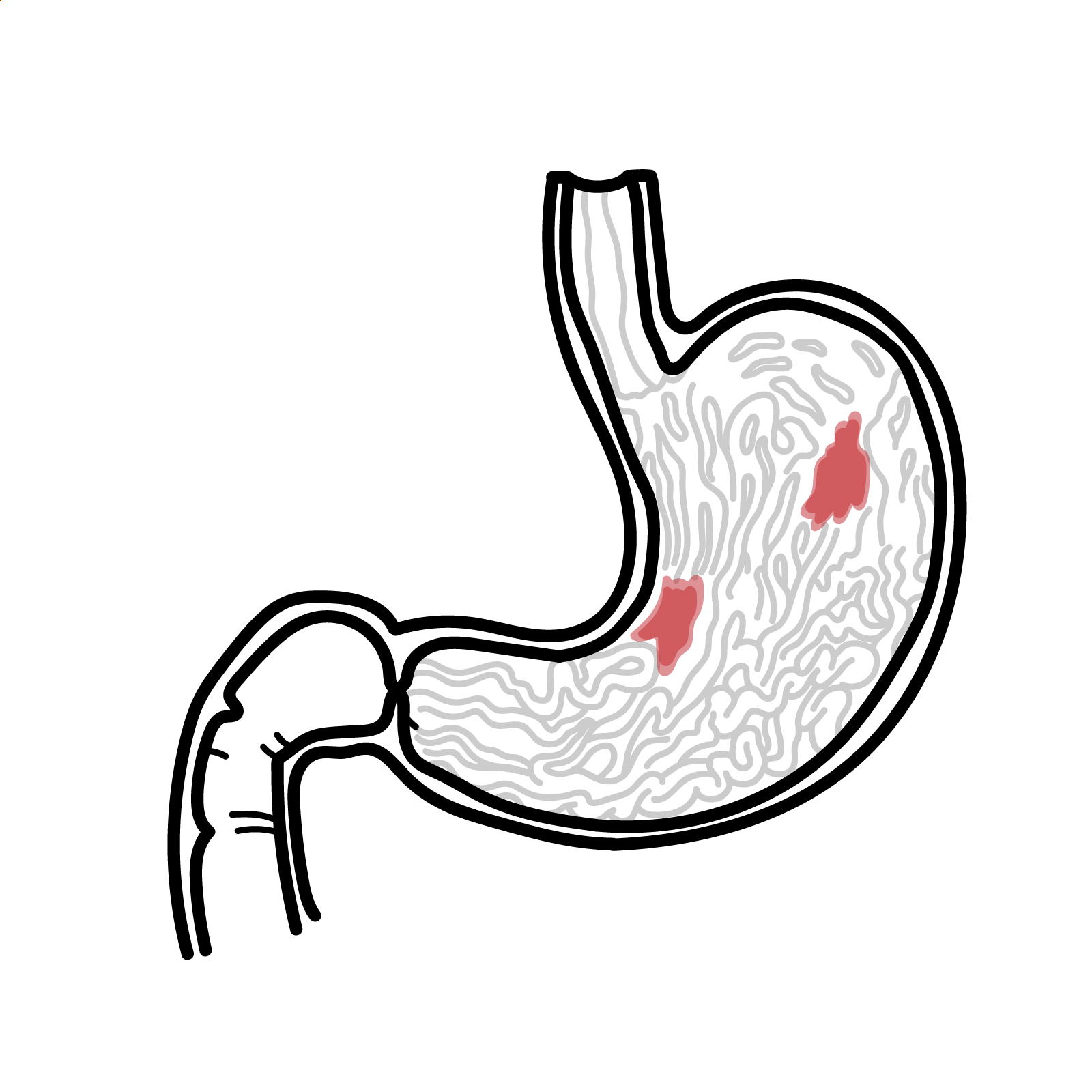
Atrophic gastritis is a chronic inflammatory disorder in which normal gastric glands are progressively lost and replaced by fibrosis or metaplastic epithelium. It is most commonly caused by chronic Helicobacter pylori infection or autoimmune destruction of oxyntic glands. The disease is often clinically silent but is important because it can cause iron and vitamin B12 deficiency and forms part of the pathway to gastric neoplasia.
Atrophic gastritis is a histopathological diagnosis. Endoscopic appearances may suggest atrophy, but biopsies are required to confirm the diagnosis, establish its distribution and severity, and identify intestinal metaplasia or dysplasia.
Definition
- Atrophic gastritis
- Chronic inflammation with loss of native gastric glands, with or without replacement by metaplastic epithelium.
- Metaplastic atrophy
- Loss of native gastric glands with replacement by intestinal or pseudopyloric-type glands.
- Autoimmune gastritis
- Immune-mediated, predominantly corpus–fundus atrophy caused by destruction of acid-producing parietal cells.
- Pernicious anaemia
- A late manifestation of autoimmune gastritis characterised by intrinsic-factor deficiency, vitamin B12 deficiency and megaloblastic anaemia.
Anatomy & Physiology
The gastric mucosa differs by region:
- The antrum contains gastrin-producing G cells.
- The corpus and fundus contain oxyntic glands with parietal cells, which secrete hydrochloric acid and intrinsic factor, and chief cells, which produce pepsinogen.
- Hydrochloric acid supports protein digestion, releases dietary vitamin B12 from food and facilitates non-haem iron absorption.
- Intrinsic factor binds vitamin B12 and permits its absorption in the terminal ileum.
This regional organisation explains the contrasting patterns of disease: autoimmune gastritis is typically corpus-predominant with relative antral sparing, whereas chronic H. pylori gastritis may progress from antral-predominant inflammation to multifocal atrophy involving both antrum and corpus.1,2
Aetiology & Risk Factors
Aetiology
The two principal causes are:
- Chronic H. pylori–associated gastritis
- The most common cause worldwide.
- Persistent inflammation may progress through glandular atrophy, intestinal metaplasia, dysplasia and intestinal-type gastric adenocarcinoma—the Correa cascade.
- Atrophy is often multifocal and may involve the antrum, incisura and corpus.
- Autoimmune gastritis
- Autoimmune destruction of parietal cells produces corpus–fundus-predominant atrophy.
- Parietal-cell and intrinsic-factor antibodies may support the diagnosis but are not sufficiently accurate to establish or exclude it without compatible histology.2
- Pernicious anaemia represents late-stage disease rather than a synonym for all autoimmune gastritis.
Less common causes of gastric gland loss include previous gastric surgery, chronic bile reflux, radiation injury and rare infiltrative or inflammatory disorders. These should be considered only when the clinical and histological pattern is atypical.
Risk Factors
Risk factors for H. pylori infection and associated atrophy include:
- Birth or residence in a region with a high prevalence of H. pylori or gastric cancer
- Household crowding and socioeconomic disadvantage, particularly during childhood
- A first-degree family history of gastric cancer
- Cigarette smoking, which also increases gastric cancer risk
- Persistent H. pylori infection after unsuccessful eradication therapy
Autoimmune gastritis is associated with:
- Autoimmune thyroid disease, particularly Hashimoto thyroiditis
- Type 1 diabetes mellitus
- Vitiligo, Addison disease and other autoimmune disorders
- A personal or family history of autoimmune disease
Long-term proton pump inhibitor therapy can cause hypergastrinaemia, but it is not sufficient by itself to diagnose atrophic gastritis. Histological gland loss and its distribution remain decisive.
Pathophysiology
H. pylori–associated atrophic gastritis
- H. pylori colonises the gastric mucous layer and causes chronic active gastritis.
- Persistent inflammation damages gastric glands and may extend from the antrum to the corpus.
- Native glands are lost and replaced by fibrosis or intestinal metaplasia.
- Continuing inflammation and accumulated molecular alterations may lead to dysplasia and intestinal-type gastric adenocarcinoma.
Autoimmune gastritis
- T-cell-mediated injury and autoantibodies target parietal-cell components, including the gastric H⁺/K⁺-ATPase.
- Loss of parietal cells causes hypochlorhydria or achlorhydria and reduced intrinsic-factor production.
- Reduced acid impairs iron absorption; reduced intrinsic factor eventually causes vitamin B12 deficiency.
- Loss of gastric acid removes negative feedback on antral G cells, causing hypergastrinaemia.
- Persistent gastrin stimulation produces enterochromaffin-like cell hyperplasia and may lead to type 1 gastric neuroendocrine tumours.2
Iron deficiency may appear before vitamin B12 deficiency in autoimmune gastritis. Acid loss impairs non-haem iron absorption early, whereas clinically important vitamin B12 deficiency develops only after body stores have been depleted.
Clinical Manifestations
Many patients are asymptomatic, and the diagnosis is made during endoscopy or the investigation of anaemia.
Gastrointestinal features
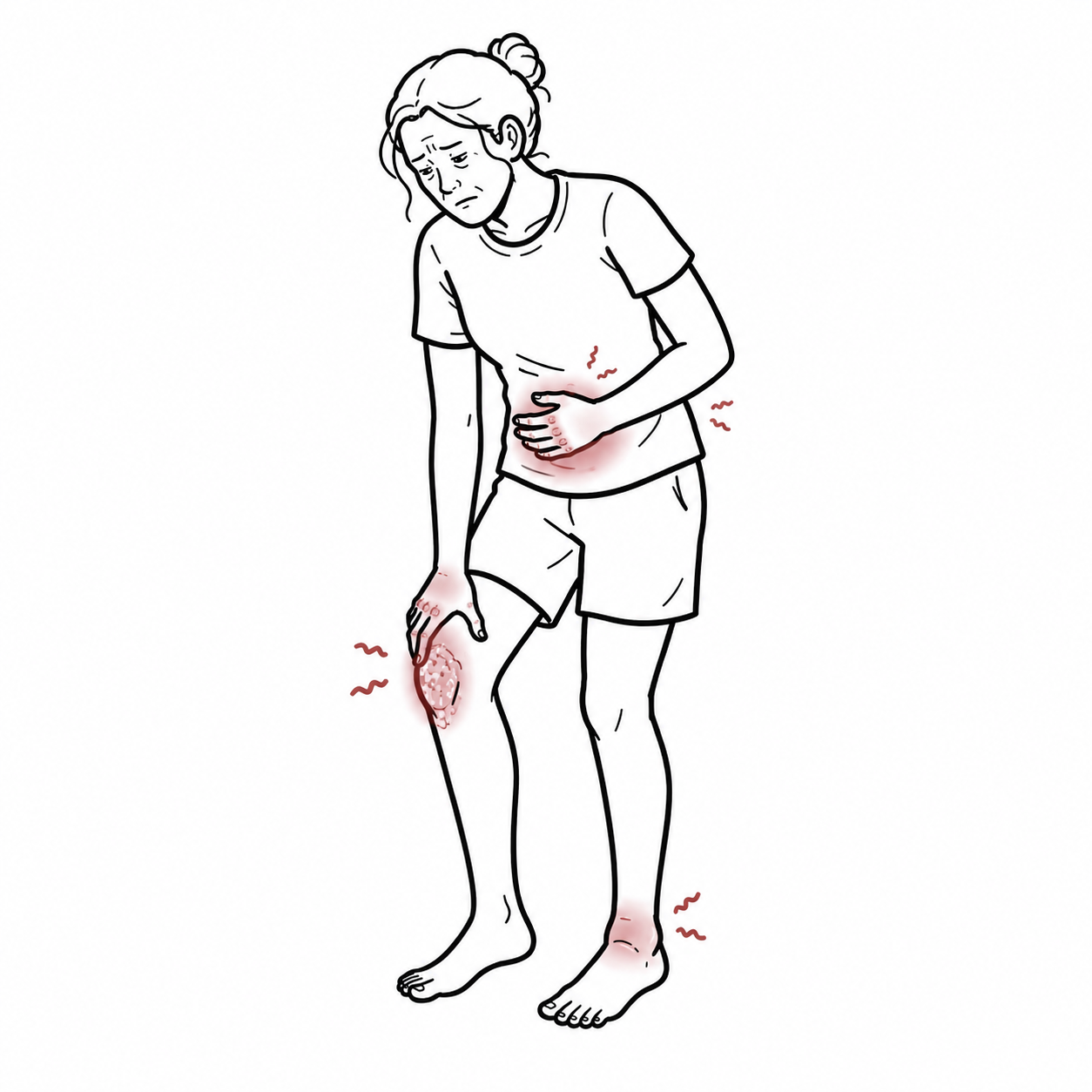
- Dyspepsia, epigastric discomfort or early satiety
- Nausea, bloating or postprandial fullness
- Symptoms may be absent despite advanced histological disease
Iron deficiency
- Fatigue, reduced exercise tolerance and exertional dyspnoea
- Pallor
- Microcytic or normocytic anaemia
- Recurrent or unexplained iron deficiency, particularly if oral iron response is poor
Vitamin B12 deficiency and pernicious anaemia
- Fatigue and pallor
- Glossitis
- Macrocytosis or megaloblastic anaemia
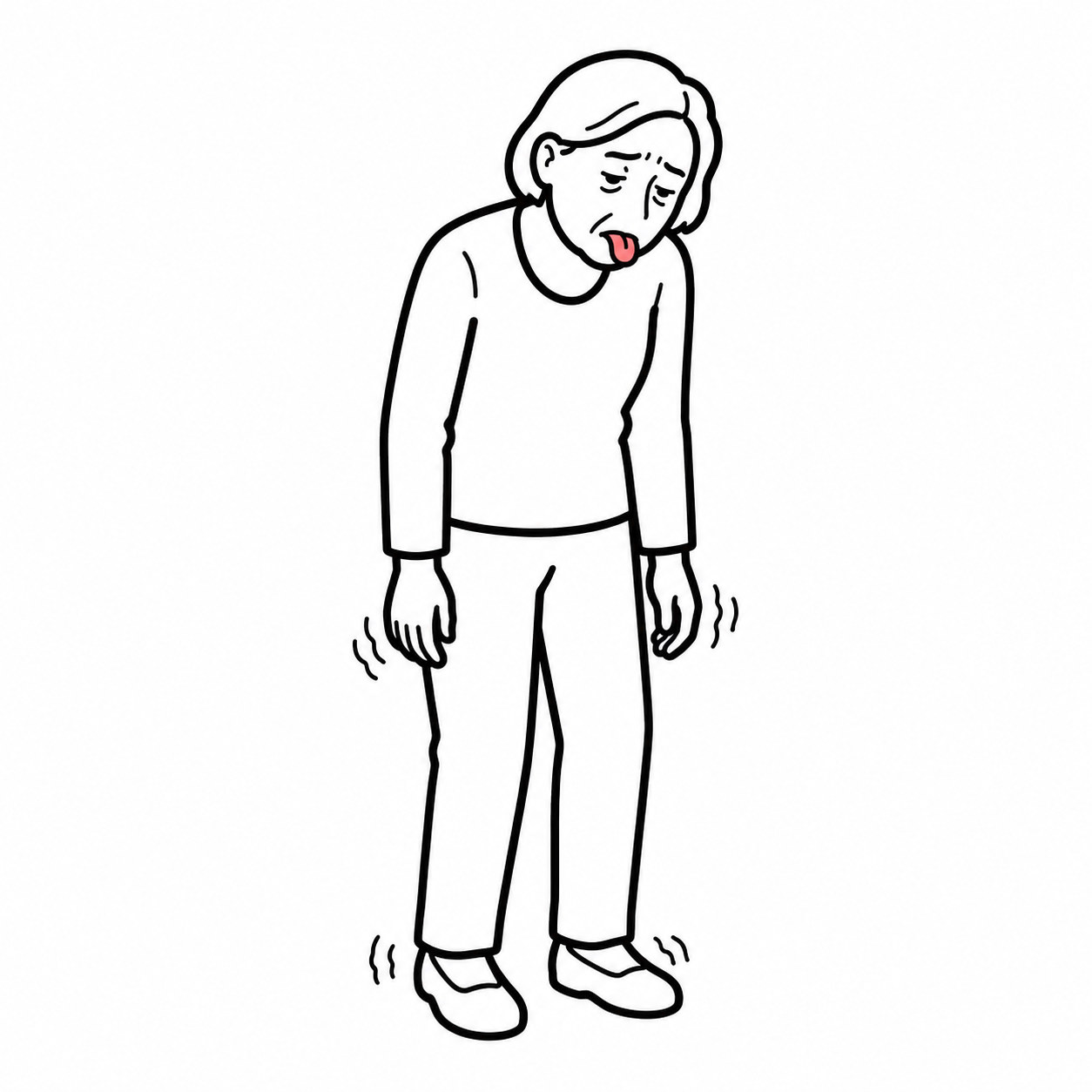
- Paraesthesia, impaired vibration or proprioception, gait disturbance and cognitive change
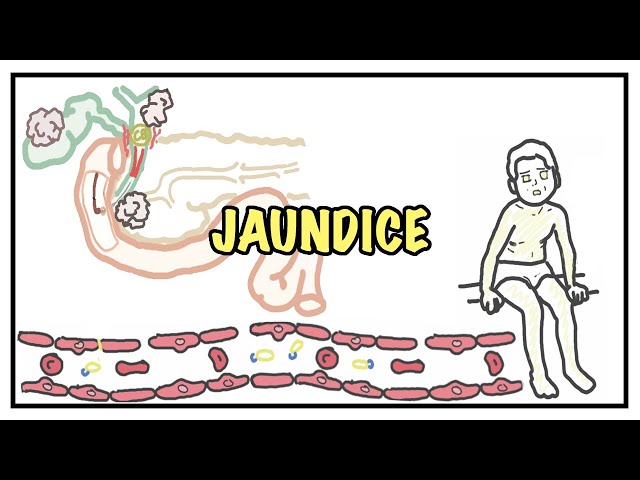
- Mild jaundice from ineffective erythropoiesis in severe deficiency

A normal mean corpuscular volume does not exclude vitamin B12 deficiency. Coexisting iron deficiency can mask macrocytosis, and neurological injury may occur without anaemia.
Diagnosis
The diagnosis requires upper gastrointestinal endoscopy with histological confirmation of gastric gland loss. The extent, severity and distribution of atrophy—not a positive antibody test alone—determine the diagnosis and help stratify neoplastic risk.
Upper gastrointestinal endoscopy and biopsy
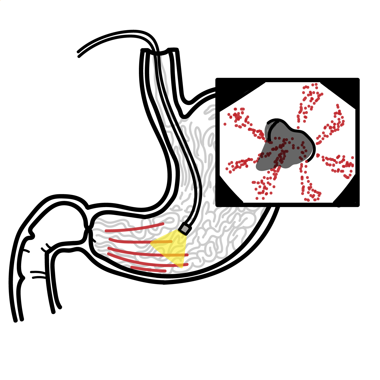
High-quality endoscopy should carefully inspect the gastric mucosa using high-definition white-light endoscopy with image enhancement where available. Endoscopic features may include:
- Pallor and thinning of the mucosa
- Loss or flattening of rugal folds
- Increased visibility of submucosal vessels
- Intestinal metaplasia or focal mucosal irregularity
- Polyps, nodules or lesions suspicious for dysplasia or cancer
Biopsies should include targeted samples from visible abnormalities and systematic topographic sampling.
Do not rely on a single antral biopsy. Atrophic gastritis is a patchy disease, and inadequate mapping can miss corpus involvement or incorrectly classify a high-risk patient as having limited disease.
Histopathology
Features include:
- Presence and severity of glandular atrophy
- Anatomical distribution: antrum/incisura, corpus or both
- Intestinal metaplasia and, where reported, complete versus incomplete type
- Active or chronic H. pylori gastritis
- Dysplasia or neoplasia
- Features supporting autoimmune gastritis, such as corpus-predominant oxyntic gland loss, pseudopyloric or intestinal metaplasia and enterochromaffin-like cell hyperplasia

Determine the cause
Test for active H. pylori infection in every patient with atrophic gastritis. Histology, urea breath testing or stool antigen testing may be used according to the clinical setting. Serology does not reliably distinguish active from previous infection. Treat confirmed infection and document eradication with a non-serological test.
If autoimmune gastritis is suspected, request:
- Anti-parietal-cell antibodies: more sensitive but less specific
- Anti-intrinsic-factor antibodies: less sensitive but more specific for pernicious anaemia
Antibody results support, but do not replace, compatible gastric histology.
Differential diagnoses
| Differential | Distinguishing features |
|---|---|
| Non-atrophic H. pylori gastritis | Chronic inflammation without histological gland loss |
| Reactive or chemical gastropathy | Foveolar hyperplasia and chemical injury pattern; usually limited inflammation |
| Proton pump inhibitor effect | Hypergastrinaemia and fundic gland changes without the characteristic mapping-biopsy pattern of atrophy |
| Coeliac disease or ileal disease | Alternative causes of iron or vitamin B12 deficiency; gastric histology does not show diagnostic atrophy |
| Previous gastric or bariatric surgery | Anatomical and absorptive cause apparent from history |
Corpus-predominant atrophy with relative antral sparing, hypergastrinaemia, iron or vitamin B12 deficiency and supportive autoantibodies strongly suggests autoimmune gastritis. Antral and corpus involvement instead favours multifocal H. pylori–associated disease, although overlap can occur.
Treatment
Treatment addresses the underlying cause, replaces deficiencies and provides risk-stratified endoscopic surveillance. Established gland loss is not reliably reversible, so management aims to prevent progression and detect complications early.
Eradicate H. pylori
- Treat all patients with confirmed active H. pylori infection using an appropriate guideline-based eradication regimen.
- Confirm eradication with a urea breath test, stool antigen test or appropriate biopsy-based testing.
- Testing should occur after treatment, allowing an adequate interval after antibiotics and withholding proton pump inhibitors when clinically safe to reduce false-negative results.
Eradication reduces ongoing inflammation and gastric cancer risk, although pre-existing advanced atrophy or intestinal metaplasia may not regress.2,3
Replace nutritional deficiencies
- Iron deficiency: provide oral or intravenous iron according to severity, tolerance and response; investigate other causes of blood loss or malabsorption when appropriate.
- Vitamin B12 deficiency: replace promptly. Parenteral therapy is preferred initially when deficiency is severe, neurological manifestations are present or malabsorption is established; high-dose oral replacement may be effective in selected patients with reliable follow-up.
- Monitor the full blood count, iron indices and vitamin B12 response. Neurological symptoms require urgent treatment and may recover incompletely if therapy is delayed.
Do not delay vitamin B12 replacement while awaiting antibody results when deficiency is accompanied by neurological manifestations. Folate alone may improve the anaemia while allowing neurological injury to progress.
Endoscopic surveillance
Surveillance should be based on histological extent, severity and individual risk rather than applied to every patient:
- Advanced atrophy—including severe atrophy, involvement of both antrum and corpus, or OLGA/OLGIM stage III–IV: high-quality surveillance endoscopy approximately every 3 years is recommended or suggested by major guidelines.
- Mild disease limited to the antrum, without additional risk factors: routine surveillance is generally not recommended.
Surveillance recommendations are supported mainly by observational evidence rather than randomised trials. A three-year interval is a common framework for advanced disease, but the final plan should account for baseline endoscopy quality, age, comorbidity and whether the patient would be fit for treatment if neoplasia were detected.
Complications & Prognosis
Complications
- Iron deficiency with or without anaemia
- Vitamin B12 deficiency and pernicious anaemia
- Irreversible peripheral neuropathy, posterior-column dysfunction or cognitive effects from delayed vitamin B12 treatment
- Gastric intestinal metaplasia and dysplasia
- Intestinal-type gastric adenocarcinoma, particularly with extensive or advanced H. pylori–associated atrophy
- Type 1 gastric neuroendocrine tumours in autoimmune gastritis due to chronic hypergastrinaemia
- Associated autoimmune disorders, particularly autoimmune thyroid disease
Prognosis
Atrophic gastritis is chronic. Correction of iron and vitamin B12 deficiency usually improves haematological manifestations, but established neurological damage may be incomplete or irreversible. H. pylori eradication reduces continued inflammatory injury and neoplastic risk, but advanced atrophy and intestinal metaplasia may persist.2,3
Risk of progression is greatest with:
- Extensive atrophy involving the corpus as well as the antrum
- Severe histological change or OLGA/OLGIM stage III–IV
- Incomplete-type intestinal metaplasia
- Persistent H. pylori infection
- A first-degree family history of gastric cancer
- Smoking and demographic risk associated with high-incidence regions
- Dysplasia, which requires specialist endoscopic assessment and management
References
- Shah SC, Piazuelo MB, Kuipers EJ, Li D. AGA clinical practice update on the diagnosis and management of atrophic gastritis: expert review. Gastroenterology. 2021;161(4):1325–1332.e7. doi:10.1053/j.gastro.2021.06.078
- Morgan DR, Corral JE, Li D, et al. ACG Clinical Guideline: diagnosis and management of gastric premalignant conditions. Am J Gastroenterol. 2025;120(4):709–737. doi:10.14309/ajg.0000000000003350
- Dinis-Ribeiro M, Libânio D, Uchima H, et al. Management of epithelial precancerous conditions and early neoplasia of the stomach (MAPS III): European Society of Gastrointestinal Endoscopy, European Helicobacter and Microbiota Study Group and European Society of Pathology Guideline update 2025. Endoscopy. 2025;57(5):504–554. doi:10.1055/a-2529-5025







Members only discussions coming soon…