

Gastric Antral Vascular Ectasia (GAVE)
Overview
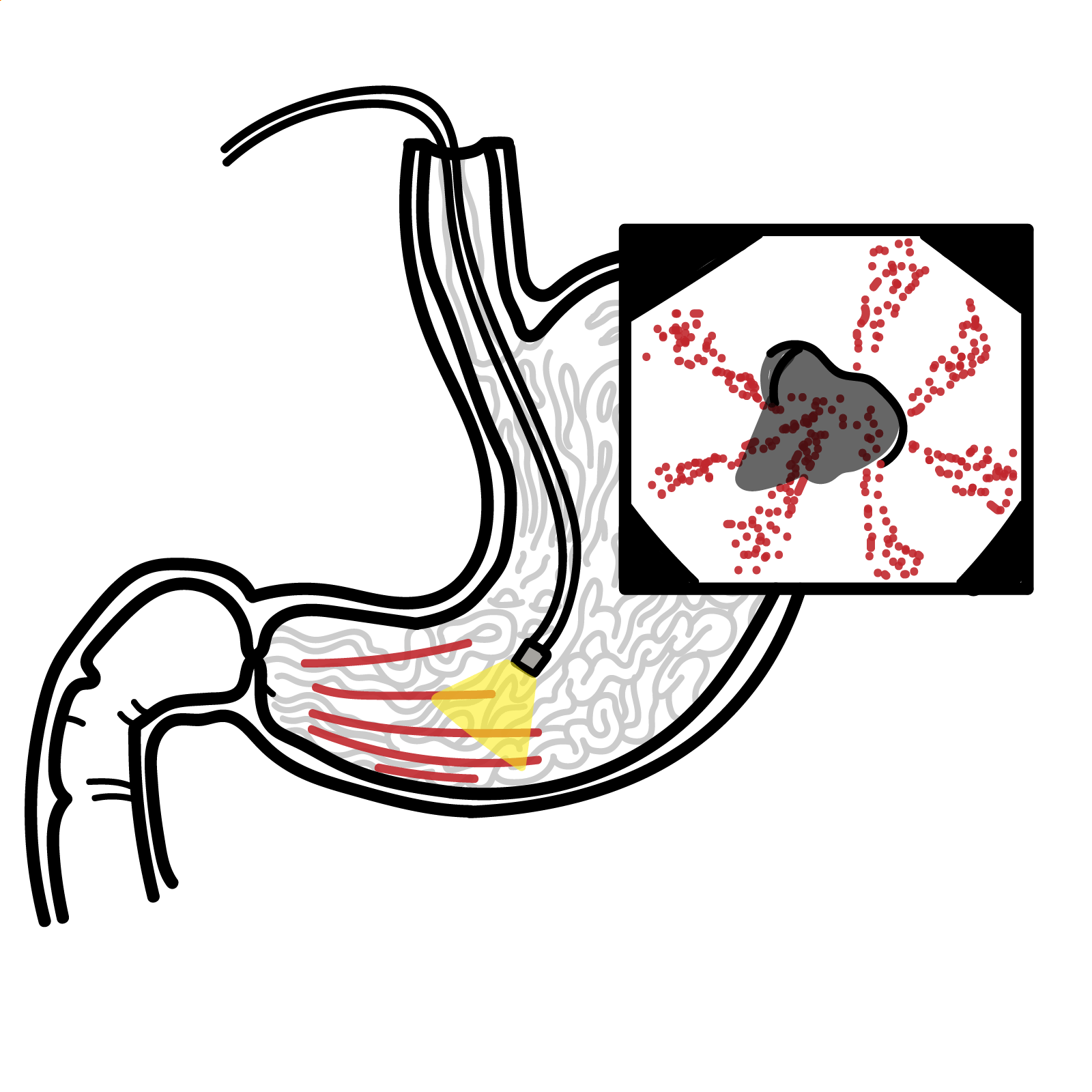
Gastric antral vascular ectasia (GAVE, “watermelon stomach”) is a rare but important cause of non-variceal upper gastrointestinal bleeding, often presenting with chronic iron-deficiency anaemia or recurrent overt bleeding in older adults, particularly women [1–4]. It is frequently associated with systemic conditions such as liver cirrhosis (~30% of cases) and autoimmune/connective-tissue disease (especially systemic sclerosis). Although GAVE is often seen in patients with portal hypertension, it is pathophysiologically distinct from portal hypertensive gastropathy. Untreated, GAVE can lead to transfusion-dependent anaemia, hemodynamically significant upper GI haemorrhage, and procedure-related morbidity.[1–4,7]
Definition
- Gastric antral vascular ectasia (GAVE): Localised dilatation and ectasia of mucosal vessels in the gastric antrum producing characteristic endoscopic patterns (linear “watermelon” or diffuse “honeycomb”/nodular) and causing chronic or acute GI bleeding.
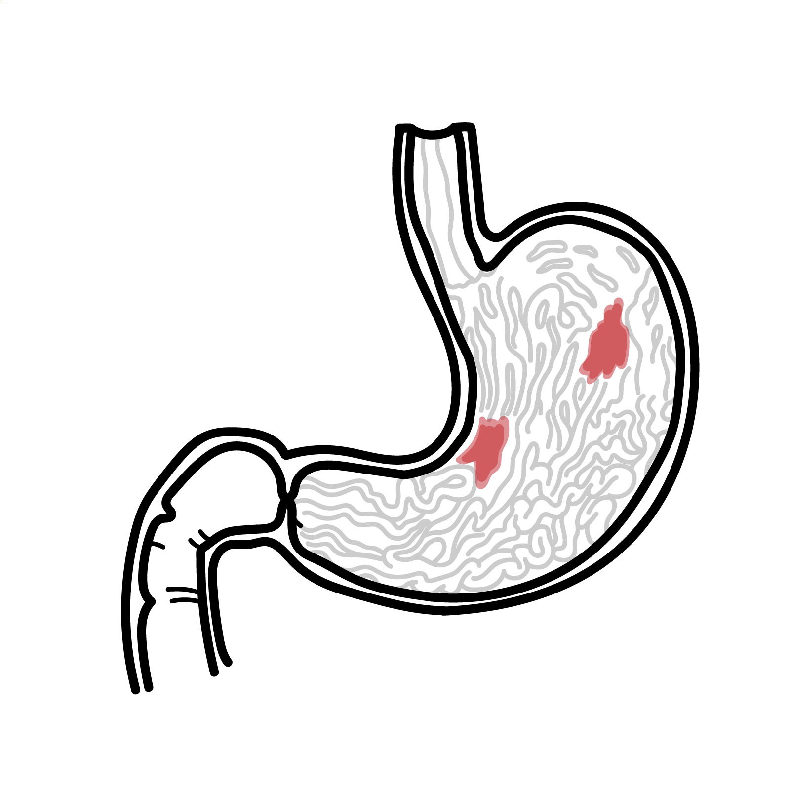
- Portal hypertensive gastropathy (PHG): Distinct portal-hypertension–related mucosal abnormality involving fundus/body with a mosaic “snakeskin” pattern and red spots; may coexist with GAVE but has different pathophysiology and management.
- Angiodysplasia/angioectasia: Dilated, thin-walled mucosal or submucosal vessels in GI tract that predispose to bleeding; GAVE is a specific antral angiodysplastic lesion with unique histology and distribution.
- Systemic sclerosis–associated GAVE: Subtype occurring in systemic sclerosis, often early in disease, with iron-deficiency anaemia and sometimes severe GI bleeding; associated with specific autoantibodies (e.g. anti–RNA polymerase III).
Anatomy & Physiology
- Gastric regions
- Antrum is the distal stomach between incisura and pylorus; functions as a pump and grinder.
- Contains thick circular muscle and prominent longitudinal folds, creating high mechanical stress during peristalsis.
- Vascular supply & drainage
- Arterial supply from right and left gastric and gastroepiploic arteries; submucosal arterioles give rise to dense mucosal capillary plexus.
- Venous drainage via submucosal veins → left and right gastric veins → portal venous system; pressure changes in portal system influence antral venous congestion.
- Mucosal defence & blood-flow regulation
- Gastric mucosal blood flow is regulated by prostaglandins, nitric oxide, and neurohumoral factors in response to acid, food, and mechanical stimuli.
- Adequate microcirculation is essential for epithelial repair; chronic congestion or shearing can remodel capillaries and venules.
- Antral motility
- Strong phasic contractions mix chyme and drive it through pylorus; repetitive shearing forces act along longitudinal folds where vessels run.
- Neurohormonal modulators (gastrin, VIP, prostaglandins) affect both motility and mucosal blood flow.
The antrum has intense motility and portal-system venous drainage, predisposing it to mechanical and hemodynamic injury.
Aetiology & Risk Factors
Aetiology (multifactorial, incompletely understood)
- Chronic mechanical stress from strong antral peristalsis → repeated mucosal prolapse and trauma along longitudinal folds, promoting vascular ectasia and fibromuscular hyperplasia.
- Abnormal antral motility and gastric emptying, including “antral pump” dysfunction, possibly mediated by neurohormonal factors.
- Microvascular injury and remodeling driven by mucosal ischemia–reperfusion, venous congestion, and altered mucosal repair.
- Autoimmune-mediated microangiopathy in systemic sclerosis and other autoimmune diseases (e.g. anti–RNA polymerase III–positive SSc).[5]
- Contribution of portal hypertension in a subset of patients (especially cirrhotics), but GAVE can occur without portal hypertension and often persists despite portal-pressure reduction.[1,9,11]
- Possible hormonal influences (e.g. hypergastrinaemia, prostaglandins, vasoactive peptides) altering mucosal blood flow and angiogenesis.[1,3,6]
- Older age (typically >60–70 years).
- Female sex (female:male ~2:1).
- Liver cirrhosis and portal hypertension of any aetiology (~30% of GAVE patients).
- Autoimmune/connective-tissue disease: systemic sclerosis, primary biliary cholangitis, SLE, Sjögren’s, Raynaud’s phenomenon.
- Chronic kidney disease (especially dialysis-dependent).
- Cardiovascular disease and chronic heart failure.
- Metabolic disorders: type 2 diabetes mellitus, NAFLD/NASH-related cirrhosis.
- Bone-marrow transplantation, chronic immunosuppression.
“elderly woman with comorbid cirrhosis or autoimmune disease + iron-deficiency anaemia” → GAVE (or PHG in cirrhosis).
In a cirrhotic patient with chronic anaemia despite beta-blockers/TIPS and minimal PHG, actively look for GAVE—treating the portal pressure alone may not control bleeding.
Pathophysiology
- Predisposing milieu
- Systemic factors (cirrhosis, autoimmune disease, CKD, metabolic and vascular disease) alter hemodynamics, microvasculature, and repair capacity.
- Antral hypermotility and mucosal prolapse increase mechanical shear at the distal antrum.
- Microvascular injury & congestion
- Repetitive mucosal trauma and venous congestion cause focal capillary and venule dilation in lamina propria.
- Increased mucosal blood flow and stasis favour microthrombi formation.
- Structural remodeling
- Lamina propria develops:
- Marked capillary/venule ectasia with fibrin thrombi.
- Fibrohyalinosis and spindle-cell (smooth-muscle/fibroblast) proliferation → fibromuscular hyperplasia.
- These changes produce elevated, friable longitudinal ridges or diffuse nodular/“honeycomb” mucosa.
- Lamina propria develops:
- Clinical bleeding
- Fragile ectatic vessels rupture with minor trauma or acid exposure → chronic occult bleeding (most common) or episodic overt haematemesis/melena.
- Recurrent microbleeds cause iron-deficiency anaemia; severe lesions or superimposed coagulopathy can lead to acute massive haemorrhage.
Histology triad for GAVE vascular ectasia + fibrin thrombi + fibromuscular (spindle-cell) proliferation in lamina propria.
Clinical Manifestations
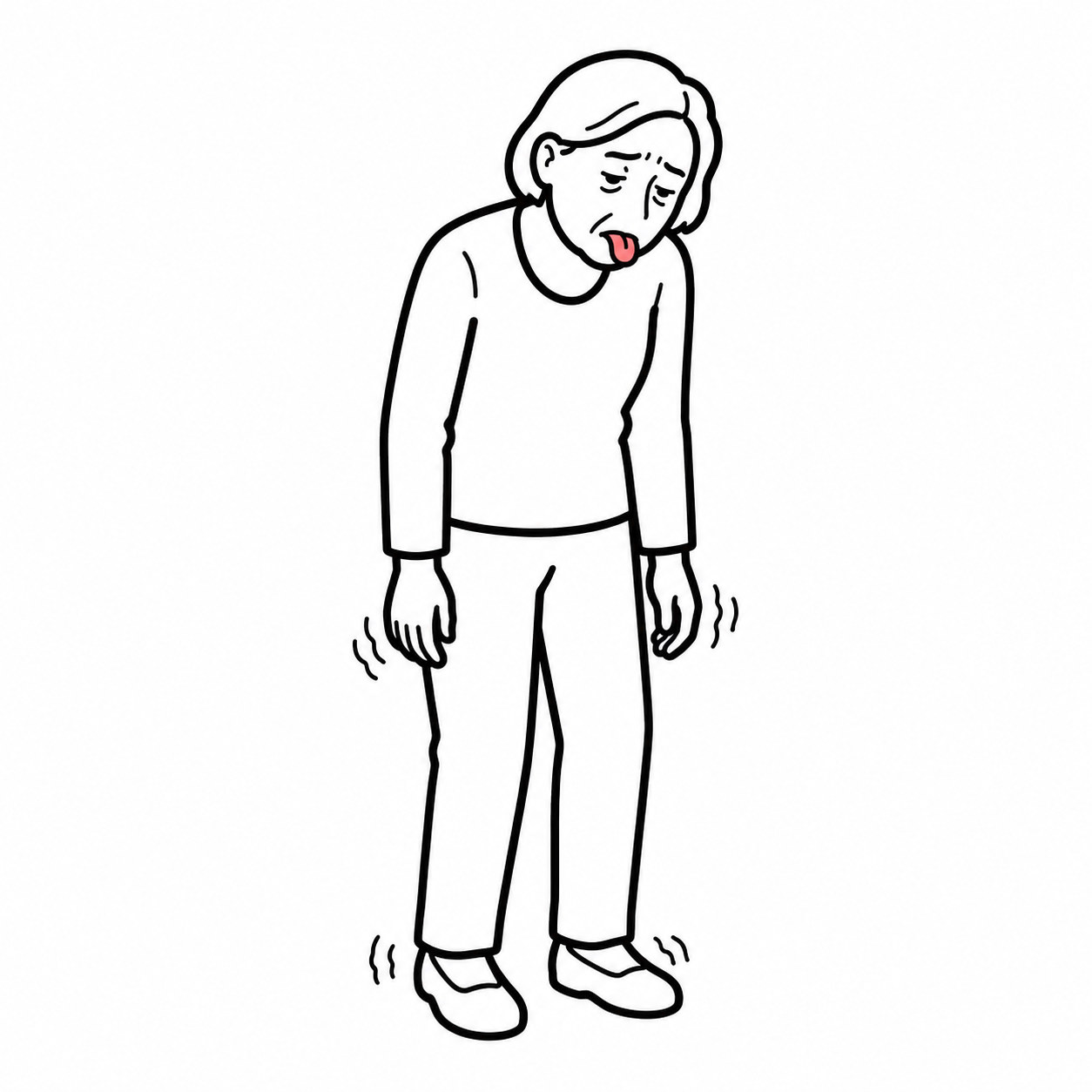
- Chronic iron-deficiency anaemia (most common presentation)
- Overt upper GI bleeding: melena, haematemesis, “coffee-ground” vomiting (less frequent)
- Recurrent need for blood transfusions or IV iron despite PPI therapy.
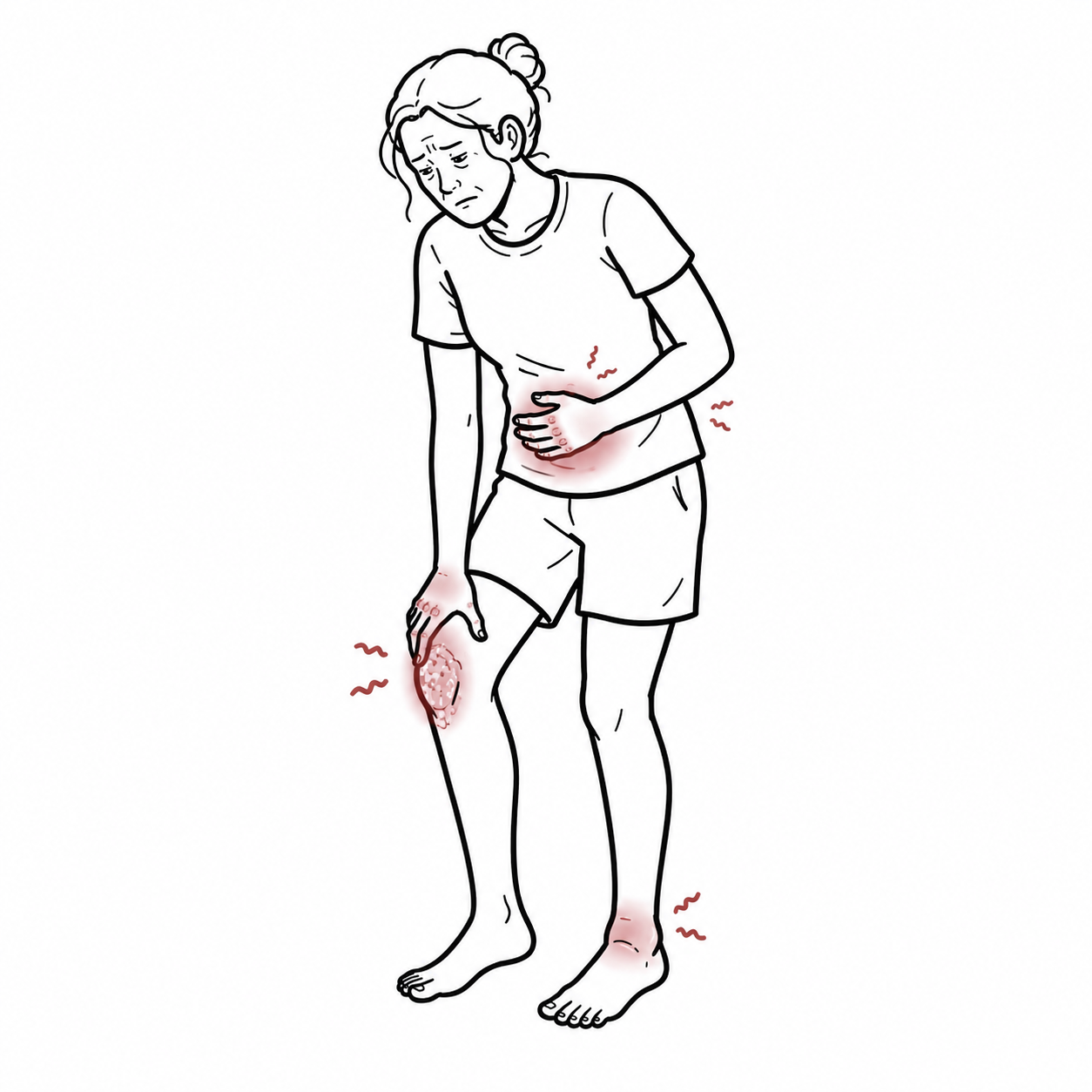
- Non-specific upper GI symptoms: dyspepsia, early satiety, epigastric discomfort
- Symptoms/signs from associated conditions
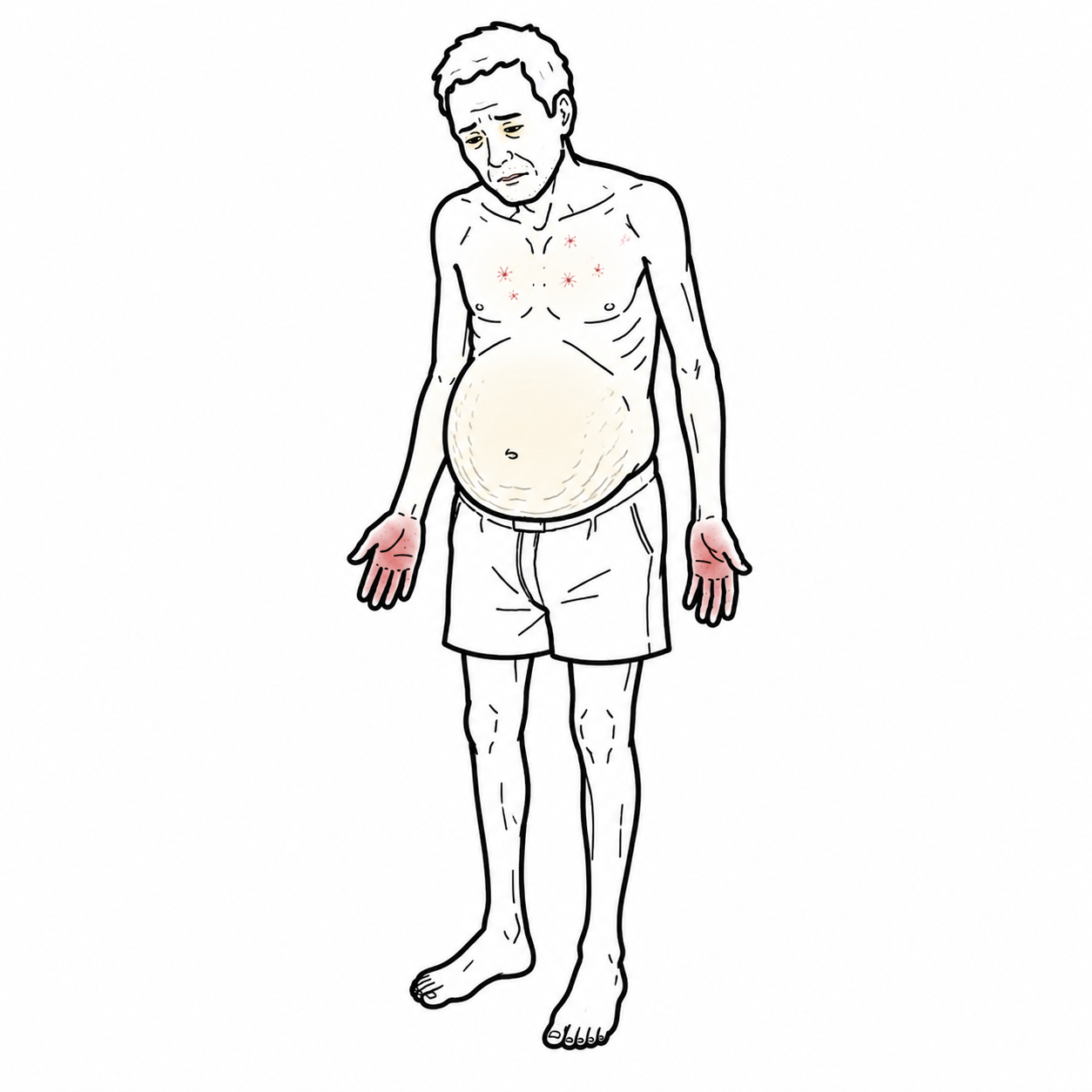
- features of cirrhosis (ascites, jaundice, encephalopathy)
- systemic sclerosis (sclerodactyly, telangiectasia, Raynaud’s).
In systemic sclerosis or other CTDs, new severe iron-deficiency anaemia—even without obvious bleeding—should trigger early gastroscopy specifically looking for GAVE.
Diagnosis
- FBC: microcytic hypochromic anaemia, sometimes normocytic in mixed disease.
- Iron studies: low ferritin, low transferrin saturation (iron-deficiency pattern).
- Coagulation profile, liver and renal function, autoimmune serology (context dependent).
Upper GI endoscopy (gold standard)
-
- Characteristic antral pattern of flat or slightly raised erythematous lesions:
- Linear/striped (“watermelon stomach”) – red stripes radiating from pylorus along folds.
- Diffuse/punctate (“honeycomb/diffuse GAVE”) – multiple red spots diffusely involving antrum, sometimes proximal extension.
- Nodular GAVE – polypoid/nodular lesions overlapping with hyperplastic polyps.
- Lesions confined predominantly to antrum (may rarely extend proximally).
- Absence of typical PHG mosaic pattern in corpus/fundus.
- Characteristic antral pattern of flat or slightly raised erythematous lesions:
- Histology – when diagnosis uncertain
- Mucosal capillary and venule ectasia in lamina propria.
- Fibrin thrombi within ectatic vessels.
- Fibrohyalinosis and spindle-cell (smooth-muscle/fibroblast) proliferation.
- Minimal inflammation.
| Condition | Typical site | Endoscopic pattern | Histology | Response to portal-pressure reduction (beta-blocker/TIPS) |
| GAVE | Antrum (± pylorus) | Linear “watermelon” stripes, diffuse “honeycomb”, or nodular lesions | Ectatic mucosal vessels with fibrin thrombi + fibromuscular hyperplasia (GAVE score >3) | Usually minimal |
| Portal hypertensive gastropathy (PHG) | Fundus & body | Mosaic “snakeskin” pattern with red/black spots | Non-specific congestion; no classic GAVE triad | Improves with reduced portal pressure |
| Gastric antral angiodysplasia (isolated) | Focal anywhere, including antrum | Isolated red spots or patches, not organised as stripes | Dilated vessels; lacks fibrohyalinosis pattern | Variable |
Classification
Endoscopic morphological classification
- Linear/striped GAVE (“watermelon type”) – longitudinal red stripes radiating from pylorus along antral folds; commonly described in non-cirrhotic and autoimmune patients.
- Diffuse/punctate GAVE (“honeycomb/ring pattern”) – innumerable small red spots or rings diffusely involving antrum, especially in cirrhosis and portal hypertension.
- Nodular GAVE – polypoid or nodular lesions with overlapping histologic features of gastric hyperplastic polyps; important because it may be mistaken for neoplasia.
Cirrhotic GAVE tends to be diffuse/ring-like, while non-cirrhotic (especially autoimmune) GAVE more often shows classic watermelon stripes, though overlap exists.
Treatment
General principles
- Control acute bleeding
- Correct anaemia and iron deficiency
- Reduce recurrent bleeding and transfusion needs
- Treat underlying systemic disease.
Supportive / medical
- Resuscitation in acute bleeding: IV fluids, blood transfusions, correction of coagulopathy as per standard UGIB protocols.
- Iron replacement: oral or IV iron to correct deficiency; often needed long-term.
- Proton-pump inhibitors (PPIs): widely used to promote mucosal healing after ablation, although they do not treat GAVE per se.
- Blood transfusion
- Pharmacologic options with limited evidence (generally adjunctive/rescue)
- Octreotide (especially in portal-hypertensive settings; benefit inconsistent).
- Estrogen–progesterone therapy, tranexamic acid, or thalidomide described in case series; reserved for refractory cases due to adverse-effect profiles.
Endoscopic therapy – first-line definitive treatment
- Argon plasma coagulation
- Endoscopic band ligation
- Radiofrequency ablation
Surgical / radiologic options
- Antrectomy or total gastrectomy
- Portal-pressure reduction (TIPS, surgical shunts, NSBBs)
- Standard for PHG and varices, but inconsistent or minimal effect on GAVE; not primary therapy for GAVE, though may help coexisting PHG.
Management of underlying disease
- Optimise cirrhosis care (alcohol abstinence, viral suppression, metabolic control).
- Systemic sclerosis or other autoimmune diseases as per rheumatology guidance.
Complications & Prognosis
- Chronic transfusion-dependent iron-deficiency anaemia → iron overload, alloimmunisation, transfusion reactions.
- Acute massive upper GI bleeding → haemodynamic instability, ICU admission, increased mortality risk.
- Procedure-related: post-APC or RFA ulceration, pain, rare perforation or antral stenosis; post-EBL ulcer bleeding; anaesthesia/sedation risk.
- Surgical: anastomotic leak, infection, nutritional deficiencies, perioperative death
- Ongoing burden of frequent hospitalisations, endoscopies, and transfusions.
- Many patients have multiple comorbidities (liver disease, CKD, cardiovascular disease), but in-hospital mortality from GAVE-related bleeding is relatively low when managed appropriately
- Despite treatment, up to ~60% may remain at least intermittently transfusion-dependent, particularly those with advanced cirrhosis or diffuse disease.
- Long-term outcome largely driven by underlying conditions (e.g. severity of cirrhosis or systemic sclerosis) rather than GAVE alone.
- Favorable prognostic indicators: non-cirrhotic status, good response to endoscopic therapy with stabilised haemoglobin, fewer comorbidities.
- Poor prognostic indicators: decompensated cirrhosis, diffuse/ring-like GAVE requiring frequent transfusions, failure of multiple endoscopic modalities, need for surgery.
References
- Fuccio L, Mussetto A, Laterza L, Eusebi LH, Bazzoli F. Diagnosis and management of gastric antral vascular ectasia. World J Gastrointest Endosc. 2013;5(1):6-13.
- Cavallaro A, Tarantino I, Luca A, et al. Recurrent gastric antral vascular ectasia: a single center experience. Front Surg. 2024;11:1356409.
- Kichloo A, El-Amir Z, Dahiya DS, et al. Gastric antral vascular ectasia: trends of hospitalizations and outcomes in the USA. Gastroenterology Res. 2021;14(4):244-52.
- Patel U, Desai R, Desai J, et al. Predictors of blood transfusion and in-hospital outcomes in patients with gastric antral vascular ectasia: a nationwide population-based analysis. Ann Transl Med. 2019;7(3):46.
- Parrado RH, Furst DE, Maranian P, Clements PJ. Gastric antral vascular ectasia in systemic sclerosis: a review. Int J Rheumatol. 2015;2015:762546.
- Aryan M, Clark K, Broitman E, et al. The misclassification of gastric antral vascular ectasia. Gastroenterol Rep (Oxf). 2022;10(4):goac040.
- Hsu WH, Ghimire S, Lai HC, et al. Insights into the management of gastric antral vascular ectasia. Ther Adv Chronic Dis. 2018;9(6):135-46.
- Peng M, He Q, Yang J, et al. Endoscopic treatment for gastric antral vascular ectasia. Ther Adv Gastroenterol. 2021;14:20406223211039696.
- Ripoll C, Garcia-Tsao G. Management of portal hypertensive gastropathy and gastric antral vascular ectasia. Dig Liver Dis. 2011;43(5):345-51.
- Ward EM, Raimondo M, Rosser B, Wallace MB, Hoffman B, Woodward T. Prevalence and natural history of gastric antral vascular ectasia in patients undergoing orthotopic liver transplantation. J Clin Gastroenterol. 2004;38(10):898-903.
- Smith E, Stolk M, Stanley A, et al. Clinical characterization of gastric antral vascular ectasia. Am J Med. 2016;129(11):1173-e7.
- Fortuna L, Costa F, Zippi M, et al. Gastric antral vascular ectasia: a case report and review of the literature. Int J Surg Case Rep. 2022;99:107617.
- Louissaint J, Zhong J, Keshishian J, et al. Endoscopic versus histopathologic agreement in the diagnosis of gastric antral vascular ectasia. Am J Gastroenterol. 2017;112(Suppl 1):S588.
- Naidu H, Chidambaram S, Bright T, Wong T. Gastric antral vascular ectasia. Endoscopy. 2014;46(1):78-9.
- Fang Z, Li Y, Zhang X, et al. A delayed gastric antral vascular ectasia: a case report and review. Medicine (Baltimore). 2024;103(52):e38247.
- Fuccio L, Mussetto A, Laterza L, et al. Histological score systems for diagnosis of gastric antral vascular ectasia. World J Gastrointest Endosc. 2013;5(1):6-13 (tables).
- Abdelmoneim RSE, Abdallah AM, El-Sayed WF, et al. The classification of gastric antral vascular ectasia in cirrhotic patients. Egypt Liver J. 2022;12:73.
- Hirsch BS, Kedia P, Kumar A, et al. Endoscopic band ligation versus argon plasma coagulation in the treatment of gastric antral vascular ectasia. Clin Endosc. 2021;54(6):847-54.
- Garg A, Shukla A, et al. Endoscopic band ligation versus argon plasma coagulation in gastric antral vascular ectasia: a randomized trial. Gastrointest Endosc. 2025;XX:XX-XX.
- Cavallaro A, et al. Recurrent gastric antral vascular ectasia and outcomes of band ligation versus thermal therapy: meta-analysis subset. Front Surg. 2024;11:1356409.






Members only discussions coming soon…