

Wrist Pain
Overview
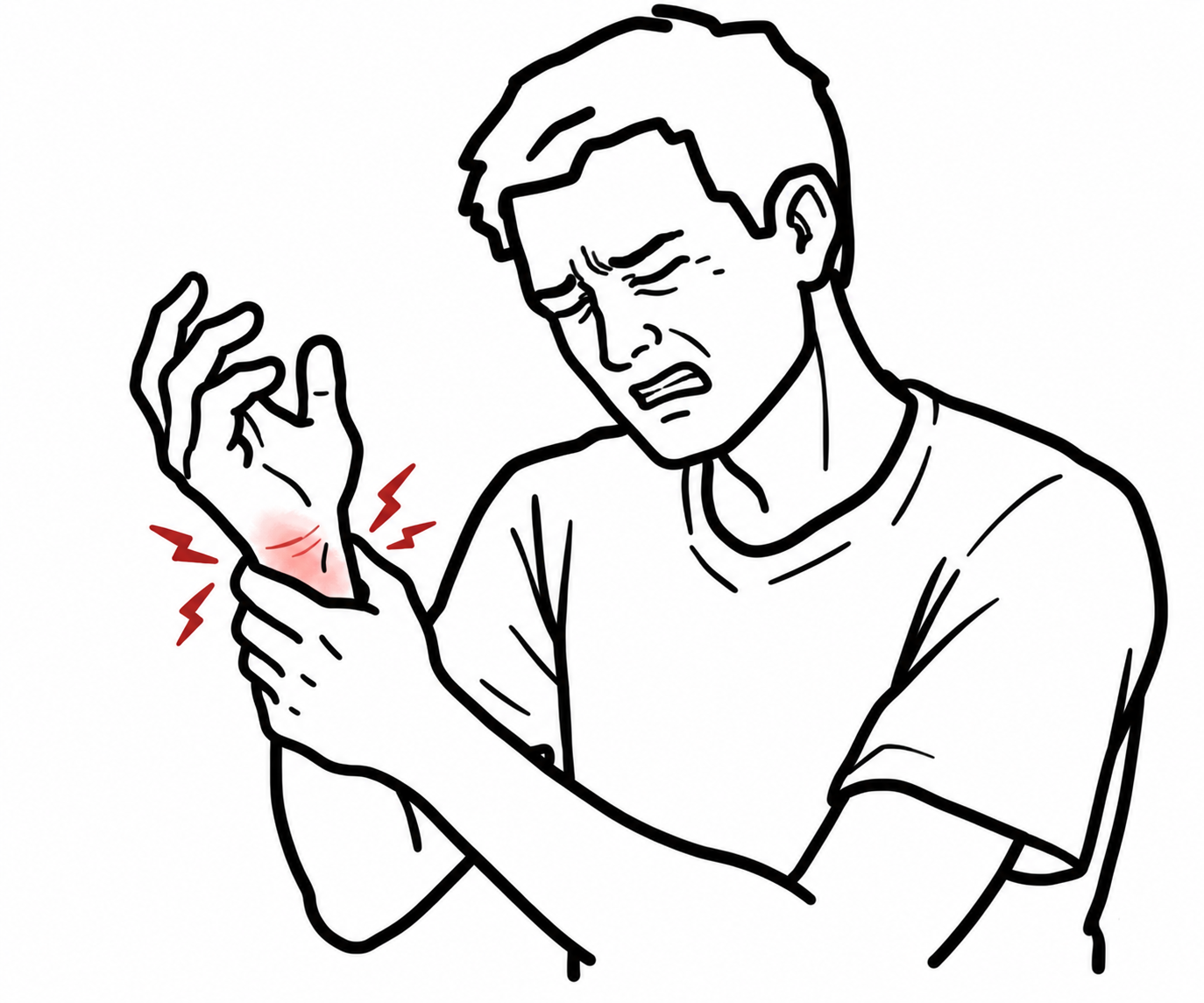
Wrist pain is discomfort originating from the radiocarpal, distal radioulnar, or midcarpal joints, or their surrounding tendons, ligaments, and nerves.
Topographical Classification
- Radial Wrist: Scaphoid fracture, De Quervain tenosynovitis, osteoarthritis of the first carpometacarpal joint, superficial radial nerve neuropathy.
- Ulnar Wrist: Triangular fibrocartilage complex injury, extensor carpi ulnaris tendinopathy, ulnar abutment syndrome, distal radioulnar joint instability.
- Dorsal Wrist: Ganglion cyst, extensor tendinopathy, scapholunate ligament dissociation, Kienböck disease.
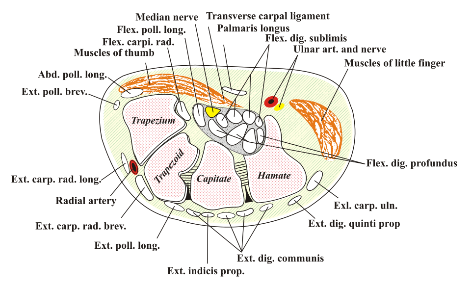
- Volar Wrist: Carpal tunnel syndrome, flexor carpi radialis tendinopathy, volar ganglion.
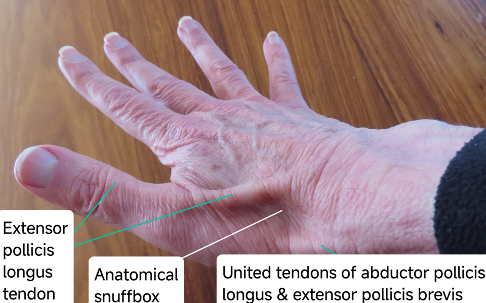
Importantly, fall onto an outstretched hand with anatomical snuffbox tenderness is a scaphoid fracture until proven otherwise, regardless of initial normal plain radiography.
The scaphoid receives its arterial blood supply retrograde from distal to proximal via branches of the radial artery. Fractures through the proximal pole carry an exceptionally high risk of avascular necrosis and non-union if not promptly immobilized.
Triage & Approach
Step 1: Emergency Triage & Surgical Red Flags
- Open Fracture / Gross Deformity / Neurovascular Compromise:
- Diminished radial pulse, cool/pale hand, capillary refill time > 2 seconds, median nerve sensory loss -> Immediate reduction, splinting, urgent orthopedic hand surgery consult.
- Acute High-Energy Trauma + Median Nerve Paresthesia:
- Suspect Lunate or Perilunate Dislocation (surgical emergency: requires immediate closed/open reduction to prevent irreversible median nerve ischemia).
- Fever + Erythema + Rapidly Progressive Severe Wrist Pain:
- Suspect Septic Arthritis or Infectious Flexor Tenosynovitis (Kanavel cardinal signs: flexed posture, symmetric enlargement, tenderness along flexor sheath, severe pain on passive extension).
Step 2: Traumatic Fall onto Outstretched Hand Assessment
- Palpate the three clinical scaphoid landmarks:
- Tenderness in the anatomical snuffbox.
- Tenderness over the scaphoid tubercle (volar wrist base).
- Pain with axial compression of the thumb.
- If any of these 3 signs are positive:
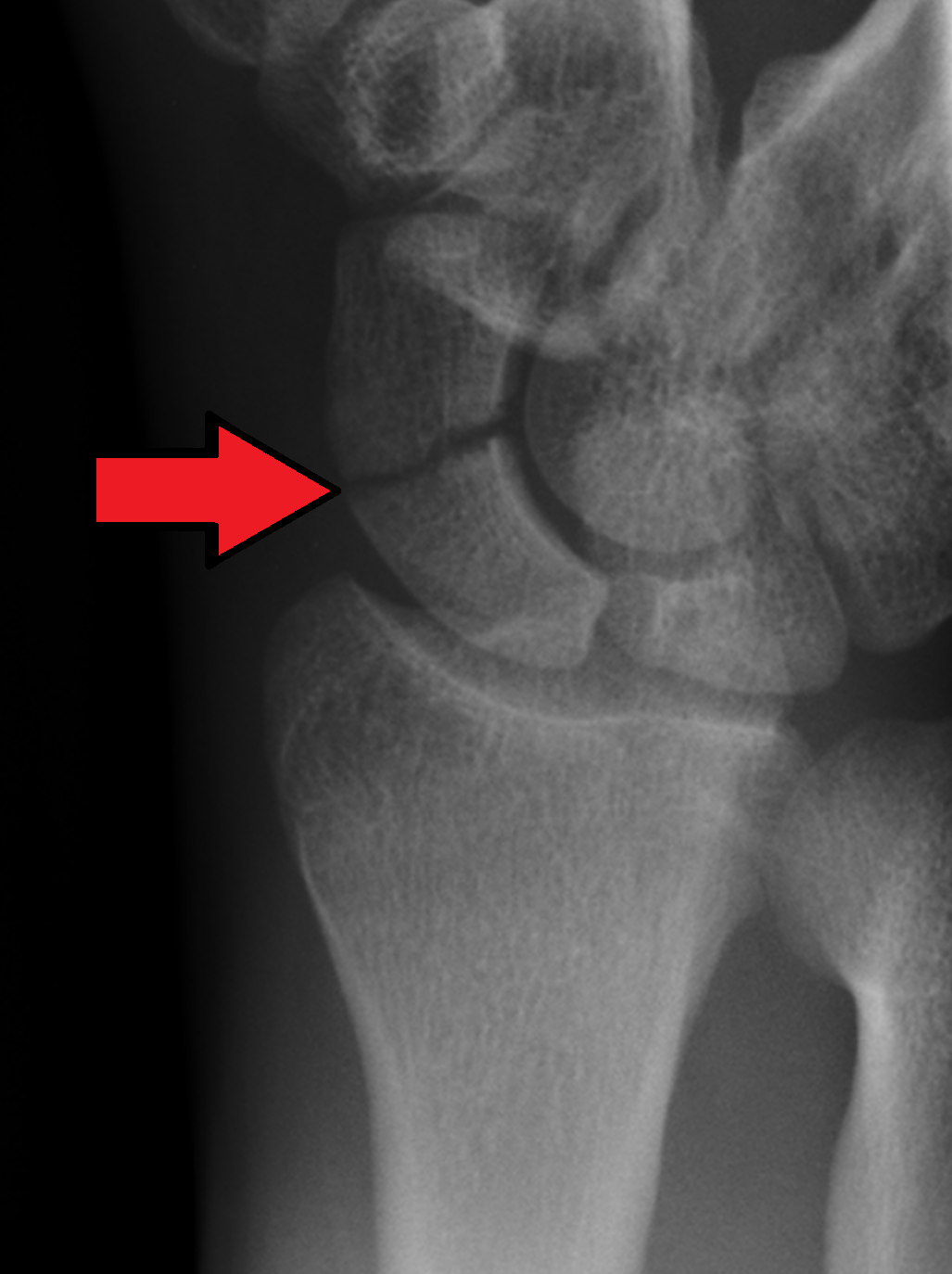
- Obtain 4-view wrist radiography (including dedicated scaphoid views).
- Fracture visible: Cast immobilization or surgical fixation based on displacement.
- Radiographs normal: Treat as Suspected Scaphoid Fracture (immobilize in a thumb spica splint, repeat clinical review and imaging in 10–14 days, or obtain early non-contrast magnetic resonance imaging).
Step 3: Non-Traumatic Branching by Anatomic Location
- Radial Pain: Perform Finkelstein test (pain over radial styloid with thumb tucked in fist during ulnar deviation -> De Quervain tenosynovitis).
- Volar Paresthesia: Median nerve distribution (thumb, index, middle, radial half of ring finger) waking patient at night -> Carpal tunnel syndrome (positive Phalen and Tinel tests).
- Ulnar Pain: Worse with forearm pronation/supination and loading (clicking, instability) -> Triangular fibrocartilage complex tear.
Plain radiographs miss up to 15–20% of acute scaphoid fractures on the day of injury. Never discharge a patient with anatomical snuffbox tenderness without a protective splint; failure to immobilize an occult fracture leads to non-union and scaphoid non-union advanced collapse.
Differential Diagnosis
Traumatic & Bone/Ligamentous Pathology
- Scaphoid Fracture:
- Distinguishing features: Fall onto an outstretched hand; maximal tenderness in the anatomical snuffbox; high risk of non-union at the proximal pole.
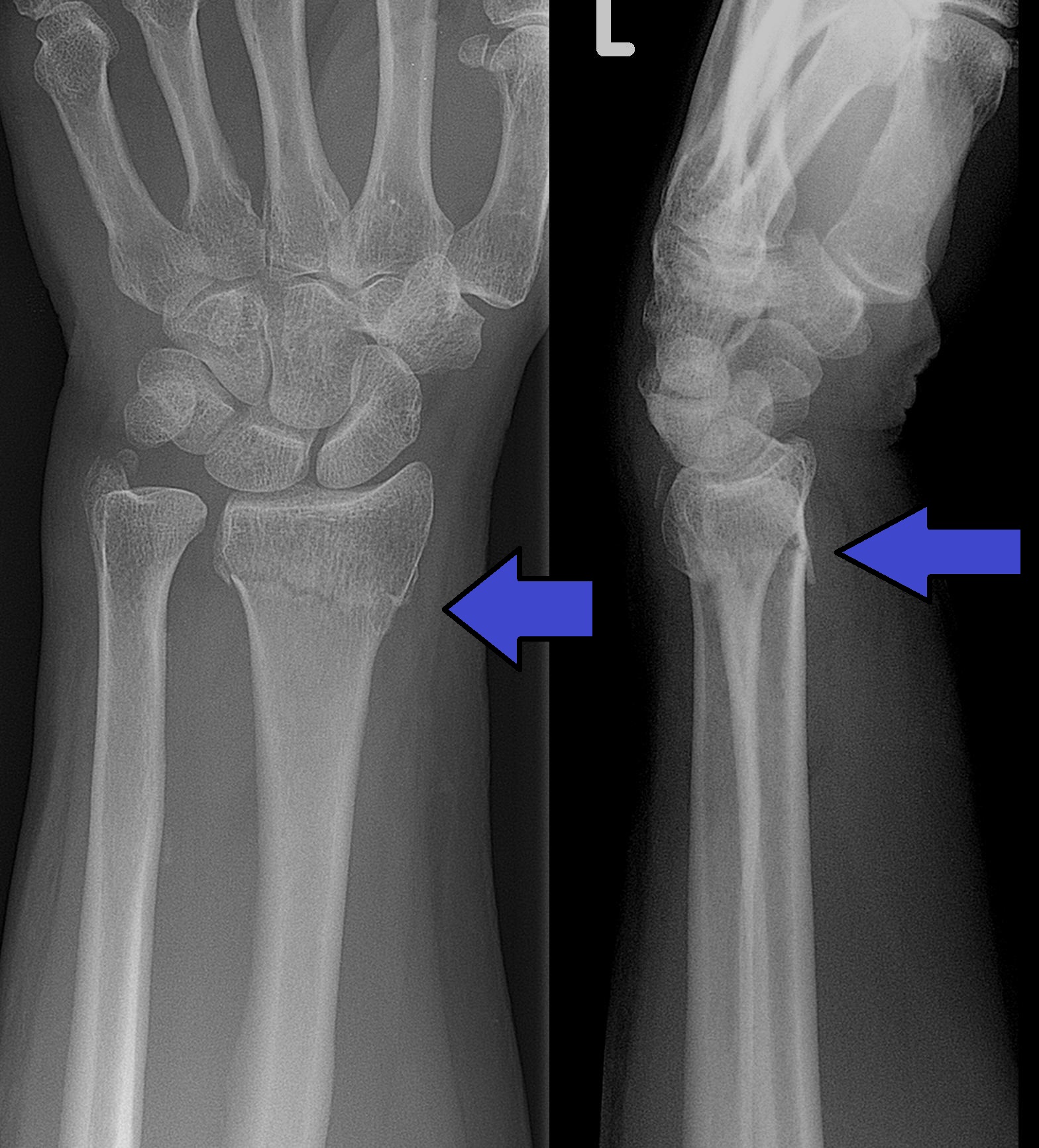
- Distal Radius Fracture (Colles / Smith / Barton):
- Distinguishing features: Classic “dinner fork” (dorsal displacement) or “garden spade” (volar displacement) deformity; focal tenderness over the distal radial metaphysis.
- Scapholunate Ligament Dissociation:
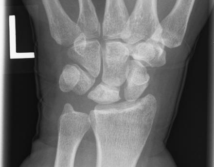
- Distinguishing features: Dorsal radial wrist pain; positive Watson shift test; widening of the scapholunate interval > 3 mm on posteroanterior radiography (pathognomonic “Terry Thomas sign”).
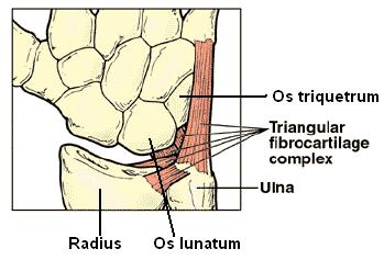
- Triangular Fibrocartilage Complex (TFCC) Tear:
- Distinguishing features: Ulnar-sided wrist pain, clicking, or catching; exacerbated by forced ulnar deviation and forearm rotation; tenderness in the soft spot between the ulnar styloid and pisiform (fovea sign).
- Kienböck Disease:
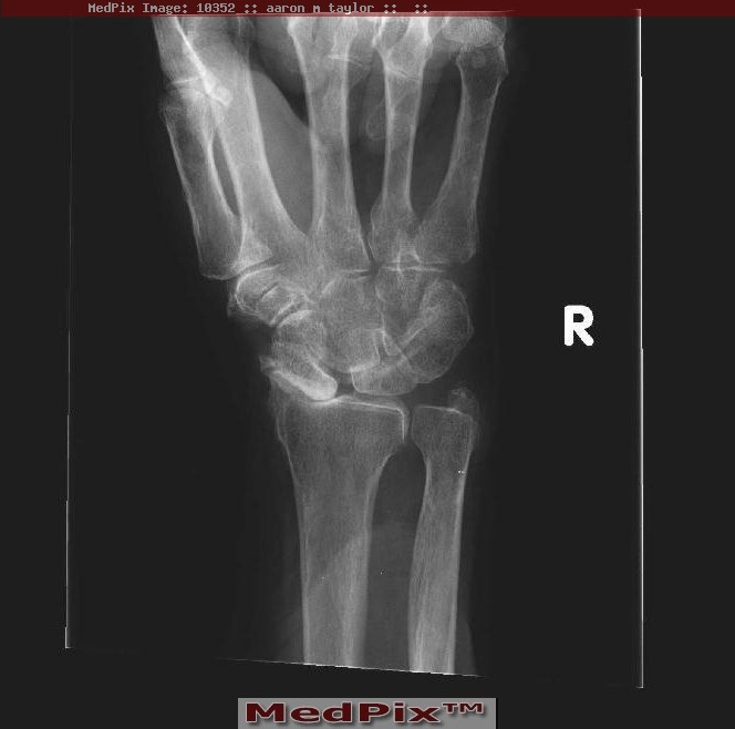
- Distinguishing features: Avascular necrosis of the lunate bone; young adults with insidious dorsal central wrist pain, stiffness, and decreased grip strength; sclerosis/collapse of lunate on imaging.
Soft Tissue & Entrapment Neuropathies
- De Quervain Tenosynovitis:
- Distinguishing features: Stenosing tenosynovitis of the first dorsal extensor compartment (abductor pollicis longus and extensor pollicis brevis); common in new parents lifting infants; severe pain over radial styloid reproduced by the Finkelstein maneuver.
- Carpal Tunnel Syndrome:
- Distinguishing features: Median nerve compression deep to flexor retinaculum; nocturnal numbness/tingling in the radial 3.5 digits; thenar muscle atrophy in chronic cases; positive Phalen test (wrist flexion for 60 seconds) and Tinel sign.
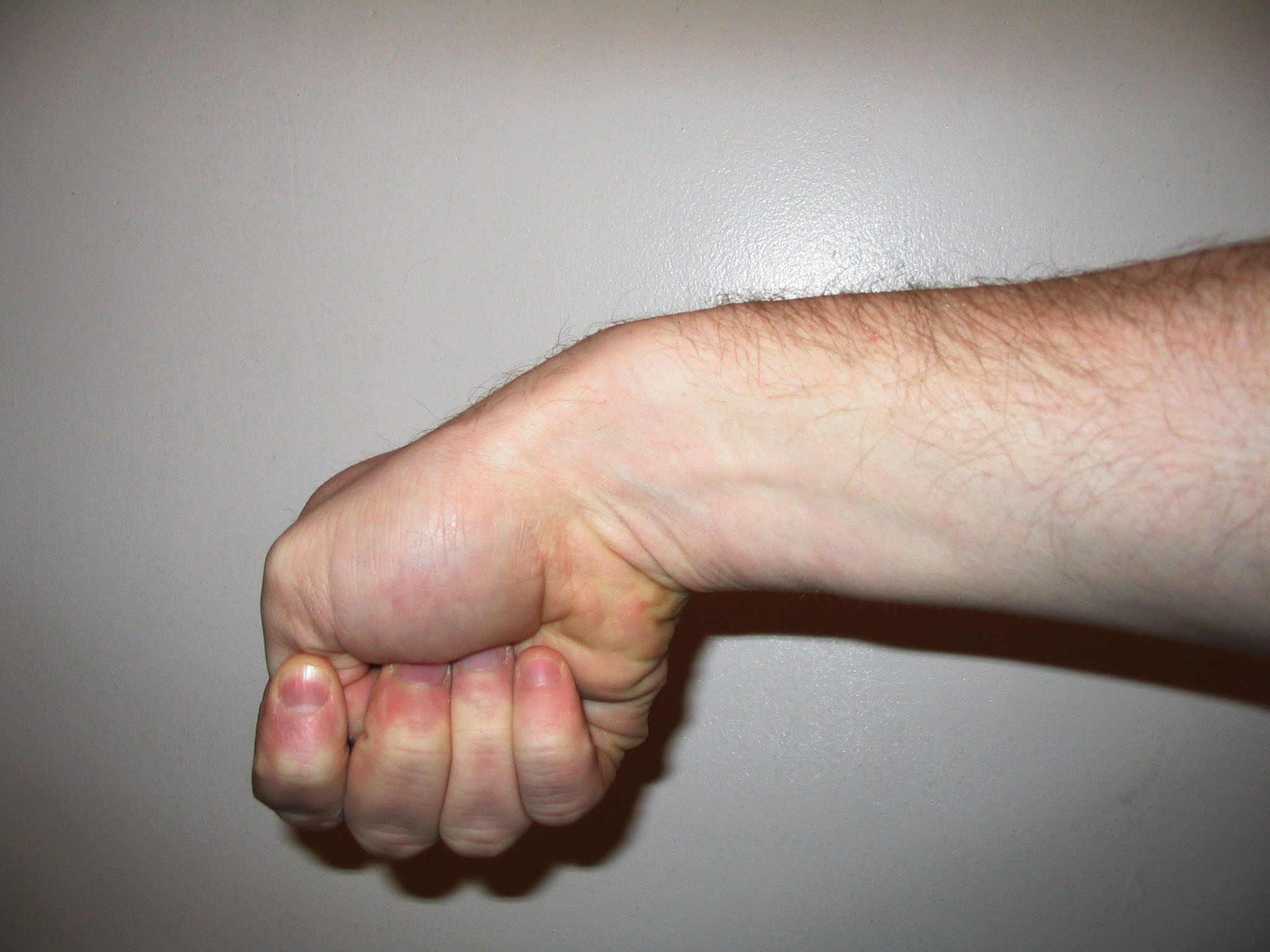
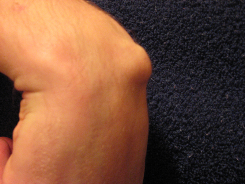
- Ganglion Cyst:
- Distinguishing features: Smooth, well-circumscribed, transilluminating mass; most commonly dorsal over the scapholunate joint (~70%) or volar over the radial artery (~20%).
Inflammatory & Degenerative Arthropathies:
- First Carpometacarpal Osteoarthritis (Basal Thumb OA):
- Distinguishing features: Pain at base of thumb aggravated by pinching/twisting (opening jars); localized tenderness, square-shaped hand deformity, positive grind test.
- Crystal Arthropathy (Gout / Pseudogout):
- Distinguishing features: Sudden onset severe monoarthritis with intense erythema and swelling; pseudogout (calcium pyrophosphate deposition) commonly involves the radiocarpal joint with chondrocalcinosis visible on plain film.
- Look for punctate calcification along the TFCC or scapholunate ligament (Chondrocalcinosis)
The “Terry Thomas sign” refers to a gap of > 3 mm between the scaphoid and lunate on an AP wrist radiograph (named after the famous British comedian with a prominent gap between his front teeth). It signifies a complete rupture of the scapholunate interosseous ligament.
Investigations
First-Line / Radiographic Workup
- Plain Wrist Radiography (Standard Views)
- Posteroanterior (PA), Lateral, and Oblique views: Evaluate distal radius alignment, carpal arcs of Gilula, and lunate alignment.
- Dedicated Scaphoid Bone X-ray
- Clenched Fist PA View: Accentuates scapholunate ligament diastasis.
- Inflammatory Markers & Arthrocentesis: Full blood count, C-reactive protein, and ultrasound-guided aspiration if septic arthritis or crystal arthropathy is suspected.
Targeted / Advanced Imaging Strategy
- Magnetic Resonance Imaging
- Modality of choice for occult scaphoid fractures (detects marrow edema within hours of injury), avascular necrosis (Kienböck disease), and soft tissue tears (scapholunate ligament and TFCC disruption).
- High-Resolution Ultrasound
- Dynamic, rapid assessment of De Quervain tenosynovitis (thickened first extensor retinaculum and sheath fluid), occult dorsal/volar ganglion cysts, and median nerve cross-sectional area at the carpal tunnel inlet (> 10 mm² indicates carpal tunnel syndrome).
- Computed Tomography
- Best for evaluating complex intra-articular distal radius fractures, bony bridging in scaphoid non-union, and carpal coalition.
- Nerve Conduction Studies / Electromyography
- Confirms delayed distal motor and sensory conduction velocity across the flexor retinaculum in carpal tunnel syndrome.
Early MRI (within 24–72 hours of injury) has superseded the traditional practice of blindly casting all suspected scaphoid fractures for 2 weeks. A normal early MRI definitively excludes a scaphoid fracture, avoiding unnecessary immobilization and time off work.
Critical Management
Scaphoid Fractures
- Non-Displaced Waist / Distal Fractures: Below-elbow cast or rigid thumb spica splint for 6–8 weeks, with serial radiographic confirmation of union.
- Displaced (> 1 mm), Angulated, or Proximal Pole Fractures: High non-union rate -> Orthopedic referral for percutaneous or open headless compression screw fixation (e.g., Herbert screw).
Infectious Flexor Tenosynovitis (Kanavel Signs Present)
- Emergency Surgical Drainage: Urgent hand surgery consultation for operative flexor sheath irrigation and debridement.
- Intravenous Antibiotics: Empirical IV Flucloxacillin 2 g q6h (or Cefazolin; add Vancomycin if methicillin-resistant Staphylococcus aureus is suspected).
De Quervain Tenosynovitis
- First-Line: Thumb spica splinting (leaving the interphalangeal joint free) + oral non-steroidal anti-inflammatory drugs + activity modification for 4–6 weeks.
- Second-Line: Ultrasound-guided corticosteroid injection into the first dorsal compartment sheath (cure rate > 70–80%; take care to avoid superficial radial nerve injury and subcutaneous skin hypopigmentation).
- Refractory: Surgical decompression of the first dorsal extensor compartment.
Carpal Tunnel Syndrome
- Conservative: Nocturnal neutral wrist splinting for 6–12 weeks + local corticosteroid injection.
- Surgical Decompression: Open or endoscopic carpal tunnel release indicated for constant numbness, thenar muscle weakness/atrophy, or failure of conservative measures.
When injecting corticosteroids for De Quervain tenosynovitis, inject into the tendon sheath rather than directly into the tendon tissue to avoid tendon rupture, and take care to identify an anatomical septum separating the abductor pollicis longus and extensor pollicis brevis, which can cause treatment failure if not both bathed in steroid.


Members only discussions coming soon…