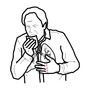

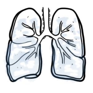
A chest X-ray is a radiographic imaging technique that uses low-dose ionising radiation to produce a two-dimensional image of the thoracic cavity, including the lungs, heart, and bones. It is essential for diagnosing respiratory, cardiac, and skeletal conditions, guiding treatment decisions. Commonly, it detects pneumonia, a frequent and potentially serious lung infection.


Please confirm you want to block this member.
You will no longer be able to:
Please allow a few minutes for this process to complete.