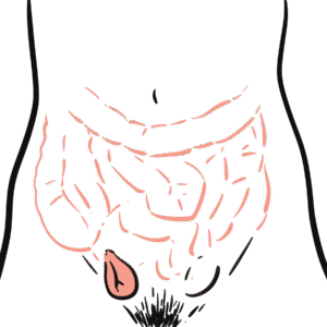
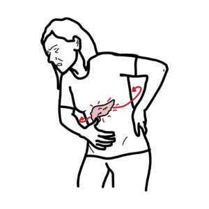
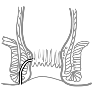
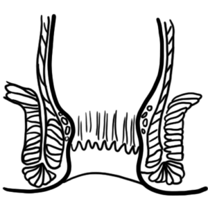
0:00 Hello, in this video, we're going to talk about hernia, specifically, inguinal 0:07 hernia. 0:08 We're going to look at the signs and symptoms, the risk factors, and I guess 0:14 how it actually 0:15 happens and how do we differentiate between the different types of inguinal her 0:20 nias. 0:20 So hernia itself is defined as a protrusion, bulge, or projection of an organ 0:26 or part of 0:27 an organ through the body wall that normally contains it. 0:31 And there are different types of hernias, and it can be divided mainly into 0:35 internal 0:36 and external. 0:37 We will mainly be talking about external hernias. 0:40 Now, external hernias include inguinal hernia, which is the most common of all 0:45 these, which 0:45 is 80%. 0:46 Then you have incisional hernia, 10%, femoral hernia, 5%, umbilical hernia, 4%, 0:54 epigastric 0:55 hernia, less than 1%, and then you have others. 0:58 So the inguinal hernia and the femoral hernia are known together as groin hern 1:03 ias, because 1:04 they occur around the groin area. 1:07 So let us actually talk about the inguinal hernia, because that's what we're 1:12 going to 1:12 talk about mainly here, because it is the most common. 1:16 So what we actually see in an inguinal hernia is the intestine, which is the 1:20 organ, actually 1:22 bulging out or pushing out of the wall that normally contains it, which is the 1:26 abdominal 1:27 wall in this case. 1:30 So the abdominal wall is actually comprised of, from the outside, the skin, the 1:36 subcutaneous, 1:37 and then you have the abdominal muscles, and there are three abdominal muscles 1:43 here. 1:43 From the outside, it's the external oblique, the internal oblique, and then you 1:47 have the 1:47 transverse abdominis. 1:49 So again, an inguinal hernia is essentially the bulge or protrusion of an organ 1:55 , this being 1:55 the intestine, through the wall that normally contains it, which is the 2:02 abdominal wall. 2:03 So what are some signs and symptoms of someone who has an inguinal hernia? 2:09 Well, obviously it depends on the severity and how much it's actually bulging 2:13 out and 2:13 if it's strangulated. 2:16 But the signs and symptoms altogether are, obviously, there's a visible lump, 2:21 that's 2:22 a sign, a heavy discomfort around the gut, you can have pain and aching on 2:29 exertion, 2:30 so maybe like lifting up something, you can experience constipation, and I 2:36 guess you can 2:37 also say that the lump enlarges upon coughing as a sign. 2:44 So there are some risk factors for someone to develop an inguinal hernia. 2:48 Well, these include a history of hernia, older age, male sex, being caucasian, 2:55 having 2:56 a chronic cough, chronic constipation, I guess a weak abdominal wall, and then 3:04 also smoking 3:05 as a risk factor. 3:08 So again, inguinal hernia is you have the bulging of the small intestine here, 3:13 through 3:13 the abdominal wall. 3:15 So let's look at this from an anterior view. 3:18 So here we have the groin area. 3:20 Here's a linear album, which makes up part of the abdominal muscle, right? 3:25 And then here you have the abdominal muscles from the outside, you have the 3:29 external oblique, 3:29 which has been opened up, which has been sort of opened up as you can see here. 3:34 And then you have the internal oblique, and then below the internal oblique you 3:37 have the 3:37 trans-visalis abdominis fascia. 3:41 And here we are actually just pulling on the spermatic cord. 3:46 It's been retracted. 3:47 So we're pulling over it. 3:48 And because we're pulling it, we can actually see below the spermatic cord, or 3:52 next to it, 3:54 we have important artery, which is actually a landmark. 3:59 Keep note of this. 4:00 And the artery is the inferior epigastric artery. 4:06 And the inferior epigastric artery is an important landmark because it sort of 4:11 differentiates 4:12 between the different types of inguinal hernia, and we'll talk about that as we 4:17 go. 4:18 And then here, I guess above the penis area, you have the conjoined tendon. 4:25 And it's sort of medial to the inferior epigastric artery. 4:28 Now let's look at the same image. 4:30 So we're looking at the muscle layers, the inferior epigastric artery and the s 4:34 permatic 4:35 cord. 4:36 Let's look at this image, but from a different angle, let's cut a cross-section 4:41 of the body 4:41 and look at it from the top. 4:45 So here you have your small intestine or intestines. 4:50 And then we have the peritoneum, which actually holds all your sort of covers, 4:55 is the inner 4:56 lining of the digest abdominal cavity. 5:00 And then the next layer you have the trans-visalis abdominis with the fascia. 5:04 And then you have the internal oblique, external oblique. 5:08 And then you have the external oblique aponeurosis, and then you have the sub 5:11 cutaneous, and then 5:12 you have the skin. 5:14 So these are the layers from the inside of the abdominal wall to the outside. 5:20 And then you have the spermatic cord, which originates behind the trans-visalis 5:23 fascia. 5:24 It goes down and it goes through all these layers and down to the scrotum. 5:29 So it travels immediately and down to the scrotum. 5:32 And then medial to the spermatic cord, behind the trans-visalis fascia, you 5:37 have the epigastric 5:39 artery, which again is an important landmark, and it's medial. 5:43 And then you have also the conjoined tendon here. 5:48 So I hope that made sense. 5:50 Just remember these layers, and remember the spermatic cord, how it travels med 5:56 ially, 5:57 and then also the inferior epigastric artery. 6:00 So let's talk about inguinal hernia. 6:01 And again, what we will see is that we will see these intestines. 6:06 They will actually go through, sort of bulge through these abdominal walls. 6:13 So now I'm just going to draw the inguinal hernia, as we saw before. 6:21 And here's the intestine, which is bulging, sort of through the abdominal wall, 6:25 pushing 6:25 against the abdominal wall. 6:27 And here we have the same image of the right side of the sort of groin area. 6:35 And then we have hernias, the inguinal hernias. 6:37 And there are two types of inguinal hernia. 6:41 We have what's known as a indirect. 6:43 So an indirect inguinal hernia is the one that sort of pushes, bulges through 6:48 the abdominal 6:49 wall, and actually goes into the scrotum. 6:53 So it sort of follows the spermatic cord pathway. 6:58 And then you have a direct inguinal hernia, which is just a hernia that sort of 7:05 bulges 7:06 through the abdominal wall and doesn't actually go down the scrotum. 7:10 So what differentiates these two types of inguinal hernias, the direct and 7:15 indirect, 7:16 is the important landmark that we talked about, which is the inferior epigast 7:21 ric artery. 7:28 And here we have the inferior epigastric artery. 7:33 And essentially, if the inguinal hernia occurs medially to the inferior epigast 7:38 ric artery, 7:39 it's a direct hernia, but if it occurs, if it begins laterally, then it's an 7:44 indirect 7:45 hernia. 7:46 Now talking about indirect hernia, it follows the spermatic cord pathway 7:53 because then indirect 7:55 hernia actually travels from, originates from the deep inguinal ring, it goes 8:01 through the 8:02 deep inguinal ring, and the superficial inguinal ring. 8:09 And so, this is obviously the same path of this spermatic cord, and so the 8:14 intestine will 8:15 actually follow this pathway, follow the spermatic cord pathway, and into the 8:21 scrotum. 8:22 Now an important landmark area, important region to know, to remember for hern 8:27 ias is 8:28 known as the Hesselbach's triangle. 8:31 And so the borders of the Hesselbach's triangle is the inguinal ligament 8:37 inferiorly. 8:38 So you have their inferior epigastric lactually, and then you have the rectus 8:49 abdominis medially. 8:53 So let's look at these two types of inguinal hernias in a bit more detail. 8:58 So again, you have two types of inguinal hernias, you have the indirect ingu 9:02 inal hernia, and 9:02 you have the direct inguinal hernia. 9:05 So let's begin by talking about the indirect inguinal hernia. 9:09 Indirect inguinal hernia is the most common type of hernia. 9:13 It's where the intestines protrude at the deep inguinal ring, and it's lateral 9:20 to the 9:21 inferior epigastric artery, so it starts laterally. 9:25 So if we were to draw that same diagram that we drew, the cross section, and we 9:29 're looking 9:30 at it from the top from a superior view, you know, we have the layers, the per 9:35 itoneum, 9:35 the transosalis fascia, the internal oblique, the external oblique, and then 9:41 you have the 9:42 external oblique up on your oasis. 9:46 And here is your deep inguinal ring, essentially a hole where the spermatic 9:50 cord originates 9:50 from the peritoneum, and then you have this superficial inguinal ring where the 9:54 spermatic 9:54 cord will exit. 9:56 So I'm actually drawing this verbedic cord here in yellow, and it's not quite 9:59 right. 10:00 It should be within, it should originate from the deep inguinal ring. 10:05 And then you have the inferior epigastric artery, and remember, an indirect her 10:09 nia occurs 10:10 lateral, laterally, to the inferior epigastric artery. 10:15 And so an indirect hernia is where the intestine will go through the deep ingu 10:20 inal ring, and 10:21 then through the superficial inguinal ring and essentially bulge out. 10:27 So I hope that made sense. 10:28 And again, the indirect inguinal hernia occurs lateral to the epigastric, 10:33 inferior epigastric 10:35 artery, and essentially follows the same pathway, the spermatic cord, as shown. 10:42 So a direct hernia is where we have a protrusion medial to the inferior epigast 10:47 ric artery within 10:48 the hisselbox triangle. 10:50 And remember, we learned about the hisselbox triangle. 10:55 So hisselbox triangle is where direct hernias occur. 10:59 And the direct hernias occur as a result of weakness in the floor of the ingu 11:05 inal canal. 11:06 And it passes through the superficial ring only. 11:08 It doesn't go through the deep end superficial. 11:11 The direct inguinal hernia only goes through the superficial, and we will see 11:17 how. 11:18 So again, we're looking at this diagram from the top. 11:22 And here I'm drawing a spermatic cord in yellow. 11:28 And here we have the inferior epigastric artery. 11:33 And here is your deep inguinal ring where the spermatic cord originates, right? 11:43 And then here you have the superficial inguinal ring where the spermatic cord 11:48 also goes through. 11:50 Well in a direct inguinal hernia, it only goes through the superficial inguinal 11:55 ring. 11:56 So what essentially, what we see essentially is the intestine will go through 12:01 these layers. 12:05 And the intestine will just go through the superficial inguinal ring, medial to 12:11 the inferior 12:11 epigastric, and not go in contact with the deep inguinal ring. 12:21 So again, the direct inguinal hernia occurs medial to the inferior epigastric. 12:27 And the indirect inguinal hernia occurs laterally to the inferior epigastric. 12:31 And I hope these diagrams made sense. 12:34 Thank you for watching.