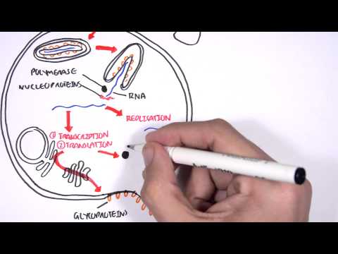
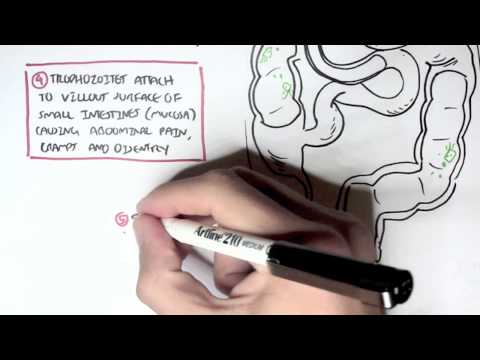
0:00 Armando Hsuregan, biology and medicine videos, please make sure to subscribe, 0:04 join the forum 0:05 and group for the latest videos, please visit Facebook, Armando Hsuregan, 0:08 please like, and 0:09 here you can also ask questions, answer questions, and post some interesting 0:12 things, including 0:12 Alex. 0:13 And you can also change the quality settings, the highest one for better 0:16 graphics. 0:17 In this video, we're going to look at the overview of microbiology. 0:21 Microbiology studies microorganisms, such as bacteria, for example. 0:26 Now microorganisms are actually present everywhere, literally, even under our 0:31 foot and on our 0:32 hair, for example. 0:33 However, very few actually cause any form of diseases, so it's okay. 0:38 And due to the development of magnifying instruments within the century, for 0:42 example, enabled scientists 0:43 to learn much more about these microorganisms. 0:48 And these magnifying instruments, for example, are there three main ones, these 0:53 are known 0:53 as the compound light microscope, the transmission electron microscope, and the 0:58 scanning electron 0:59 microscope. 1:00 What are the difference? 1:01 Well, the compound light microscope is really just the basic. 1:05 And we are able to see microorganisms through a series of lenses, and they can 1:09 actually 1:10 magnify about one, a typical one, about one thousand to two thousand times 1:16 magnification. 1:17 And we usually use a glass side to view these microorganisms. 1:21 Now an important concept to know about microscopes is what's known as 1:25 resolution. 1:26 And because microorganisms are very, very, very small, resolution is important 1:31 for microscopes 1:32 in order to separate between two microorganisms, essentially to view fine 1:37 detail of these microorganisms. 1:40 Now the next type of microscope is a transition electron microscope. 1:43 And these have much better resolution, magnification, you can say, than the 1:48 compound light microscope. 1:50 So we can see better detail of these microorganisms. 1:54 So why can we see these microorganisms in better detail? 1:57 Because the transmission electron microscope uses electrons, not light waves. 2:02 And electron waves are much more smaller, and so we can see better detail. 2:06 And typically a transmission electron microscope can magnify up to one hundred 2:10 thousand times 2:11 magnification. 2:13 And we usually view these, the image on a photographic plate over here. 2:18 However, the bad thing about this transmission electron microscope is that we 2:22 cannot actually 2:23 use living cells, whereas a compound light microscope we could. 2:28 And the final one is a scanning electron microscope. 2:31 And this is probably the coolest microscope ever, because it enables us, 2:37 enables scientists 2:39 to view these microorganisms in 3D. 2:43 And so, and it only can magnify up to ten thousand. 2:47 However, the detail of these microorganisms in three dimension is awesome. 2:55 So now let's look at some few methods of preparation. 2:58 We're concentrating on the compound light microscope typically. 3:01 And the main type of preparation method, there are few. 3:07 But the main one is that we can suspend the microorganisms in a droplet of 3:11 water. 3:11 And so we can view it in a living state, for example, the movement. 3:16 And we can also use a dye, because usually microorganisms are actually color 3:21 less. 3:21 And after we dye it, or we replace it in water, we can then observe this micro 3:27 organism using 3:28 a microscope. 3:30 Now this dyeing technique, which is to colorate microorganisms, is actually 3:34 really important 3:35 in classifying bacteria into two major groups. 3:39 And the technique we use for dyeing is called the gram stain technique. 3:42 When we use a particular dyeing, which colors the bacteria in different colors 3:48 depending 3:49 on the cell wall composition. 3:52 And so, for example, if the bacteria will be classified as gram positive, if 3:58 the color 3:58 turns purple. 3:59 However, if the bacteria turns red or pink, it will be called gram negative 4:05 bacteria. 4:07 So gram positive is what usually purple color, gram negative is red or pink 4:11 color. 4:11 Now let's learn about some people which were important in the field of microbi 4:15 ology. 4:16 The first guy is by the name of Lewis Posture, who disapproved the theory of 4:20 spontaneous 4:20 generation. 4:21 What is spontaneous generation? 4:23 Well, it's the thought that organisms arise from essentially non-living things 4:30 such as 4:31 dust, for example. 4:32 And he also said that microorganisms are responsible for food spoilage. 4:37 And then there are these two other people, these two other men, from different 4:41 countries, 4:42 from different fields. 4:44 One was my name, Ignis Semawise, and the other one is Joseph Liston, who was 4:49 actually a surgeon 4:51 or a doctor. 4:53 And essentially what they said is that infections are contagious, which was 4:58 quite obvious, and 5:00 that they proposed ways in preventing these contagious infections. 5:05 And there are some really interesting stories behind these people. 5:08 And then in 19th century there was a really important man by the name of Robert 5:14 Koch. 5:15 And he proposed a German theory of disease. 5:18 So what is this theory? 5:19 Well this theory essentially says that a disease is obtained from a particular 5:23 microorganism 5:24 or an organism. 5:26 And so there's a popular diagram to explain this situation or this theory, when 5:31 , for example, 5:32 a sick animal, you will obtain blood from a sick animal, and then we'll isolate 5:37 this, 5:38 isolate this sample. 5:41 And then we can actually, after some time, we can actually observe it under a 5:45 microscope, 5:46 for example, and we can observe microorganisms. 5:48 And so if we take the same sample that was isolated and we actually culture it 5:52 instead 5:53 and let the organism to grow, after it's grown, we can then inject this micro 5:59 organism into 6:00 a susceptible host such as a similar healthy animal. 6:05 After some time, this should cause the same disease as in the initial animal. 6:12 And so if we again obtain, if we take blood out of this animal and we isolate 6:18 it and then 6:19 we culture it and we observe it under a microscope, we should see the same 6:24 microorganism. 6:26 And using this method, Robert Koch was able to prove that anthrac is caused by 6:30 a particular 6:30 organism. 6:31 And this was a major breakthrough to find out that a particular microorganism 6:36 or organism 6:37 can cause a disease. 6:38 And so this was a major breakthrough to find out that a, that a organism or 6:41 microorganism 6:42 can actually cause a disease. 6:47 Now let's look at some types of microorganisms. 6:50 Let's look at three main categories. 6:51 And these are bacteria, viruses and eukaryotic organisms. 6:56 The eukaryotic organisms that we're going to look at include the protists and 6:59 other eukaryotes. 7:01 Let's begin by quickly looking through what a bacteria is. 7:05 Now bacteria is a prokaryote and lives freely in the environment. 7:09 We find it literally everywhere, including inside our body. 7:13 Our body actually predominantly contains bacteria. 7:18 And so the bacterias can be classified according to a structure and has three 7:23 structures. 7:24 It can be spherical bacteria, a rod shaped bacteria and also a spiral bacteria. 7:30 And a very important thing to know is that bacteria multiply very rapidly. 7:34 And so this is why if we put a smudge of our hair or whatever inside a petri 7:39 dish, we can 7:40 see that bacteria will form colonies there within the next few days. 7:46 And so we can say that bacterias multiply very rapidly. 7:49 And the next type of microorganism is the virus. 7:52 And these include viruses such as a bacteriophage, which infects particularly a 7:56 bacteria, and 7:57 also influenza virus, which is very common in humans. 8:01 Bacteria cause infections in other organisms, such as a bacteriophage in 8:05 bacteria and the 8:06 influenza virus in humans. 8:08 Now the typical structure of a virus, they essentially have an outer envelope 8:12 made up 8:13 of usually glycoproteins and an inner protein, which protects the genetic 8:17 material, either 8:18 RNA or DNA. 8:20 The virus size is very, very small from about 20 nanometers to the biggest one, 8:25 about 300 8:27 nanometers. 8:28 And so as I mentioned, viruses infect other organisms. 8:32 And so we can say that viruses influence a virus, for example, infects the 8:36 pulmonary 8:36 cells within our lungs. 8:38 And this is how they replicate. 8:41 So viruses replicate inside a cell that they had infected. 8:45 And that was all for viruses. 8:46 Let's look at the eukaryotic organisms. 8:50 Now they consist of protists and other eukaryotes. 8:53 Let's concentrate on protists first. 8:55 And there are two types of protists family that I'm going to talk about. 8:58 One of these microorganisms are known as algae, alga. 9:01 And the alga are mainly aquatic organisms, and you can see them in the sea, 9:06 such as seaweed. 9:07 This is the type of alga. 9:09 And they actually can absorb sunlight and carry out photosynthesis. 9:14 So most alga carry out photosynthesis. 9:16 They absorb carbon dioxide, sunlight, water, and they produce oxygen for humans 9:20 . 9:20 And so because they can perform photosynthesis, they contain what's called 9:25 chlorophylls. 9:27 And most alga are not actually toxic to humans, because we usually eat seaweed, 9:30 for example. 9:31 However, some types of alga secrete neurotoxins. 9:35 And so for example, if a fish in the sea were to eat an alga, which then the al 9:40 ga would secrete 9:41 neurotoxins once inside the fish, this fish would become poisonous, and it's 9:46 poisonous 9:46 to humans if we consume this fish. 9:49 The next type of protists is the protozoa. 9:53 And these are also microorganisms. 9:55 And they are actually unicellar. 9:58 And what's fascinating about them is they actually do not contain a cell wall, 10:01 but they 10:02 contain some form of membrane. 10:03 And also, they're organelles, but the insides are actually visible. 10:08 What differs them with the alga is that they do not contain chlorophylls, so 10:13 they do not 10:14 perform photosynthesis. 10:16 And another important thing to note is that they most of them live in the water 10:19 , same 10:20 with algas. 10:21 And so that was it for the protists. 10:24 Let's look at other types of eukaryotic microorganisms. 10:27 And these include the fungi. 10:29 And when we think of fungus, fungi, we think of mushrooms. 10:33 And these fungi, they are unicellar, or they could be also multicellar, and we 10:36 find them 10:37 everywhere. 10:38 And fungi are actually excellent for the environment. 10:41 They're very good for the environment, actually, and very good for humans, in 10:45 that firstly, 10:46 they provide us with food, secondly, they provide us antibiotics for certain 10:51 things, 10:51 and also for fermentation of alcoholic, et cetera, alcohol, et cetera. 10:57 And very few cause, actually, any pathogenic effects, however, they can produce 11:04 some issues 11:05 and problems for humans, such as tinna, pedis, tinna, or an antibody, for 11:09 example. 11:10 The other type of eukaryotic is what's called the microscopic parasites. 11:14 They're called microscopic, because normal parasites are quite large. 11:18 And so that is why we call these microscopic parasites. 11:21 And they include roundworms, tapeworms, and what's called flukes. 11:24 And these types of worms, they usually target our gastrointestinal tract and 11:29 make cause 11:30 some problems. 11:31 Now, I hope you enjoy this overview of microbiology. 11:36 I hope I will put up links for each of these microorganisms to look at them in 11:40 more detail, 11:41 the bacteria, the viruses, the protozoa, and fungi and microscopic parasites 11:46 especially. 11:47 Thank you.