

Acute Liver (Hepatic) Failure
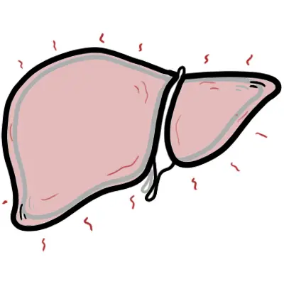
Overview
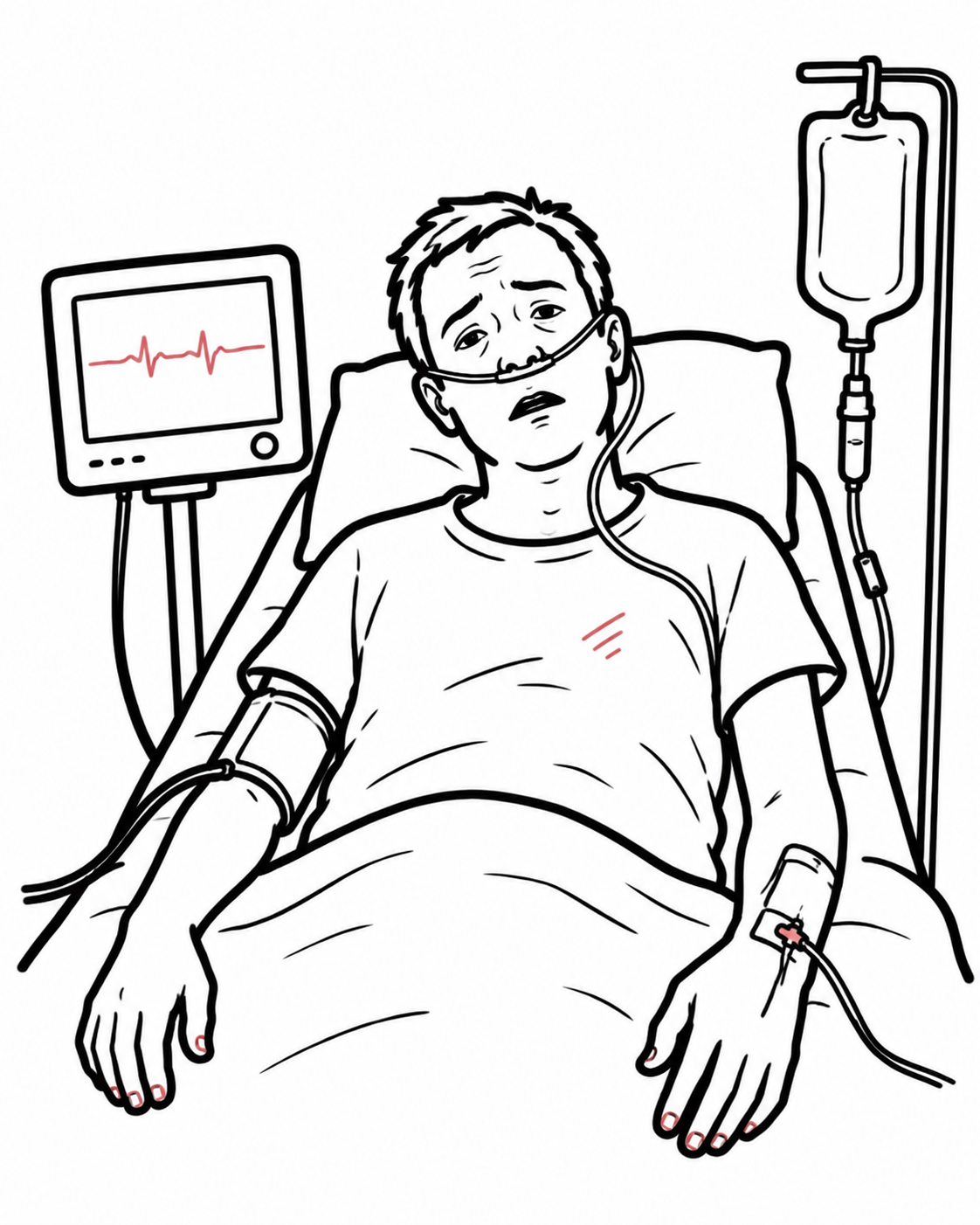
Acute liver failure (ALF) is a rare, rapidly evolving syndrome of severe hepatocellular injury with loss of hepatic function. Patients can deteriorate from apparently mild illness to encephalopathy and multiorgan failure within days. Prompt recognition, cause-specific treatment and early transplant-centre involvement are essential. These notes describe adult ALF; paediatric diagnostic criteria differ. [1–4]
Definition
Acute liver failure: acute liver injury with hepatic coagulopathy (INR ≥1.5) and any degree of hepatic encephalopathy developing within 26 weeks, generally without pre-existing cirrhosis.
Acute liver injury: acute hepatic injury without encephalopathy; severe injury with coagulopathy can progress to ALF.
Acute-on-chronic liver failure: acute deterioration in chronic liver disease, usually cirrhosis, with organ failure; a distinct syndrome.
Medical emergency: acute hepatic injury with new confusion and a rising INR requires urgent hepatology/critical-care assessment and discussion with a liver transplant centre. Do not wait for advanced coma or transplant prognostic criteria.
Anatomy & Physiology
- The portal vein and hepatic artery supply the liver; hepatic veins drain into the inferior vena cava.
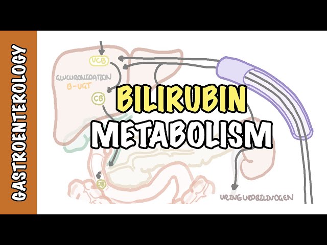
- Hepatocytes regulate glucose, synthesise coagulation proteins, process bilirubin and detoxify ammonia through the urea cycle.
- Failure of these functions explains hypoglycaemia, prolonged INR, jaundice and hyperammonaemia. Albumin may remain relatively preserved early because of its long half-life. [3,4]
Aetiology & Risk Factors
Aetiology
| Cause | Important clues |
|---|---|
| Paracetamol (acetaminophen) toxicity | Intentional overdose or repeated supratherapeutic use; include combination analgesics. A common cause in the UK/USA. |
| Idiosyncratic drug-induced liver injury | Prescription medicines, herbal remedies and dietary supplements; latency varies. |
| Viral infection | Hepatitis A, B and E; HSV is uncommon but important, especially in pregnancy or immunosuppression. HCV rarely causes ALF. |
| Autoimmune hepatitis | May present without a previous diagnosis; autoantibodies and IgG may be unremarkable in acute severe disease. |
| Hypoxic/ischaemic injury | Shock, cardiac failure or severe hypoxaemia. |
| Other causes | Wilson disease; Budd–Chiari syndrome; acute fatty liver of pregnancy/HELLP; Amanita mushroom toxins; rare malignant infiltration. Some cases remain indeterminate. [2–4] |
Risk Factors
- Risk depends on the cause: drug/toxin exposure, viral exposure or endemic-area travel, immunosuppression, pregnancy and circulatory failure.
- Obtain a detailed medication history from relatives when encephalopathy limits recall, including dose, timing, supplements and recreational drugs.
Pathophysiology
| Process | Clinical consequence |
|---|---|
| Hepatocyte necrosis and impaired regeneration | Reduced functioning liver mass and loss of synthetic/metabolic capacity. |
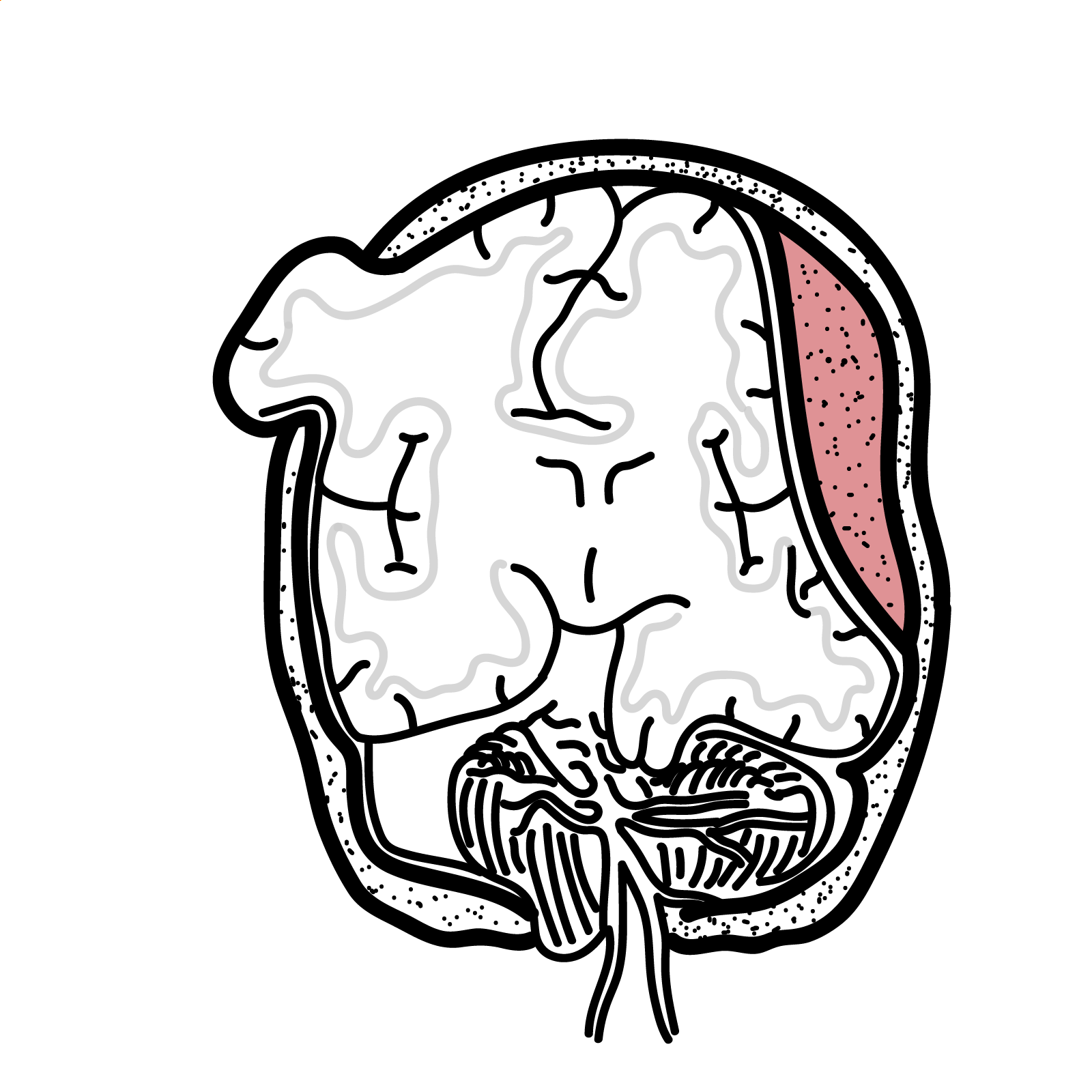
| Reduced ammonia clearance plus systemic inflammation | Astrocyte swelling and cerebral oedema; severe cases develop intracranial hypertension. |
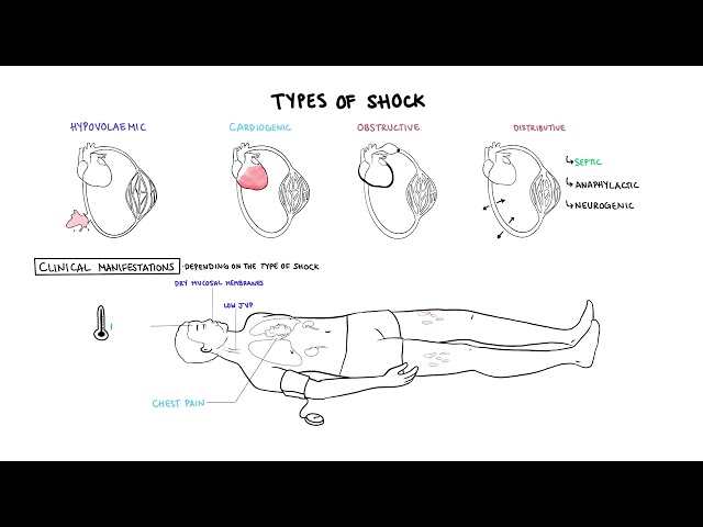
| Inflammatory mediator release and vasodilatation | Hypotension, impaired organ perfusion and multiorgan dysfunction. |
| Reduced clotting and anticoagulant protein synthesis | Altered, partly rebalanced haemostasis; INR alone does not measure bleeding risk. [3,4,7] |
Gross and Microscopic Pathology
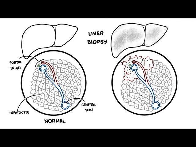
Massive or submassive hepatocellular necrosis may occur, with variable inflammation and regeneration. Histological patterns depend on the cause. A transjugular biopsy is considered when diagnostic uncertainty would change management, rather than performed routinely. [2,4]
Clinical reasoning: falling aminotransferases may represent recovery or loss of viable hepatocytes. Interpret them alongside INR, lactate, glucose, encephalopathy and organ function.
Clinical manifestation
Clinical Presentation
- Early symptoms: malaise, anorexia, nausea, vomiting and abdominal discomfort.
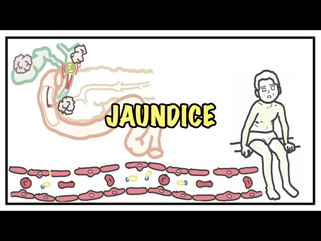
- Jaundice may develop, but can be mild or absent early, particularly in hyperacute paracetamol injury; it is not required for diagnosis.
- Encephalopathy: altered sleep, poor attention, behavioural change or confusion progressing to somnolence and coma.
- Other features: hypoglycaemia, hypotension, oliguria and manifestations of the underlying cause. [2–4]
Clinical Examination
- Assess airway, observations, hydration, neurological state and serial encephalopathy grade.
- Look for jaundice, asterixis, abdominal tenderness/hepatomegaly and ascites.
- Seek cause-specific signs: pregnancy-associated hypertension, HSV vesicles or Wilson-related haemolysis. Absence of vesicles or Kayser–Fleischer rings does not exclude those diagnoses.
- Look for evidence of chronic liver disease, but ascites alone does not establish cirrhosis.
| West Haven grade | Typical findings |
|---|---|
| I | Subtle attention/behavioural changes, sleep disturbance; generally orientated. |
| II | Lethargy, disorientation to time, inappropriate behaviour; asterixis commonly present. |
| III | Marked confusion and somnolence, but responsive to stimuli. |
| IV | Coma; asterixis cannot be elicited. [2,4] |
Diagnosis
Establish acute hepatic injury, INR ≥1.5 from hepatic dysfunction, and encephalopathy within 26 weeks. Exclude other explanations for altered consciousness, especially hypoglycaemia, sepsis, intoxication and intracranial pathology. Generally there is no established cirrhosis; fulminant Wilson disease, autoimmune hepatitis and HBV reactivation can present with an ALF phenotype despite previously unrecognised chronic disease. [1–4]
Investigations
| Investigation | Purpose / interpretation |
|---|---|
| Bedside glucose, ECG and continuous observations | Detect immediately reversible hypoglycaemia and physiological instability. |
| AST/ALT, ALP, bilirubin; PT/INR | Assess injury and synthetic function; trends are essential. |
| FBC, creatinine, electrolytes, phosphate, magnesium; blood gas and lactate | Assess haemolysis, renal injury, metabolic disturbance and perfusion. |
| Ammonia | Assess hyperammonaemia and cerebral-oedema risk; does not replace clinical encephalopathy assessment. |
| Paracetamol concentration and targeted toxicology | Test even when overdose is denied. A late undetectable concentration does not exclude toxicity. |
| Viral testing | HAV IgM; HBsAg, anti-HBc IgM and HBV DNA; HEV testing according to context. HSV PCR and other viral tests when indicated. |
| Autoimmune/Wilson work-up | ANA, smooth-muscle antibody and IgG; copper studies and haemolysis evaluation when appropriate. Ceruloplasmin alone is unreliable in ALF. |
| Pregnancy test, cultures and targeted tests | Identify pregnancy-related disease and infection; tailor testing to history. |
| Liver ultrasound with Doppler | Assess hepatic/portal vessels, outflow obstruction and liver morphology; CT/MRI if further characterisation is needed. |
| Brain CT / EEG when indicated | Assess alternative neurological pathology or suspected seizures. A normal CT does not exclude intracranial hypertension. [2–4,8] |
Wilson disease should be considered with Coombs-negative haemolysis, marked bilirubin elevation and disproportionately low ALP. These are clues, not definitive diagnostic criteria. [8]
Differential Diagnosis
- Severe acute hepatitis without encephalopathy: acute liver injury rather than established adult ALF.
- Decompensated cirrhosis/acute-on-chronic liver failure: examine previous records and evidence of chronic disease.
- Biliary obstruction: jaundice and cholestatic biochemistry; assess with imaging.
- Sepsis, hypoglycaemia, toxic/metabolic encephalopathy or intracranial disease may mimic or coexist with hepatic encephalopathy.
Classification
The O’Grady classification describes the interval from jaundice to encephalopathy. It is separate from the King’s College prognostic criteria. [5]
| Category | Interval |
|---|---|
| Hyperacute | Within 7 days |
| Acute | 8–28 days |
| Subacute | 5–12 weeks |
Hyperacute disease can have a high cerebral-oedema risk yet favourable spontaneous recovery in some causes. Subacute disease may have less cerebral oedema but poor transplant-free survival. Aetiology and the evolving clinical course matter more than timing alone. [3,5]
Treatment
Initial resuscitation and referral
- Stabilise airway, breathing and circulation; correct hypoglycaemia promptly and stop potential hepatotoxins.
- Contact a transplant centre early while investigating and treating the cause. Escalating encephalopathy, shock or organ failure requires intensive care.
- Intubate for grade III–IV encephalopathy or inability to protect the airway. Avoid unnecessary sedating medication in patients whose neurological state is being monitored. [1,2]
Cause-specific treatment
| Cause | Management |
|---|---|
| Paracetamol | Start N-acetylcysteine (NAC) immediately when toxicity is suspected; do not delay for a drug level. Continue according to clinical/biochemical response with toxicology guidance. The nomogram applies to a single ingestion with a known time, not staggered ingestion or established ALF. |
| Non-paracetamol ALF | Consider IV NAC with specialist advice. Adult trial benefit in transplant-free survival was concentrated in early encephalopathy; this must not delay transplantation. [6] |
| Drug-induced injury | Withdraw the suspected agent and seek specialist advice. |
| HBV / HSV | HBV: potent nucleos(t)ide analogue, such as entecavir or tenofovir. Suspected HSV: prompt IV aciclovir. Antiviral therapy is cause-specific. |
| Autoimmune hepatitis | Specialist-directed corticosteroids in selected patients; reassess promptly and pursue transplant evaluation when ALF develops. |
| Ischaemic injury | Treat shock, cardiac dysfunction and hypoxaemia. |
| AFLP / HELLP | Urgent obstetric and critical-care management, including prompt delivery when indicated. |
| Budd–Chiari / Wilson / mushroom poisoning | Urgent specialist pathways: anticoagulation/decompression for selected Budd–Chiari cases; transplant assessment for Wilson ALF; poison-centre advice, NAC and silibinin where available for amatoxin poisoning. [2,4] |
Supportive care and complication prevention
| Problem | Management principles |
|---|---|
| Cerebral oedema / intracranial hypertension | Head elevation, neutral neck position, avoid hypoxia/hypotension and fever. Specialist-directed hypertonic saline or mannitol when indicated. Hyperventilation is a short rescue measure, not routine prolonged treatment. |
| Hyperammonaemia / acute kidney injury | Consider early continuous renal replacement therapy (CRRT); it is preferred over intermittent dialysis in unstable ALF and can reduce ammonia. [9] |
| Encephalopathy / seizures | Serial neurological assessment; EEG if seizures suspected. Treat seizures with ICU-directed agents, often levetiracetam. Evidence for lactulose/rifaximin in ALF is inconclusive; they are not substitutes for critical care. |
| Hypotension | Careful IV fluid resuscitation; noradrenaline for persistent shock. |
| Hypoglycaemia / electrolytes | Frequent glucose checks, IV dextrose as needed, careful electrolyte replacement and fluid balance. |
| Coagulopathy | Do not routinely give FFP merely to normalise INR. Give blood products for significant bleeding or selected procedures, guided by the specialist team; vitamin K if deficiency is possible. [7] |
| Infection / nutrition | Obtain cultures and treat suspected infection promptly; routine antimicrobial prophylaxis is not recommended. Provide enteral nutrition when safe. [1,2] |
Use continuous physiological monitoring, frequent neurological/glucose checks and repeated blood tests according to instability; once-daily testing is insufficient in rapidly deteriorating disease. High-volume plasma exchange may be used in specialist centres as a bridge to recovery/transplant. Other artificial liver-support devices have uncertain survival benefit and do not replace transplantation. [4,10]
Key treatment cautions: an elevated INR does not justify routine plasma transfusion. Osmotherapy treats cerebral oedema, rather than directly lowering ammonia. Routine prolonged hyperventilation and prophylactic antiepileptics are not standard ALF treatment.
Complications and Prognosis
Complications
- Cerebral oedema, intracranial hypertension, herniation and seizures.
- Acute kidney injury, severe hyperammonaemia, hypoglycaemia, electrolyte disturbance and acidosis.
- Infection/sepsis, circulatory shock and respiratory failure.
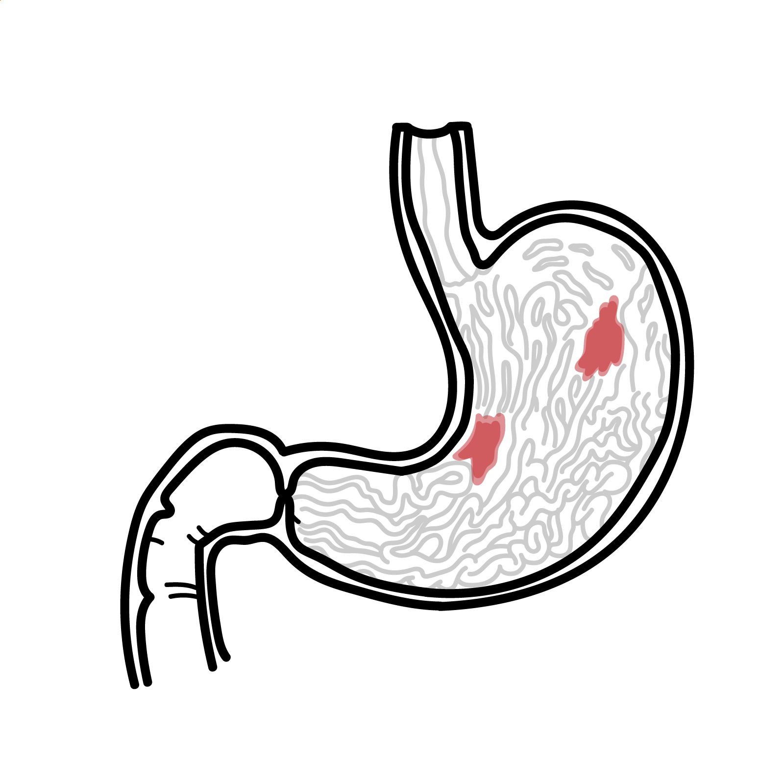
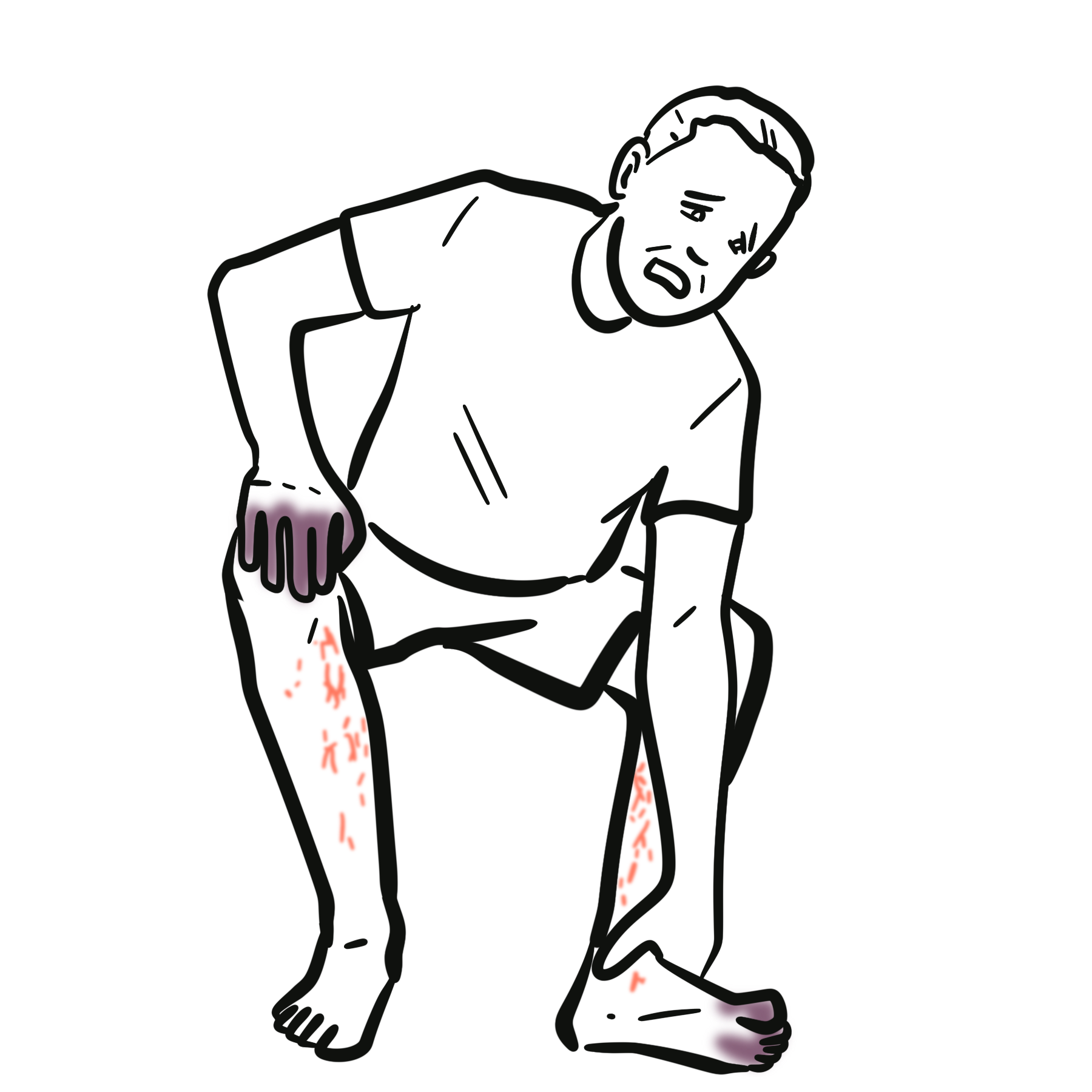
- Bleeding can occur, although clinically important haemorrhage is less common than the INR suggests. [2–4,7]
Prognosis
Outcome depends on aetiology, encephalopathy severity, synthetic dysfunction, lactate and extrahepatic organ failure. Paracetamol and ischaemic injury often have greater recovery potential than Wilson disease, idiosyncratic drug injury or subacute indeterminate disease. Prognostic models are imperfect and require repeated assessment; a single universal spontaneous-survival percentage is misleading. [3,4]
References
1. Shingina A, Mukhtar N, Wakim-Fleming J, Alqahtani S, Wong RJ, Limketkai BN, et al. Acute liver failure guidelines. Am J Gastroenterol. 2023;118(7):1128–1153. doi:10.14309/ajg.0000000000002340.
2. European Association for the Study of the Liver. EASL Clinical Practical Guidelines on the management of acute (fulminant) liver failure. J Hepatol. 2017;66(5):1047–1081. doi:10.1016/j.jhep.2016.12.003.
3. Stravitz RT, Lee WM. Acute liver failure. Lancet. 2019;394(10201):869–881. doi:10.1016/S0140-6736(19)31894-X.
4. Tujios S, Stravitz RT, Lee WM. Management of acute liver failure: update 2022. Semin Liver Dis. 2022;42(3):362–378. doi:10.1055/s-0042-1755274.
5. O’Grady JG, Schalm SW, Williams R. Acute liver failure: redefining the syndromes. Lancet. 1993;342(8866):273–275. doi:10.1016/0140-6736(93)91818-7.
6. Lee WM, Hynan LS, Rossaro L, Fontana RJ, Stravitz RT, Larson AM, et al. Intravenous N-acetylcysteine improves transplant-free survival in early stage non-acetaminophen acute liver failure. Gastroenterology. 2009;137(3):856–864.e1. doi:10.1053/j.gastro.2009.06.006.
7. Stravitz RT, Ellerbe C, Durkalski V, Schilsky M, Fontana RJ, Peterseim C, et al. Bleeding complications in acute liver failure. Hepatology. 2018;67(5):1931–1942. doi:10.1002/hep.29694.
8. Korman JD, Volenberg I, Balko J, Webster J, Schiodt FV, Squires RH Jr, et al. Screening for Wilson disease in acute liver failure: a comparison of currently available diagnostic tests. Hepatology. 2008;48(4):1167–1174. doi:10.1002/hep.22446.
9. Cardoso FS, Gottfried M, Tujios S, Olson JC, Karvellas CJ; US Acute Liver Failure Study Group. Continuous renal replacement therapy is associated with reduced serum ammonia levels and mortality in acute liver failure. Hepatology. 2018;67(2):711–720. doi:10.1002/hep.29488.
10. Larsen FS, Schmidt LE, Bernsmeier C, Rasmussen A, Isoniemi H, Patel VC, et al. High-volume plasma exchange in patients with acute liver failure: an open randomised controlled trial. J Hepatol. 2016;64(1):69–78. doi:10.1016/j.jhep.2015.08.018.




Members only discussions coming soon…