

Groin Swelling/Lump
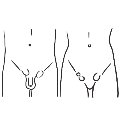
OVERVIEW
A groin swelling or lump is any palpable mass arising in the region between the lower anterior abdominal wall, inguinal ligament, proximal thigh, and external genitalia. The most common causes are an inguinal or femoral hernia and inguinal lymphadenopathy.
The immediate priority is to identify a strangulated or obstructed hernia, vascular lesion, severe infection, or malignancy. A painful irreducible hernia with vomiting or features of bowel obstruction is a surgical emergency.
Key Anatomical Landmark — Pubic Tubercle:
- Inguinal hernia: Usually arises above and medial to the pubic tubercle.
- Femoral hernia: Usually arises below and lateral to the pubic tubercle.
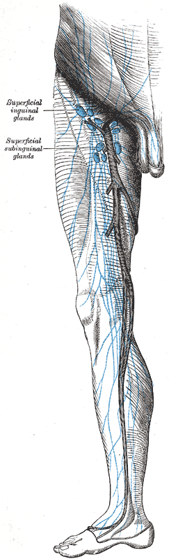
- Inguinal lymph nodes: Form discrete superficial nodules inferior to the inguinal ligament.
- Femoral arterial lesion: Located near the femoral pulse and may be pulsatile or associated with a bruit.
- Saphena varix: A compressible venous swelling at the saphenofemoral junction, inferolateral to the pubic tubercle.
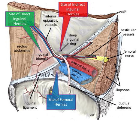
A groin swelling located below and lateral to the pubic tubercle should be considered a femoral hernia until proven otherwise, particularly in a woman. Femoral hernias have a narrow neck and a substantially higher risk of incarceration and strangulation.
APPROACH
Step 1: Identify Emergency Red Flags
- Suspected strangulated hernia:
- Sudden severe groin pain.
- Tender, tense, irreducible swelling.
- Absent cough impulse.
- Erythema, warmth, or discolouration of overlying skin.
- Fever, tachycardia, hypotension, or systemic toxicity.
- Requires immediate surgical assessment and operative management.
- Bowel obstruction:
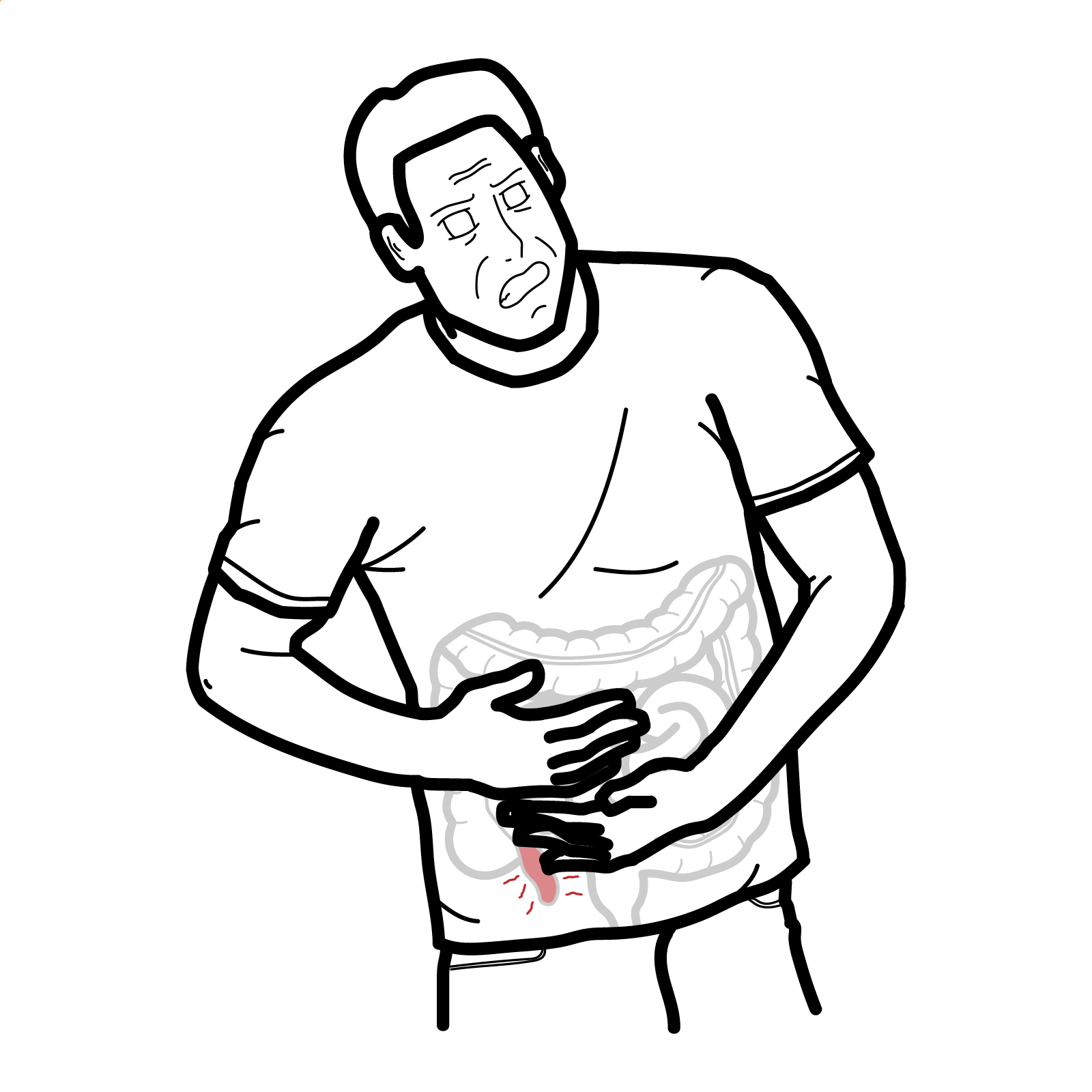
- Colicky abdominal pain, vomiting, abdominal distension, constipation, or obstipation.
- Obtain urgent surgical review and cross-sectional imaging if it will not delay definitive treatment.
- Femoral vascular emergency:
- Expanding or pulsatile groin mass, bruit, recent femoral arterial access, trauma, anticoagulant use, limb ischaemia, or haemodynamic instability.
- Do not attempt aspiration, incision, biopsy, or hernia reduction.
- Obtain urgent duplex ultrasound and vascular surgical assessment.
- Severe infection:
- Fluctuant painful swelling, cellulitis, purulent drainage, fever, or sepsis.
- Consider abscess, suppurative lymphadenitis, infected vascular lesion, hidradenitis suppurativa, or psoas abscess.
- Testicular torsion or incarcerated inguinal hernia in a child:
- Acute groin or scrotal pain, vomiting, high-riding testis, or an irreducible inguinoscrotal swelling.
- Requires immediate paediatric surgical or urological review.
Step 2: Characterise the Lump
- Relationship to posture and intra-abdominal pressure:
- Examine the patient standing and supine.
- Ask the patient to cough or perform a Valsalva manoeuvre.
- Hernias typically enlarge with standing, coughing, or straining.
- Reducibility:
- A reducible lump that disappears when supine strongly suggests a hernia or saphena varix.
- An irreducible lump may represent incarceration, lymphadenopathy, tumour, abscess, or thrombosed vascular pathology.
- Cough impulse:
- An expansile cough impulse supports a hernia.
- A cough impulse may be absent in an incarcerated hernia and is not completely specific.
- Consistency and mobility:
- Soft and lobulated: Lipoma.
- Firm and discrete: Lymph node.
- Hard, fixed, or matted: Malignancy or chronic infection.
- Fluctuant: Abscess, cyst, hydrocele, or lymphocele.
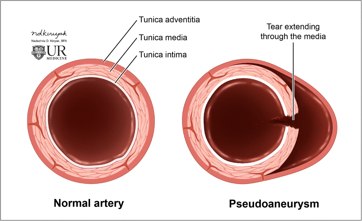
- Pulsatile: Arterial aneurysm or pseudoaneurysm.
- Tenderness and skin findings:
- Tender erythematous lesions suggest infection, strangulation, inflammation, or thrombosis.
- Examine for punctum, ulceration, sinus formation, scars, bruising, or hidradenitis.
Step 3: Examine the Drainage Territory and Associated Structures
- Examine both groins and all peripheral lymph-node regions.
- Inspect the lower abdomen, perineum, external genitalia, anus, buttocks, and both lower limbs for infection, ulceration, trauma, melanoma, or another malignant lesion.
- Perform a complete scrotal and testicular examination in males.
- Assess lower-limb pulses, perfusion, and neurological function when a vascular lesion is possible.
- Consider abdominal, pelvic, rectal, vaginal, or prostate examination according to the clinical context.
Never insert a needle into, biopsy, or forcefully manipulate an unexplained pulsatile groin lump. A femoral pseudoaneurysm may closely resemble a hernia or abscess and can cause catastrophic haemorrhage if punctured.
DIFFERENTIAL DIAGNOSIS
Hernias
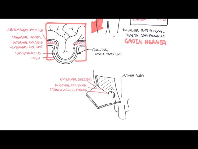
- Indirect inguinal hernia: Passes through the deep inguinal ring lateral to the inferior epigastric vessels and may extend into the scrotum. More common in younger patients but can occur at any age. Clinically distinguishing indirect from direct hernia is unreliable; definitive classification is usually anatomical or operative.
- Direct inguinal hernia: Protrudes through a weakness in the posterior inguinal wall within Hesselbach’s triangle. Usually presents as a broad-based medial bulge in an older adult.
- Femoral hernia: Lies below and lateral to the pubic tubercle, medial to the femoral vein. More common in women and frequently presents with incarceration or strangulation.
- Recurrent or incisional hernia: Occurs near a previous inguinal, femoral, vascular, transplant, or lower abdominal surgical incision.
- Obturator hernia: Usually does not produce a visible groin lump. Consider in an older, thin woman with bowel obstruction and medial thigh pain exacerbated by hip extension or rotation.
Lymphatic Causes
- Reactive inguinal lymphadenopathy: Tender, mobile nodes associated with lower-limb skin infection, wounds, genital infection, or inflammatory disease.
- Sexually transmitted infection: Inguinal nodes may accompany genital herpes, syphilis, lymphogranuloma venereum, chancroid, or other genital infections. Look for genital ulcers, urethral or vaginal discharge, rash, and sexual exposure.
- Lymphoma: Persistent, rubbery or firm, usually non-tender lymphadenopathy; may be generalised. Associated features include fever, drenching night sweats, weight loss, pruritus, hepatosplenomegaly, or abnormal blood counts.
- Metastatic malignancy: Hard, fixed, enlarging or matted nodes. Potential primary sites include melanoma and malignancies of the lower limb, vulva, penis, scrotal skin, anal canal, and lower pelvic structures.
Vascular Causes
- Femoral artery aneurysm or pseudoaneurysm: Pulsatile mass with a bruit or thrill. Pseudoaneurysm commonly follows femoral arterial catheterisation, surgery, trauma, or injection drug use. Duplex ultrasound may demonstrate characteristic “to-and-fro” flow within the pseudoaneurysm neck.
- Saphena varix: Dilatation of the proximal great saphenous vein at the saphenofemoral junction. Soft, compressible swelling that may disappear when supine and exhibit a cough impulse. May have a bluish appearance or associated lower-limb varicose veins.
- Venous thrombosis or varicosity: Tender cord-like or compressible venous swelling, sometimes associated with limb oedema.
Infectious and Inflammatory Causes
- Groin abscess / infected epidermoid cyst: Tender, erythematous, fluctuant superficial mass; may have a punctum or purulent discharge.
- Hidradenitis suppurativa: Recurrent painful nodules, abscesses, sinus tracts, and scarring in intertriginous areas.
- Psoas or iliopsoas abscess: Fever, back or flank pain, limp, hip held in flexion, and pain on passive hip extension. May track beneath the inguinal ligament and present as a groin mass.
- Tuberculous or atypical mycobacterial lymphadenitis: Chronic, relatively painless nodes that may become matted, fluctuant, or form draining sinuses.
Genitourinary and Scrotal Causes
- Undescended or ectopic testis: Groin mass with an empty ipsilateral hemiscrotum.
- Hydrocele of the cord: Smooth cystic inguinal swelling that may transilluminate and usually lacks an expansile cough impulse.
- Inguinoscrotal hydrocele: Cystic scrotal swelling; the examiner may be unable to palpate above it if it extends into the inguinal canal.
- Varicocele: “Bag of worms” above the testis, more prominent when standing or performing Valsalva.
- Epididymo-orchitis or testicular tumour: May be perceived as a lower groin swelling but should be localised to the scrotal contents on examination.
Benign and Malignant Soft-Tissue Masses
- Lipoma: Soft, mobile, painless and non-reducible; no cough impulse.
- Epidermoid or sebaceous cyst: Superficial, mobile swelling that may have a central punctum.
- Haematoma or seroma: Associated with trauma, surgery, anticoagulation, bruising, or a recent procedure.
- Soft-tissue sarcoma: Enlarging, deep, firm or fixed mass, often painless. Features of concern include size ≥5 cm, rapid growth, deep location, pain, recurrence, or fixation to surrounding structures.
A normal-sized inguinal lymph node can be palpable. The features that warrant further investigation are progressive enlargement, hard or fixed consistency, matting, unexplained persistence, associated systemic symptoms, or absence of a plausible infection in the lymphatic drainage territory.
INVESTIGATIONS
First-Line Assessment
- Focused history and examination: Onset, growth, pain, reducibility, relationship to coughing or standing, bowel symptoms, fever, weight loss, night sweats, genital symptoms, lower-limb infection, trauma, previous surgery, recent femoral vascular access, and anticoagulant use.
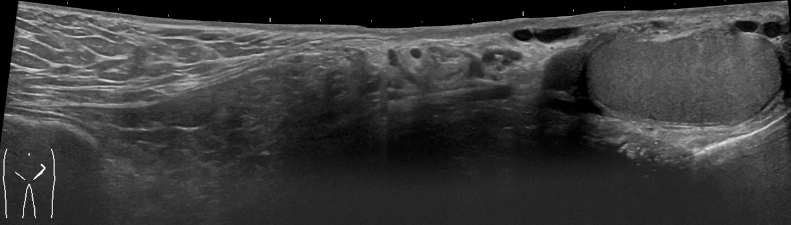
- Dynamic groin ultrasound: First-line imaging when the examination is equivocal. Perform with standing, Valsalva, and graded compression where possible. Differentiates hernia, lymph node, cyst, solid tumour, hydrocele, and vascular lesions.
- Duplex Doppler ultrasound: Mandatory when the lump is pulsatile or a pseudoaneurysm, saphena varix, thrombosis, or another vascular lesion is suspected.
- Laboratory investigations when indicated: FBC, CRP/ESR, renal and liver function, blood cultures if febrile or septic. STI NAAT and serology when genital infection or sexual exposure is suspected. LDH, blood film, HIV testing, tuberculosis testing, or other directed investigations for unexplained lymphadenopathy.
Targeted Imaging and Tissue Diagnosis
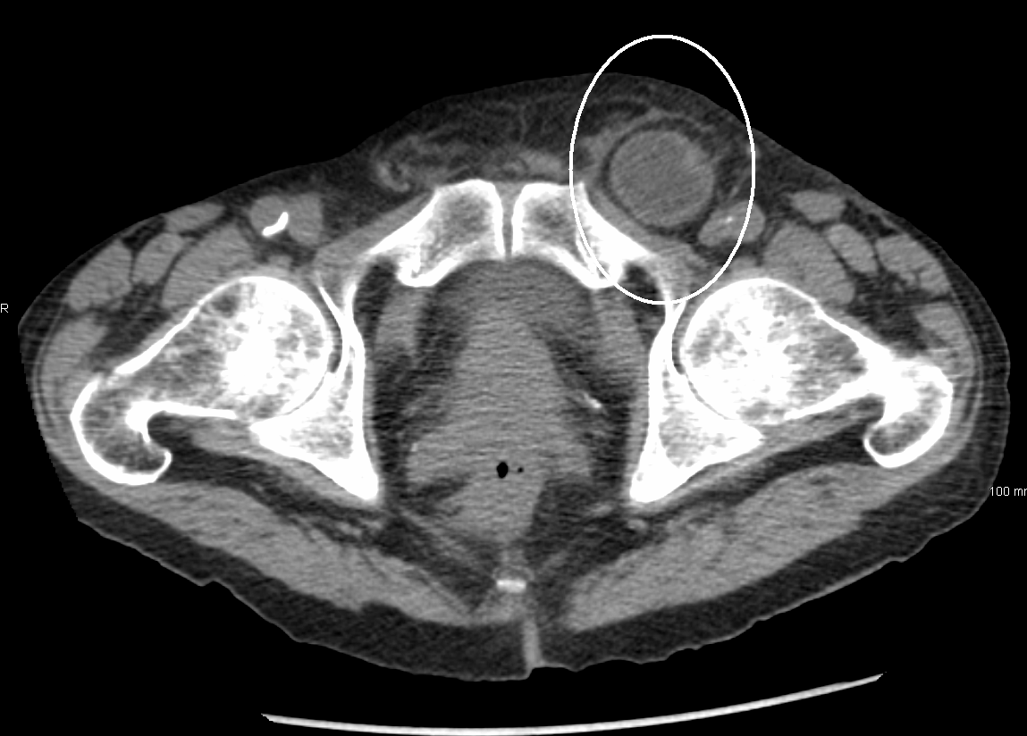
- CT abdomen and pelvis with intravenous contrast: Appropriate for suspected bowel obstruction, strangulation, occult or recurrent hernia, deep infection, psoas abscess, malignancy, or unclear complex anatomy.
- MRI pelvis or groin: Useful for an occult hernia when ultrasound is negative but suspicion remains, and for defining a soft-tissue tumour or relationship to neurovascular structures.
- CT angiography: Consider for an expanding pseudoaneurysm, complex vascular anatomy, active haemorrhage, distal ischaemia, or procedural planning.
- Lymph-node biopsy: Persistent or suspicious lymphadenopathy generally requires tissue diagnosis. Core-needle biopsy may establish metastatic disease. Excisional biopsy is preferred when lymphoma is suspected because lymph-node architecture is required.
- Soft-tissue mass biopsy: Arrange specialist imaging and sarcoma-service referral before biopsy of a suspicious deep or enlarging mass. Biopsy trajectory must be planned so it can be removed during definitive surgery.
Do not routinely biopsy a suspected soft-tissue sarcoma before specialist referral. An incorrectly placed biopsy tract can contaminate uninvolved compartments and compromise subsequent limb-sparing surgery.
CRITICAL MANAGEMENT
Strangulated or Obstructed Hernia
- Keep the patient nil by mouth.
- Obtain large-bore intravenous access and commence fluid resuscitation.
- Provide analgesia and antiemetic therapy.
- Correct electrolyte abnormalities and insert a nasogastric tube when significant vomiting or bowel obstruction is present.
- Administer broad-spectrum intravenous antibiotics when strangulation, bowel ischaemia, perforation, or sepsis is suspected.
- Arrange immediate operative exploration and hernia repair, with bowel resection if non-viable intestine is identified.
- Do not delay surgery to obtain imaging when the clinical diagnosis of strangulation is clear.
Incarcerated Hernia Without Features of Strangulation
- Obtain urgent surgical assessment.
- Gentle manual reduction may be considered only by an experienced clinician when strangulation is not suspected.
- Provide analgesia, place the patient supine, and use slow sustained pressure rather than forceful manipulation.
- Following successful reduction, observe the patient and arrange timely definitive surgical repair.
- Persistent pain, systemic features, or concern for reduction of non-viable bowel requires urgent reassessment and imaging or operation.
Elective Groin Hernia Management
- Symptomatic inguinal hernia: Offer elective surgical repair.
- Asymptomatic or minimally symptomatic inguinal hernia in men: Watchful waiting may be reasonable following shared decision-making, provided the patient can recognise symptoms of incarceration and access urgent care.
- Women with a groin hernia: Timely repair is generally recommended because of the increased likelihood of an occult femoral hernia and greater strangulation risk.
- Femoral hernia: Refer for timely surgical repair even if symptoms are limited.
- Mesh-based open or laparo-endoscopic repair is used in most adults, with the approach tailored to sex, hernia type, recurrence, previous surgery, anaesthetic risk, surgeon expertise, and patient preference.
Vascular Lesions
- Femoral pseudoaneurysm: Obtain urgent vascular assessment. Management may include observation for selected small stable lesions, ultrasound-guided compression, ultrasound-guided thrombin injection, endovascular treatment, or open repair.
- Haemodynamic instability, rapid expansion, infection, neurological deficit, distal limb ischaemia, skin compromise, or associated arteriovenous fistula: Requires urgent vascular intervention.
- Saphena varix: Manage associated venous insufficiency and refer for vascular assessment when symptomatic or diagnostically uncertain.
Lymphadenopathy, Infection and Tumour
- Treat an identified local bacterial infection and reassess the nodes after clinical resolution.
- Drain a confirmed abscess when indicated and provide antibiotics according to severity and local guidance.
- Treat STIs and ensure appropriate partner notification and management.
- Refer persistent or suspicious lymphadenopathy for imaging and tissue diagnosis.
- Refer a suspected sarcoma through an urgent specialist pathway before biopsy or excision.
- Do not perform an unplanned “shell-out” excision of an unexplained deep or enlarging groin mass.
Watchful waiting applies only to carefully selected patients with minimally symptomatic inguinal hernias—principally men. It is not appropriate for a femoral hernia, a painful or enlarging hernia, or any hernia with incarceration, obstruction, or strangulation.
REFERENCES
- Stabilini C, van Veenendaal N, Aasvang E, et al. Update of the international HerniaSurge guidelines for groin hernia management. BJS Open. 2023;7(5):zrad080. doi:10.1093/bjsopen/zrad080.
- HerniaSurge Group. International guidelines for groin hernia management. Hernia. 2018;22(1):1–165. doi:10.1007/s10029-017-1668-x.
- American College of Radiology. ACR Appropriateness Criteria®: hernia [Internet]. Reston (VA): American College of Radiology. Available from: https://acsearch.acr.org/docs/3158169/Narrative/
- LeBlanc KE, LeBlanc LL, LeBlanc KA. Inguinal hernias: diagnosis and management. Am Fam Physician. 2013;87(12):844–848.
- American College of Cardiology. Postcatheterization femoral pseudoaneurysms [Internet]. Washington (DC): ACC; 2019. Available from: https://www.acc.org/latest-in-cardiology/ten-points-to-remember/2019/06/04/10/26/postcatheterization-femoral-pseudoaneurysms
- Ilgeldiev S, Kleif J, Gögenur I, et al. Differential diagnoses of inguinal swellings: a case series of atypical diagnoses. J Surg Case Rep. 2024;2024(3):rjae130.
- National Institute for Health and Care Excellence. Suspected cancer: recognition and referral. NICE guideline NG12 [Internet]. London: NICE. Available from: https://www.nice.org.uk/guidance/ng12




Members only discussions coming soon…