

Umbilical and Epigastric Hernia
Overview
Umbilical and Epigastric hernias are types of Primary Ventral abdominal wall hernias. Epigastric hernias are two to three times more common in men.
Umbilical hernias are three times more likely in females. Most epigastric hernias contain preperitoneal fat, while most umbilical hernias contain omentum or bowel. These hernias are usually asymptomatic unless incarcerated, obstructed or strangulated.
A strangulated hernia is a surgical emergency due to high risk of perforation and sepsis. Treatment can be conservative for small asymptomatic hernias but is otherwise surgical.1 2
Definition
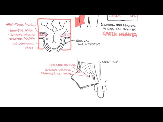
Hernia: A bulge or protrusion of an organ or tissue through an abnormal opening within the anatomic structure that normally confines it.
Ventral abdominal wall hernia: hernia through frontal wall of the abdomen; divided into Incisional and Spontaneous.
Incisional: occurs at site of prior abdominal surgical incision, which has resulted in weakening of the wall.
Spontaneous (Primary): commonly occur near midline of the abdomen, without prior surgical trauma; most common are Umbilical and Epigastric.
Umbilical hernia: defect in the umbilical ring.
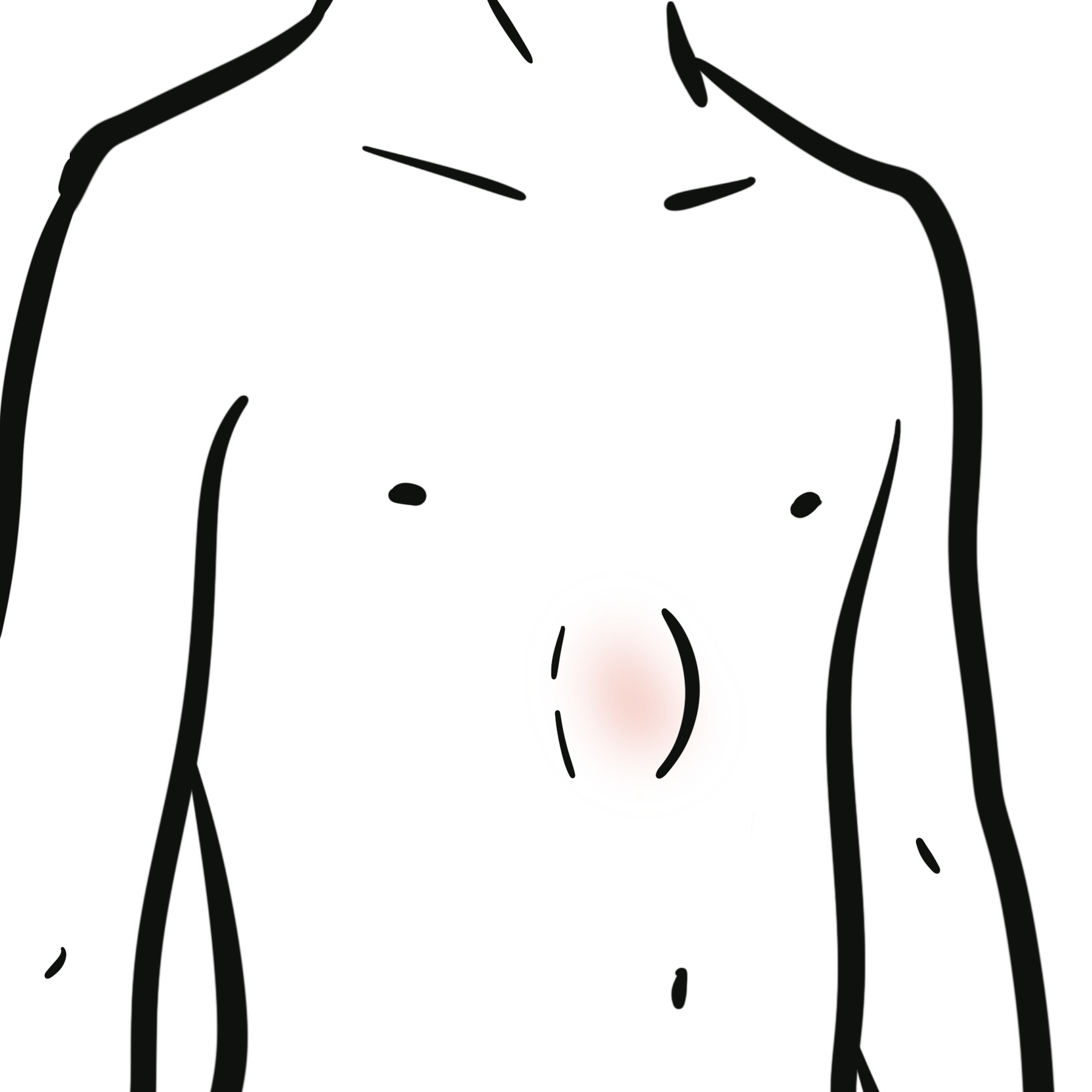
Epigastric hernia: defect in linea alba between xiphisternum and umbilicus.
Paraumbilical hernia: defect adjacent to the umbilicus
Reducible: can be pushed back into place easily (either manually or spontaneously)
Incarcerated: contents are trapped within hernia sac, can NOT be returned into the abdominal cavity.
Obstructed: incarcerated hernia containing bowel that causes intestinal obstruction.
Strangulated: compromised blood supply causing ischaemia and necrosis.
Anatomy
Abdominal wall
- Encloses abdominal cavity
- Can be divided into Anterolateral and Posterior wall
Anterolateral wall layers
- Skin
- Superficial fascia – composition depends on location
- Above umbilicus – single sheet of connective tissue
- Below umbilicus – divided into:
- Camper’s fascia: fatty superficial layer
- Scarpa’s fascia: membranous deep layer
- Muscles
- Lateral wall
- External oblique
- Internal oblique
- Transversus abdominis
- Anterior wall
- Pyramidalis
- Rectus abdominis
- Lateral wall
- Transversalis fascia
- Extraperitoneal fat
- Peritoneum
Rectus abdominis
- Linea alba: divides the muscle into two
- Area of weakness predisposed to epigastric hernia formation
- Linea semilunaris: represents the lateral border
- Rectus sheath: formed by the aponeuroses of three flat muscles and encloses rectus abdominis and pyramidalis
- Anterior sheath: formed by external oblique and half of internal oblique
- Posterior sheath: formed by half of internal oblique and transversus abdominis
- Arcuate line: point where posterior layer of rectus sheath ends – midway between umbilicus and pubic symphysis – the posterior aponeuroses move to the anterior rectus sheath
Umbilical ring – natural weakness of abdominal wall, where the Umbilical vessels and Urachus passed through during foetal life.
Spigelian hernia is a ventral abdominal wall hernia that develops along Linea semilunaris in the Spigelian fascia.
Aetiology & Risk factors
| Epigastric hernia risk factors | Umbilical hernia risk factors |
| Increased intra-abdominal pressure: Chronic cough, constipation, heavy lifting, obesity | |
| Smoking | Abdominal distension |
| Chronic steroid use | Ascites |
| Diabetes | Pregnancy |
| Older age | Female sex |
| Male sex | — |
Pathophysiology
Classification
Umbilical hernia
Origin:
- Congenital: occurs in infants when umbilical ring fails to close; most close spontaneously in the first 2 to 5 years of life
- Acquired: occur in adulthood due to increased intra-abdominal pressure
Epigastric hernia
Contents:
- Adipose: consists solely of preperitoneal fat (most common)
- Uninhabited peritoneal sac: empty peritoneal sac with no abdominal organs
- Complete: contains abdominal viscera such as omentum and bowel
Ventral wall hernias (both Umbilical and Epigastric)
Defect size:
- Small (<1cm)
- Medium (1-4cm)
- Large (>4cm)
Clinical state:
- Reducible: contents can be pushed back into abdominal cavity
- Incarcerated: contents cannot be pushed back into abdominal cavity
- Strangulated: blood supply is compromised1 3
Clinical Manifestation
Most are asymptomatic, unless obstructed or strangulated
Patient might be able to hear bowel sounds from the hernia if there is bowel involvement
Small epigastric hernias may be painful – preperitoneal fat becomes trapped within narrow defect causing local nerve irritation
Red flag symptoms suggesting strangulation:
- Increasing pain
- Erythema
- Discolouration of skin over hernia
- Hard or tender lump
- Systemic symptoms: high fever, fatigue, rapid heart rate
Red flag symptoms suggesting obstruction:
Strangulated hernia is dangerous due to high risk of perforation and sepsis, early recognition and treatment is crucial.
Hernias with bowel involvement might cause partial obstruction, which then lead to patient having to manually reduce hernia to properly empty bowel.
Examination
General inspection – midline swelling or mass
- Observe hernia when patient is:
- Standing
- Lying supine
- Coughing
- Determine location – umbilical or epigastric
Palpation – assess:
- Reducibility
- Tenderness
- Cough impulse: expansion with coughing suggests hernia
Differential diagnosis
| Lipoma | Benign tumour of adipose tissue |
| Sebaceous cyst | Benign keratin-filled cyst arising from a blocked hair follicle or epidermis |
| Rectus sheath haematoma | Collection of blood within the rectus sheath |
| Rectus abdominis diastasis | Separation of the rectus abdominis muscles caused by widening of the linea alba without a fascial defect |
| Desmoid tumour | A benign but locally invasive fibroblastic tumour arising from musculoaponeurotic tissues |
| Urachal anomalies | Congenital abnormalities resulting from incomplete closure of the embryological urachus |
| Umbilical endometriosis | Endometrial tissue within the umbilicus |
| Metastatic umbilical nodule (Sister Mary Joseph node) | Umbilical metastasis from an intra-abdominal or pelvic malignancy |
Investigations
Diagnosis is primarily clinical and imaging is not routinely required
Imaging can be used
- Ultrasound – when physical examination is inconclusive; however, it has limited effectiveness in patients with significant obesity or large hernias
- CT – useful for preoperative planning when assessing defect size in patients without palpable hernia or with obesity3
Treatment
Conservative management
Used for: Asymptomatic hernias (very low risk of strangulation) and for patients with Significant surgical risk
Includes:
- Weight reduction
- Smoking cessation
- Management of chronic cough
- Constipation treatment
Surgical management
For hernias that are symptomatic, obstructed, incarcerated or strangulated
Repair can be open or laparoscopic
Two types of repair:
| Primary suture repair | Mesh repair |
| Used for small defects (<1-2cm) | Used for larger defects |
| Higher risk of recurrence | Lower risk of recurrence |
| High tension | Minimal tension |
| Lower risk of infection | Higher risk of infection |
For strangulated hernia – resection of necrotic bowel might be necessary3
Complications
Untreated hernia
- Incarceration
- Strangulation
- Bowel obstruction
- Perforation and sepsis
Post-operative
- Recurrence
- Seroma
- Haematoma
- Adhesions
- Mesh infection or migration3
References
- Brooks DC. Overview of abdominal wall hernias in adults. In: Rosen M, Chen W, editor. [Internet]. [updated 2026 Apr 16; cited 2026 Jun 15]. Available from: UpToDate
- Conze J, Klinge U, Schumpelick V. Hernias. Surgical Treatment: Evidence-Based and Problem-Oriented [Internet]. Munich: Zuckschwerdt; 2001 [cited 2026 Jun 15]. Available from: https://www.ncbi.nlm.nih.gov/books/NBK6888/
- Holt AC, Bamarni S, Leslie SW. Umbilical hernia [Internet]. Treasure Island (FL): StatPearls Publishing; 2026 Jan- [cited 2026 Jun 15]. Available from: https://www.ncbi.nlm.nih.gov/books/NBK459312/




Members only discussions coming soon…