

Angioedema (allergic or idiopathic)

Overview
Angioedema is a transient, localised swelling of the deeper dermis, subcutaneous tissue or submucosa caused by increased vascular permeability. It commonly affects the lips, eyelids, tongue, face, extremities or genitalia. Gastrointestinal and upper-airway involvement may also occur.1
Allergic angioedema is usually mast-cell-mediated and often occurs with urticaria, pruritus or other features of anaphylaxis. Recurrent episodes without an identifiable trigger may be classified as idiopathic after medicines, hereditary or acquired C1-inhibitor deficiency, and other causes have been appropriately excluded.1,2
Tongue, pharyngeal or laryngeal swelling can rapidly compromise the airway. Angioedema accompanied by respiratory compromise, hypotension or other systemic allergic features must be treated as anaphylaxis.3
Definition
- Angioedema
- Transient, localised swelling of deep dermal, subcutaneous or submucosal tissue caused by increased vascular permeability.
- Mast-cell-mediated angioedema
- Angioedema caused by mast-cell mediators, including histamine; it may be allergic or spontaneous and often occurs with urticaria or pruritus.
- Allergic angioedema
- Mast-cell-mediated angioedema occurring after exposure to a specific allergen, usually with a compatible temporal relationship.
- Idiopathic angioedema
- Recurrent angioedema for which no specific cause is identified after an appropriate clinical assessment and targeted investigation.
- Bradykinin-mediated angioedema
- Angioedema driven by bradykinin rather than mast-cell mediators; it typically occurs without urticaria and responds poorly to antihistamines, corticosteroids and adrenaline.
Anatomy & Physiology
Small vessels within the deep dermis and submucosal tissues regulate the movement of fluid between the circulation and interstitial space. Mast-cell mediators such as histamine cause vasodilation and increased endothelial permeability.
Angioedema involves deeper tissue than urticaria. Consequently, the swelling is less sharply demarcated, may feel tight or painful rather than itchy, and usually resolves more slowly than an individual urticarial wheal.
The tongue, pharynx and larynx are clinically important because relatively small increases in tissue volume can significantly narrow the upper airway.
Classification
| Mechanism | Typical features | Common causes |
|---|---|---|
| Mast-cell-mediated | Rapid onset; urticaria, pruritus or flushing may occur; usually responds to antihistamines | Food, medicine or venom allergy; chronic spontaneous urticaria; idiopathic histaminergic angioedema |
| Bradykinin-mediated | Usually no urticaria or pruritus; slower evolution; abdominal attacks may occur; poor response to antihistamines, corticosteroids or adrenaline | Angiotensin-converting enzyme inhibitor use; hereditary or acquired C1-inhibitor deficiency |
| Uncertain or idiopathic | Recurrent swelling without an identified cause after appropriate evaluation | Idiopathic histaminergic or non-histaminergic angioedema |
The presence of urticaria supports mast-cell-mediated disease but is not absolute. Some patients with mast-cell-mediated angioedema have no wheals, while coincidental urticaria can occur in a patient with hereditary angioedema.2
Aetiology & Risk Factors
Aetiology
Allergic angioedema may be triggered by:
- Foods, particularly in immunoglobulin E-mediated food allergy.
- Medicines, including antibiotics and perioperative agents.
- Insect venom.
- Latex.
- Allergen immunotherapy.
- Cross-reactive raw fruits or vegetables in pollen-food allergy syndrome.
Non-allergic mast-cell activation may occur with:
- Non-steroidal anti-inflammatory drugs.
- Opioids and other medicines capable of directly activating mast cells.
- Acute infections.
- Chronic spontaneous urticaria.
- Physical or inducible urticarias.
Idiopathic angioedema is diagnosed when no external trigger, medicine or underlying disorder is identified. Episodes that improve with regular non-sedating H1-antihistamines are often described as idiopathic histaminergic angioedema.
Risk Factors
Risk is increased by:
- Previous angioedema, urticaria or anaphylaxis.
- Known food, medicine, venom or latex allergy.
- Chronic spontaneous urticaria.
- Atopic disease.
- Non-steroidal anti-inflammatory drug exposure.
- Angiotensin-converting enzyme inhibitor use.
- A family history of recurrent angioedema.
- Oestrogen exposure in some forms of hereditary angioedema.
- Lymphoproliferative or autoimmune disease associated with acquired C1-inhibitor deficiency.
Pathophysiology
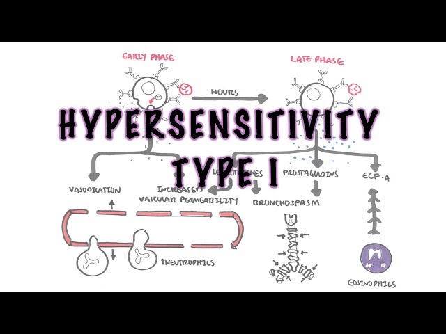
In allergic angioedema, allergen-specific immunoglobulin E cross-links receptors on mast cells and basophils. The resulting release of histamine, leukotrienes, prostaglandins and other mediators increases vascular permeability, allowing plasma to enter the interstitial tissues.
Chronic spontaneous urticaria with angioedema is also mast-cell-driven, although a reproducible external allergen is usually absent. Autoimmune mast-cell activation contributes in some patients.1
Bradykinin-mediated angioedema follows excessive bradykinin generation or impaired degradation. Bradykinin activates endothelial B2 receptors and increases vascular permeability. This mechanism explains why antihistamines, corticosteroids and adrenaline are generally ineffective in hereditary, acquired C1-inhibitor-deficient and angiotensin-converting enzyme inhibitor-associated angioedema.2
Urticaria, pruritus, rapid onset and a clear allergen exposure favour mast-cell-mediated angioedema. Recurrent abdominal swelling, absence of wheals, slower progression and failure to respond to antihistamines should prompt investigation for bradykinin-mediated disease.
Clinical Manifestations
Cutaneous and Mucosal Swelling
Common sites include:
- Lips and perioral tissues.
- Eyelids and face.
- Tongue and oral mucosa.
- Hands and feet.
- Genitalia.
The swelling is usually asymmetric and non-pitting. Mast-cell-mediated episodes may be accompanied by:
- Urticaria.
- Pruritus.
- Flushing.
- A sensation of warmth.
- Local discomfort or tightness.
Upper-Airway Involvement
Warning features include:
- Tongue or floor-of-mouth swelling.
- Voice change or hoarseness.
- Dysphagia or odynophagia.
- Drooling.
- Throat tightness.
- Stridor.
- Increasing work of breathing.
- Inability to speak normally or handle secretions.
Features Suggesting Anaphylaxis
Angioedema may form part of anaphylaxis when accompanied by:
- Bronchospasm, wheeze or respiratory compromise.
- Hypotension, collapse or altered consciousness.
- Persistent severe gastrointestinal symptoms after likely allergen exposure.
- Rapid involvement of multiple organ systems.3
Features Suggesting Bradykinin-Mediated Disease
- Recurrent swelling without urticaria or pruritus.
- Attacks evolving over several hours and lasting for days.
- Recurrent severe abdominal pain, vomiting or bowel-wall oedema.
- Laryngeal attacks.
- Family history or onset during childhood or adolescence.
- Angiotensin-converting enzyme inhibitor exposure.
- Poor response to antihistamines, corticosteroids or adrenaline.2
Diagnosis & Investigations
Angioedema is primarily a clinical diagnosis. The immediate priority is to assess the airway and determine whether the presentation meets clinical criteria for anaphylaxis. Investigations must not delay emergency treatment.3
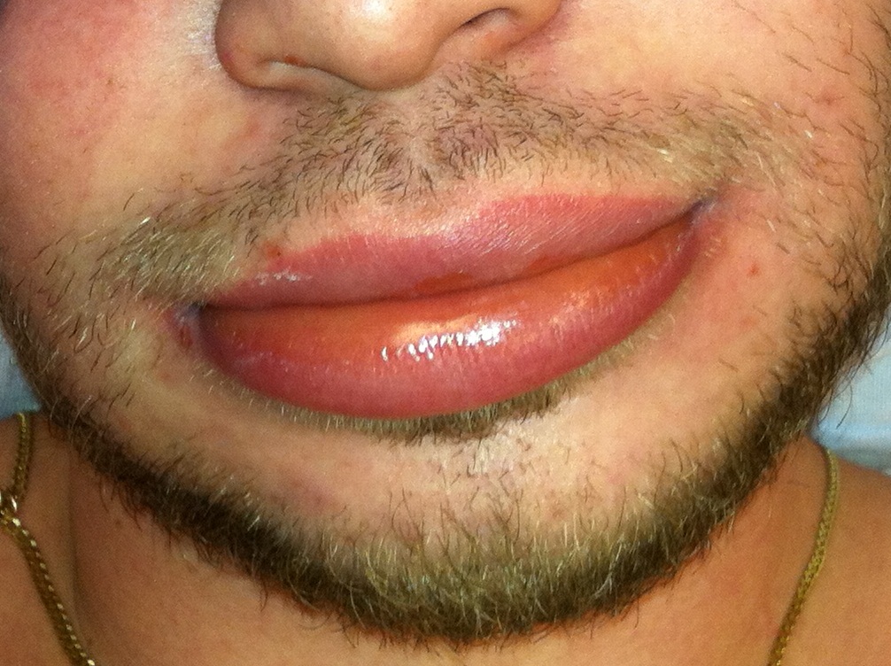
Angioedema involving the lips. Photograph: James Heilman, MD; CC BY-SA 3.0.
What to notice
Detailed image description
Source page: https://commons.wikimedia.org/wiki/File:AngioedemaFra.JPG Original asset: https://upload.wikimedia.org/wikipedia/commons/7/78/AngioedemaFra.JPG Creator: James Heilman, MD. Licence: Creative Commons Attribution-ShareAlike 3.0 Unported, https://creativecommons.org/licenses/by-sa/3.0/ Modifications: none. Retrieved 2026-08-16.
Clinical Assessment
Establish:
- Onset, progression and duration of swelling.
- Anatomical sites involved.
- Presence of urticaria, pruritus or flushing.
- Respiratory, cardiovascular and gastrointestinal symptoms.
- Food, medicine, venom, latex or occupational exposure.
- All medicines, particularly angiotensin-converting enzyme inhibitors and non-steroidal anti-inflammatory drugs.
- Previous episodes and response to antihistamines or adrenaline.
- Family history and age at first episode.
- Recurrent unexplained abdominal attacks.
- Associated infection, autoimmune disease or lymphoproliferative disease.
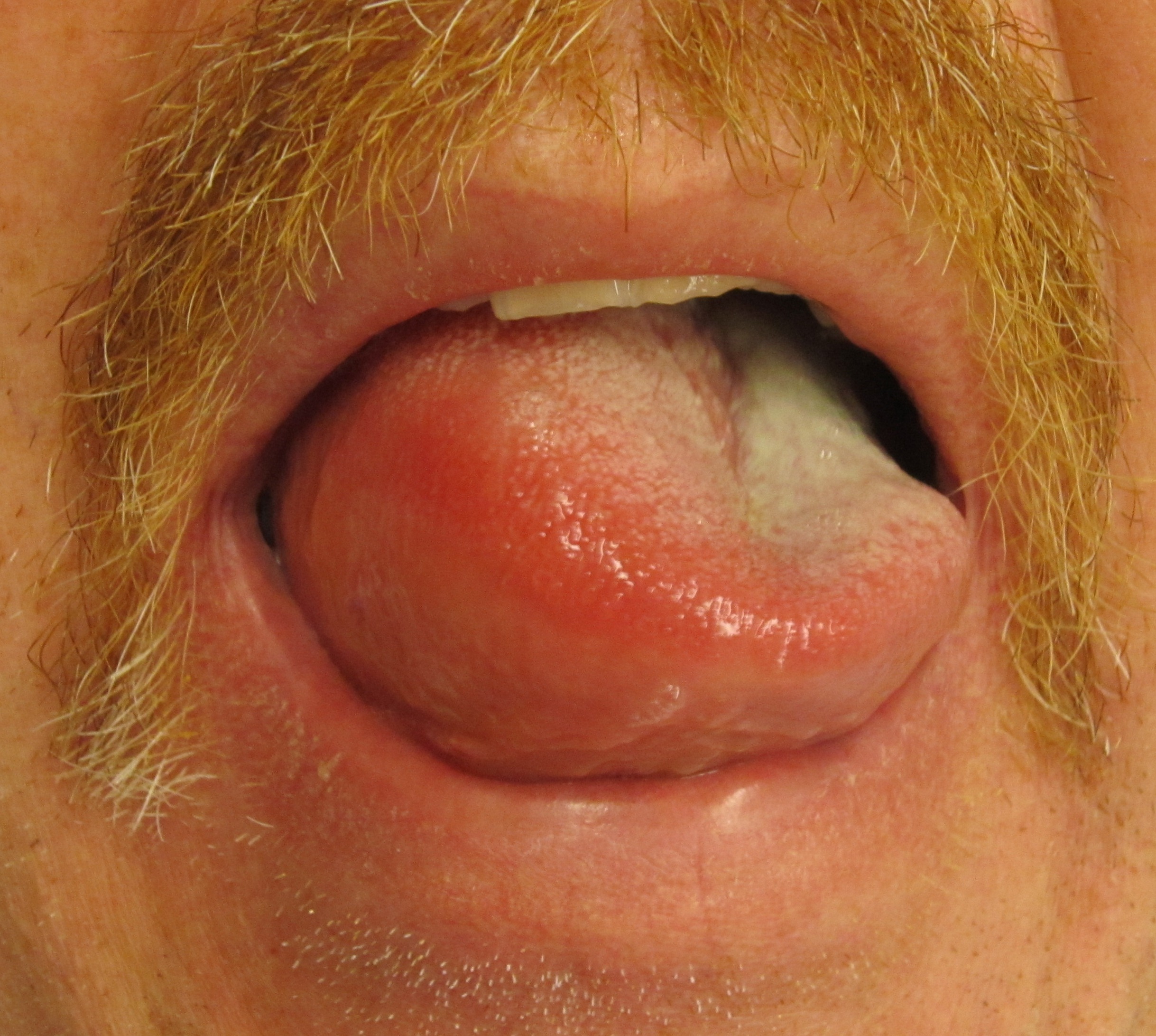
Angioedema of the tongue. Progressive tongue or oropharyngeal swelling requires urgent airway assessment. Photograph: James Heilman, MD; CC BY-SA 3.0.
What to notice
Detailed image description
Source page: https://commons.wikimedia.org/wiki/File:Angioedema2013.JPG Original asset: https://upload.wikimedia.org/wikipedia/commons/5/5c/Angioedema2013.JPG Creator: James Heilman, MD. Licence: Creative Commons Attribution-ShareAlike 3.0 Unported, https://creativecommons.org/licenses/by-sa/3.0/ Modifications: none. The source page records consent for publication. Retrieved 2026-08-16.
Investigations for Suspected Allergic Angioedema
Routine testing is usually unnecessary after a single, mild episode with an obvious trigger.
When a specific immediate allergy is suspected, targeted assessment may include:
- Allergen-specific immunoglobulin E testing.
- Skin-prick testing under specialist supervision.
- Baseline serum tryptase when recurrent or severe systemic reactions raise concern for a mast-cell disorder.
- Acute serum tryptase after suspected anaphylaxis, interpreted against a later baseline.
Broad, untargeted allergy panels are discouraged because sensitisation does not necessarily establish clinical allergy.
Recurrent Angioedema Without Wheals
Measure:
- Complement C4.
- C1-inhibitor antigenic level.
- C1-inhibitor functional activity.
Abnormal results should usually be confirmed on a separate sample. A normal C4 result alone does not reliably exclude hereditary angioedema when clinical suspicion is strong.2
Consider C1q when acquired C1-inhibitor deficiency is suspected, particularly with later onset, no family history or an associated lymphoproliferative disorder. Specialist genetic assessment may be required when the phenotype strongly suggests hereditary angioedema despite normal C1-inhibitor testing.2
Important Differentials
- Anaphylaxis.
- Hereditary or acquired angioedema.
- Angiotensin-converting enzyme inhibitor-associated angioedema.
- Contact dermatitis.
- Cellulitis or erysipelas.
- Orofacial infection.
- Superior vena cava obstruction.
- Nephrotic, cardiac, hepatic or thyroid-related generalised oedema.
- Melkersson–Rosenthal syndrome.
- Morbihan disease.
- Factitious swelling.
A normal appearance between attacks does not exclude angioedema. Recurrent swelling without wheals—especially with abdominal attacks, a family history or poor antihistamine response—requires assessment for bradykinin-mediated disease.
Treatment
Immediate Assessment
- Assess airway, breathing, circulation and mental status.
- Call for emergency assistance if airway involvement or anaphylaxis is possible.
- Stop or remove the suspected trigger when feasible.
- Monitor oxygen saturation, blood pressure and cardiac rhythm in significant reactions.
- Prepare early for advanced airway management when tongue, pharyngeal or laryngeal swelling is progressing.
Anaphylaxis or Threatened Airway
Give intramuscular adrenaline promptly into the anterolateral thigh when angioedema forms part of anaphylaxis. The recommended healthcare dose is 0.01 mg/kg of 1 mg/mL adrenaline, up to 0.5 mg, repeated every 5–15 minutes when required.3
Provide supplemental oxygen, intravenous access and fluid resuscitation as clinically indicated. Antihistamines may improve cutaneous symptoms but must not delay or replace adrenaline. Corticosteroids are not first-line treatment for anaphylaxis.3
Antihistamines treat histamine-mediated skin symptoms; they do not treat shock or airway compromise. When anaphylaxis is suspected, intramuscular adrenaline is the first-line treatment.
Isolated Mast-Cell-Mediated Angioedema
For angioedema without anaphylaxis or airway compromise:
- Use a regular second-generation, non-sedating H1-antihistamine.
- Avoid the identified allergen or aggravating medicine.
- Avoid sedating antihistamines when a suitable non-sedating alternative is available.
- Consider a short corticosteroid course only selectively; repeated or prolonged corticosteroid treatment should be avoided.
Recurrent Idiopathic Angioedema
Management generally follows the chronic spontaneous urticaria pathway:
- Regular standard-dose second-generation H1-antihistamine.
- If inadequately controlled, specialist-guided dose escalation up to fourfold the standard dose.
- Omalizumab for persistent mast-cell-mediated disease despite high-dose antihistamine therapy.
- Ciclosporin may be considered by specialists for severe refractory disease.1
Treatment response should be documented. Persistent attacks despite appropriately dosed antihistamines should prompt reconsideration of the mechanism and diagnosis.
Trigger Management and Follow-Up
- Permanently discontinue a suspected angiotensin-converting enzyme inhibitor and assess for bradykinin-mediated disease.
- Avoid confirmed allergens and provide a written emergency plan.
- Refer for allergy assessment after anaphylaxis or when the trigger remains uncertain.
- Prescribe and teach adrenaline autoinjector use when clinically indicated.
- Review aggravating medicines, including non-steroidal anti-inflammatory drugs.
- Refer recurrent angioedema without wheals to allergy or clinical immunology.
Complications & Prognosis
Complications
- Upper-airway obstruction and hypoxic injury.
- Anaphylactic shock.
- Recurrent emergency presentations.
- Unnecessary dietary or medicine avoidance after unstructured allergy testing.
- Adverse effects from repeated corticosteroid treatment.
- Delayed diagnosis of hereditary or acquired angioedema.
- Anxiety, impaired quality of life and fear of recurrence.
Prognosis
Most isolated mast-cell-mediated episodes resolve without permanent tissue injury. Prognosis depends on whether the trigger can be identified and avoided and whether recurrent disease responds to prophylactic antihistamine therapy.
Idiopathic histaminergic angioedema often improves with regular second-generation antihistamines, although the duration of disease is variable. Failure to respond should prompt diagnostic reassessment rather than indefinite escalation of ineffective therapy.
Upper-airway angioedema can be fatal. Patients with previous airway involvement, anaphylaxis, recurrent unexplained episodes or suspected bradykinin-mediated disease require specialist follow-up and an individualised emergency plan.
References
- Zuberbier T, Abdul Latiff AH, Abuzakouk M, Aquilina S, Asero R, Baker D, et al. The international EAACI/GA²LEN/EuroGuiDerm/APAAACI guideline for the definition, classification, diagnosis, and management of urticaria. Allergy. 2022;77(3):734–766. doi:10.1111/all.15090.
- Maurer M, Magerl M, Betschel S, Aberer W, Ansotegui IJ, Aygören-Pürsün E, et al. The international WAO/EAACI guideline for the management of hereditary angioedema—the 2021 revision and update. Allergy. 2022;77(7):1961–1990. doi:10.1111/all.15214.
- Cardona V, Ansotegui IJ, Ebisawa M, El-Gamal Y, Fernandez Rivas M, Fineman S, et al. World Allergy Organization anaphylaxis guidance 2020. World Allergy Organ J. 2020;13(10):100472. doi:10.1016/j.waojou.2020.100472.






Members only discussions coming soon…