

Femoroacetabular Impingement Syndrome
Overview
Femoroacetabular impingement syndrome (FAIS) is a motion-related clinical disorder of the hip in which symptoms and clinical signs occur in association with characteristic femoral and/or acetabular morphology. The accepted diagnostic concept is a triad of symptoms, clinical signs and imaging findings; bone morphology alone does not establish the syndrome.
Patients commonly present with motion-related or position-related hip or groin pain, often provoked by hip flexion, rotation, squatting, running, pivoting or prolonged sitting. Clicking, catching, stiffness, restricted movement and reduced sporting or occupational function may also occur.
FAIS is clinically important because repeated mechanical conflict may contribute to acetabular labral and articular cartilage injury. Cam morphology is also associated with later hip osteoarthritis, although progression is not inevitable and it remains uncertain whether treating FAIS prevents future osteoarthritis.
Definition
- Femoroacetabular impingement syndrome (FAIS)
- A motion-related clinical disorder of the hip diagnosed when characteristic symptoms, clinical signs and relevant imaging findings are all present.
- Cam morphology
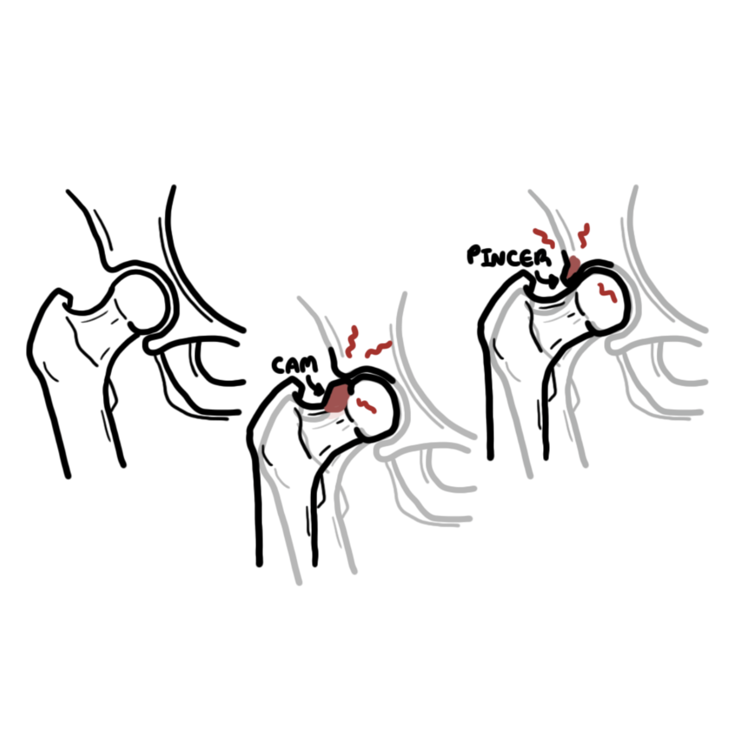
- A non-spherical prominence or reduced concavity at the femoral head–neck junction that can reduce clearance during hip motion.
- Pincer morphology
- Focal or global acetabular overcoverage that can increase contact between the acetabular rim and femoral neck.
- Acetabular labrum
- A fibrocartilaginous rim attached to the acetabulum that contributes to hip stability, load distribution and maintenance of the joint fluid seal.
Anatomy & Physiology
The hip is a ball-and-socket synovial joint formed by the femoral head and acetabulum. The acetabular labrum is attached to the acetabular rim and helps deepen the socket, distribute load, maintain the fluid seal and contribute to joint stability.
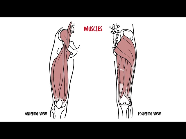
The femoral head is normally approximately spherical, with a concave head–neck junction that provides clearance during hip flexion and rotation. Acetabular orientation and coverage determine how much of the femoral head is contained by the socket. Hip motion is further influenced by femoral version, pelvic position and dynamic control from the gluteal, deep rotator, adductor and trunk muscles.
FAIS occurs when hip morphology and movement combine to produce symptomatic premature contact between the proximal femur and acetabular rim.1
Aetiology & Risk Factors
Aetiology
FAIS reflects an interaction between osseous morphology and movement rather than a single isolated lesion. The morphology may be predominantly femoral (cam), acetabular (pincer), or mixed.
- Cam morphology: reduced sphericity or a bony prominence at the femoral head–neck junction reduces clearance during hip motion.
- Pincer morphology: focal or global acetabular overcoverage increases contact between the acetabular rim and femoral neck.
- Mixed morphology: cam and pincer features coexist.
The same morphology may be asymptomatic in one person and symptomatic in another. Symptoms depend on the interaction between morphology, joint loading, range of motion, activity exposure, associated chondrolabral injury and individual pain and functional factors.1,2
Risk Factors
- Cam or pincer morphology in combination with provocative hip loading.
- Participation in activities requiring repeated deep hip flexion, rotation, cutting or pivoting.
- High-level sporting exposure during skeletal development, particularly in sports with repeated hip loading, which is associated with development of cam morphology.
- Reduced hip motion, impaired hip or trunk strength, and altered movement strategies that increase symptomatic loading.
- Coexisting labral or articular cartilage injury.
Pathophysiology
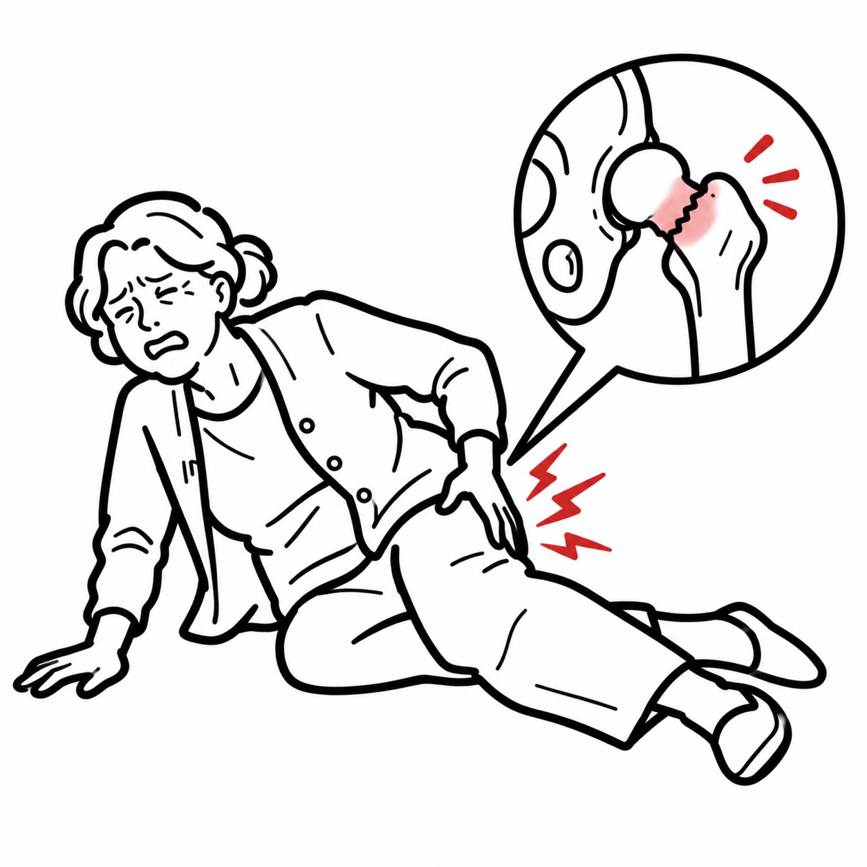
During hip flexion and rotation, reduced clearance between the femoral head–neck junction and acetabular rim can produce repeated mechanical contact. In cam-type morphology, the non-spherical femoral head–neck junction may enter the acetabulum and generate shear at the anterosuperior chondrolabral junction. With pincer morphology, acetabular overcoverage may produce more direct compression of the labrum against the femoral neck.
Repeated loading can lead to labral irritation or tearing, chondrolabral separation and articular cartilage injury. Pain and protective movement patterns may then contribute to further restriction of hip motion, weakness and loss of function. Structural damage and symptoms are not perfectly correlated; therefore management should target the patient’s clinical syndrome rather than the radiographic shape alone.1,2
Why are deep squatting, prolonged low sitting and pivoting commonly provocative? Hip flexion brings the femoral head–neck junction closer to the acetabular rim; adduction and internal rotation can further reduce clearance in a susceptible hip, increasing contact and reproducing symptoms.
Clinical Manifestations
Symptoms are usually gradual in onset and are commonly related to movement or sustained hip positions.1,2
Symptoms
- Anterior hip or groin pain; some patients describe pain using a “C-sign” around the anterior and lateral hip.
- Pain with deep squatting, lunging, running, kicking, pivoting or directional change.
- Pain after prolonged sitting or when entering or leaving a low car seat.
- Hip stiffness or restricted movement.
- Clicking, catching or locking, which may occur with associated labral pathology but is not diagnostic by itself.
- Reduced sporting performance, walking tolerance or occupational function.
- Less commonly, pain referred to the lateral hip, buttock, thigh or knee.
Clinical Examination
- Reduced hip flexion and/or internal rotation, particularly when the hip is flexed.
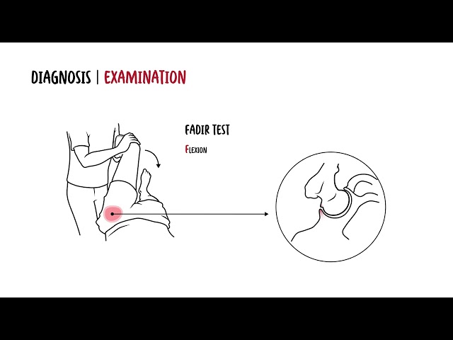
- Reproduction of familiar pain with flexion-adduction-internal rotation (FADIR).
- Pain or restriction with other hip provocation tests such as FABER.
- Reduced hip muscle strength or endurance.
- Altered pelvic, trunk or lower-limb control during functional tasks such as squatting or single-leg loading.
- Antalgic gait when symptoms are more severe.
The FADIR test is useful as a provocative or screening manoeuvre but is not sufficiently specific to diagnose FAIS by itself. Examination should also consider lumbar spine, sacroiliac, abdominal and extra-articular hip sources when the presentation is atypical.
Diagnosis & Investigations
FAIS is diagnosed when the clinical triad of appropriate symptoms, positive clinical signs and relevant imaging findings is present.1 No single symptom, examination manoeuvre or radiographic measurement is independently diagnostic.
Cam or pincer morphology on imaging is not the same as femoroacetabular impingement syndrome. FAIS requires concordant symptoms, clinical signs and imaging findings; asymptomatic morphology alone should not be labelled as the syndrome.1
Plain Radiography
Plain radiography is the usual first-line imaging investigation. A reproducible anteroposterior pelvis view together with a dedicated lateral hip view can assess cam morphology, acetabular coverage and version, dysplasia, joint-space narrowing and alternative osseous pathology.
Radiographic features should be interpreted in clinical context. Measurements such as the alpha angle can characterise morphology but should not be used in isolation to diagnose FAIS.
MRI and MR Arthrography
MRI is used when further assessment of the labrum, articular cartilage and other intra-articular or peri-articular structures is required. Dedicated hip MRI protocols can also better characterise cam morphology and femoral torsion. MR arthrography may be considered in selected settings, although high-quality non-contrast MRI can provide substantial chondrolabral information.
CT Scan
Diagnostic Injection
An image-guided intra-articular local anaesthetic injection may help determine whether pain is arising from the hip joint when the pain source remains uncertain. Symptomatic improvement supports an intra-articular pain generator but does not independently establish FAIS.
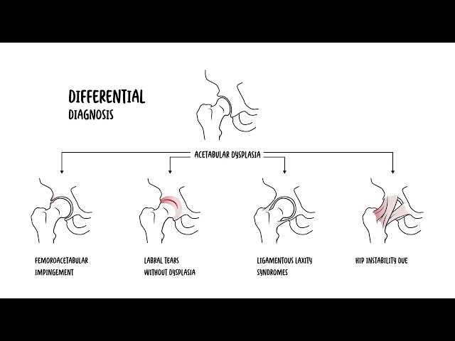
Differential Diagnoses
- Hip osteoarthritis.
- Developmental dysplasia of the hip or hip instability.
- Acetabular labral pathology without FAIS.
- Stress fracture or other occult bone injury.
- Osteonecrosis of the femoral head.
- Greater trochanteric pain syndrome or gluteal tendinopathy.
- Iliopsoas-related pain or snapping hip syndrome.
- Adductor-related or other groin pain.
- Lumbar radiculopathy or referred lumbosacral pain.
- Inflammatory, infective or neoplastic hip pathology when suggested by the clinical context.
Pain may coexist with acetabular labral injury or chondral damage, particularly with longstanding mechanical impingement.
Classification
FAIS is commonly described according to the predominant morphology. These categories describe anatomy rather than symptom severity.
| Pattern | Primary morphology | Typical mechanical effect |
|---|---|---|
| Cam | Reduced femoral head–neck offset or non-spherical prominence | Shear and compression at the anterosuperior chondrolabral junction during flexion and rotation. |
| Pincer | Focal or global acetabular overcoverage | Earlier contact between the acetabular rim and femoral neck, with labral compression. |
| Mixed | Coexisting cam and pincer morphology | Combined femoral and acetabular contributors to symptomatic contact. |
Treatment
Treatment aims to reduce pain, restore function and allow return to desired activities. Management is individualised according to symptom severity, functional goals, morphology, associated tissue injury, rehabilitation response and the presence or absence of osteoarthritis.
Non-operative Management
Structured non-operative management is appropriate as initial treatment for many patients and should be active rather than limited to rest.
- Education about FAIS and the difference between symptoms and imaging morphology.
- Temporary modification of activities or positions that repeatedly provoke symptoms, while maintaining general activity where possible.
- Progressive hip and lower-limb strengthening.
- Trunk and lumbopelvic strengthening and control.
- Movement retraining to reduce provocative loading during sport, work and daily activities.
- Restoration of clinically relevant mobility without repeatedly forcing painful end-range impingement positions.
- Progressive return to running, sport or occupational loading.
- Simple analgesia or NSAIDs when clinically appropriate and not contraindicated.
Manual therapy may be used as an adjunct for selected mobility or pain impairments, but rehabilitation should principally address strength, movement, function and graded exposure.
Injections
Intra-articular corticosteroid injection may provide temporary symptom relief in selected patients, but it does not correct the underlying morphology and should not replace a structured rehabilitation programme. Evidence for other injectables is less established.
Surgical Management
Specialist surgical assessment may be considered when symptoms remain functionally limiting despite an adequate course of structured non-operative care, the diagnosis is secure, and imaging abnormalities correspond with the patient’s symptoms and signs. Advanced hip osteoarthritis reduces the likelihood that joint-preserving arthroscopy will provide an appropriate solution.
Hip arthroscopy typically aims to address the relevant osseous morphology and associated labral or chondral pathology. Randomised trials in selected adults with symptomatic FAIS have shown improvement with both structured physiotherapy and arthroscopic surgery; arthroscopy produced greater average short-term improvement in patient-reported hip outcomes in these trials, but surgery also carries procedural risks and not every patient benefits.
Treatment choice should therefore use shared decision-making and consider symptom burden, prior rehabilitation, activity goals, cartilage status, radiographic osteoarthritis, operative risk and patient preference.
Complications & Prognosis
Complications
- Persistent or recurrent hip and groin pain.
- Reduced hip movement and activity tolerance.
- Acetabular labral injury.
- Chondrolabral separation and articular cartilage damage.
- Reduced sporting or occupational participation.
- Progression to hip osteoarthritis in some patients, particularly where cam morphology and cartilage injury are present.
Prognosis
Many patients improve with education, activity modification and structured rehabilitation, although recovery may take several months and should be judged by pain, function, strength, movement quality and participation rather than imaging appearance alone.
Appropriately selected patients undergoing arthroscopic treatment can also experience meaningful improvement in pain and function.4,5 The long-term natural history varies, and neither conservative care nor surgery has been conclusively shown to prevent future hip osteoarthritis.
References
- Griffin DR, Dickenson EJ, O’Donnell J, Agricola R, Awan T, Beck M, et al. The Warwick Agreement on femoroacetabular impingement syndrome (FAI syndrome): an international consensus statement. Br J Sports Med. 2016;50(19):1169–1176. doi:10.1136/bjsports-2016-096743
- Enseki KR, Bloom NJ, Harris-Hayes M, Cibulka MT, Disantis A, Di Stasi S, et al. Hip pain and movement dysfunction associated with nonarthritic hip joint pain: a revision. J Orthop Sports Phys Ther. 2023;53(7):CPG1–CPG70. doi:10.2519/jospt.2023.0302
- Schmaranzer F, Kheterpal AB, Bredella MA. Best practices: hip femoroacetabular impingement. AJR Am J Roentgenol. 2021;216(3):585–598. doi:10.2214/AJR.20.22783
- Palmer AJR, Ayyar Gupta V, Fernquest S, Rombach I, Dutton SJ, Mansour R, et al. Arthroscopic hip surgery compared with physiotherapy and activity modification for the treatment of symptomatic femoroacetabular impingement: multicentre randomised controlled trial. BMJ. 2019;364:l185. doi:10.1136/bmj.l185
- Griffin DR, Dickenson EJ, Wall PDH, Achana F, Donovan JL, Griffin J, et al. Hip arthroscopy versus best conservative care for the treatment of femoroacetabular impingement syndrome (UK FASHIoN): a multicentre randomised controlled trial. Lancet. 2018;391(10136):2225–2235. doi:10.1016/S0140-6736(18)31202-9



Members only discussions coming soon…