

Liver Cirrhosis
Overview
Cirrhosis is the advanced stage of chronic liver injury characterised by widespread fibrosis, regenerative nodules and distortion of normal hepatic architecture. This causes two major consequences:
- Portal hypertension
- Reduced hepatic function
Cirrhosis may remain asymptomatic for years before progressing from a compensated to a decompensated stage.
Definition
Compensated cirrhosis: cirrhosis without major clinical complications.
Decompensated cirrhosis: development of ascites, hepatic encephalopathy or portal-hypertensive gastrointestinal bleeding; jaundice commonly accompanies advanced decompensation.
Portal hypertension: increased pressure within the portal venous system due mainly to increased resistance through the cirrhotic liver.
Ascites: pathological accumulation of fluid within the peritoneal cavity.
Hepatic encephalopathy: potentially reversible brain dysfunction caused by liver failure and/or portosystemic shunting.
Anatomy & Physiology
Aetiology & Risk Factors
- Alcohol-associated liver disease
- Metabolic dysfunction-associated steatotic liver disease—MASLD/MASH
- Chronic hepatitis B or C
- Autoimmune hepatitis
- Primary biliary cholangitis
- Primary sclerosing cholangitis
- Haemochromatosis
- Wilson disease
- Alpha-1 antitrypsin deficiency
- Chronic biliary obstruction
- Congestive cardiac disease
- Drugs and toxins
- Cryptogenic cirrhosis—cause not identified
Several causes may coexist, particularly metabolic liver disease and alcohol exposure.
Pathophysiology
- Chronic hepatocyte injury causes inflammation and activation of hepatic stellate cells.
- Stellate cells produce excess collagen and extracellular matrix.
- Progressive fibrosis forms fibrous septa and regenerative nodules.
- Distortion of the hepatic vasculature increases intrahepatic resistance, causing portal hypertension.
- Splanchnic vasodilatation reduces effective circulating arterial volume.
- This activates the renin–angiotensin–aldosterone system, sympathetic nervous system and antidiuretic hormone.
- Sodium and water retention subsequently contribute to ascites and oedema.
- Loss of functioning hepatocytes causes impaired:
- Albumin production
- Coagulation-factor production
- Bilirubin metabolism
- Drug and toxin clearance
- Immune and metabolic function.
Clinical Manifestations
Compensated cirrhosis
Patients may be asymptomatic or have:
- Fatigue and weakness
- Reduced appetite
- Unintentional weight loss
- Nausea or abdominal discomfort
- Pruritus
- Reduced libido
- Muscle loss
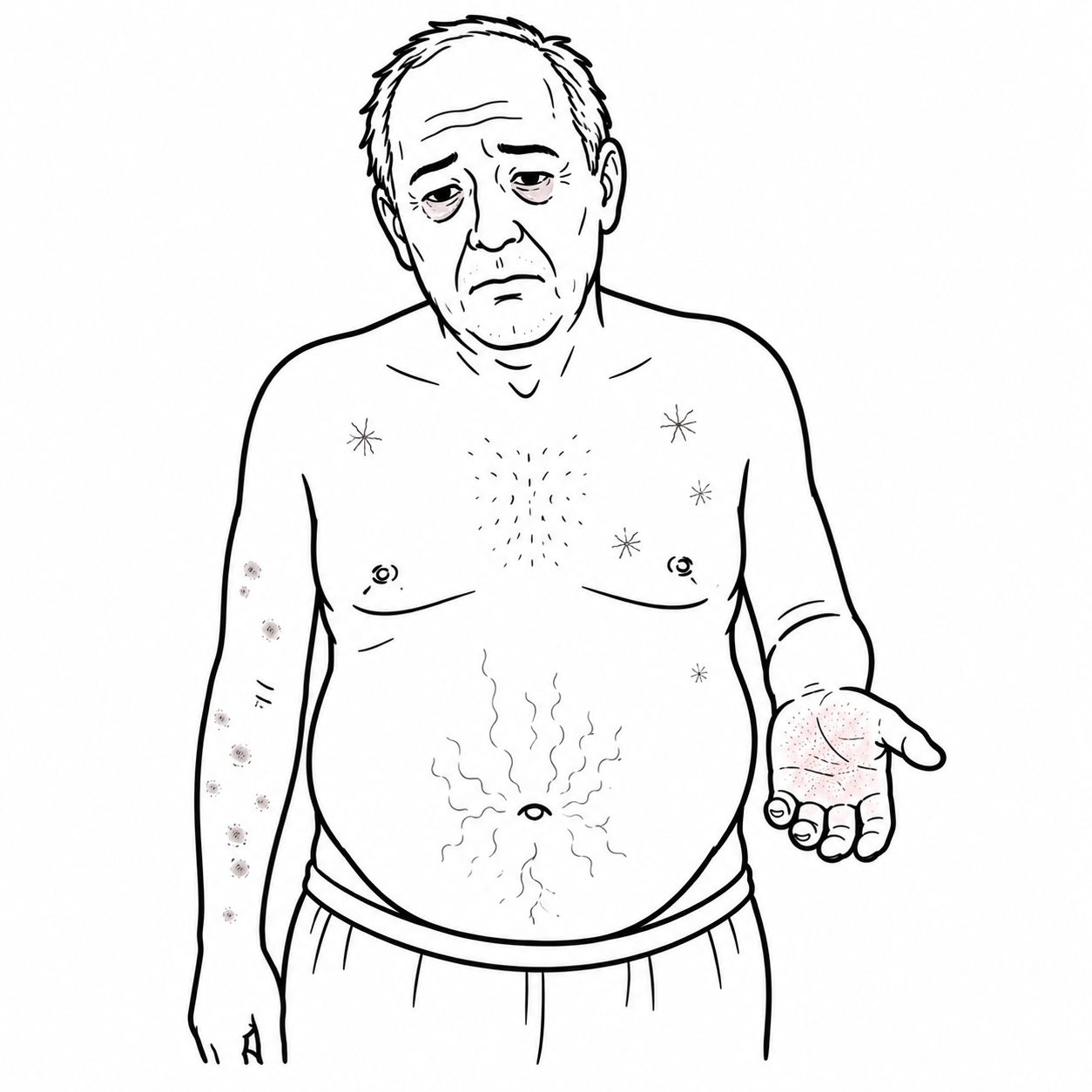
Clinical examination
- Jaundice
- Spider naevi
- Palmar erythema
- Easy bruising
- Muscle wasting
- Gynaecomastia
- Testicular atrophy
- Loss of body hair
- Hepatomegaly or a small nodular liver
- Splenomegaly
- Dupuytren contracture
- Clubbing
Decompensated cirrhosis
- Ascites and abdominal distension
- Peripheral oedema
- Variceal gastrointestinal bleeding:
- Haematemesis
- Melaena
- Haematochezia with severe bleeding
- Hepatic encephalopathy:
- Sleep-pattern reversal
- Confusion
- Personality change
- Asterixis
- Reduced consciousness
- Jaundice
- Infection or sepsis
- Reduced urine output from acute kidney injury or hepatorenal syndrome.
New or worsening ascites requires diagnostic paracentesis.
Investigations
- FBC:
- Thrombocytopenia from portal hypertension and hypersplenism
- Anaemia from bleeding, nutritional deficiency or hypersplenism
- Leukopenia
- Liver enzymes:
- Synthetic liver function:
- Low albumin
- Prolonged INR
- Raised bilirubin
- UEC and creatinine:
- Hyponatraemia
- Acute kidney injury
- Hepatorenal syndrome
- Glucose, calcium and phosphate
- CRP and cultures if infection is suspected
Normal ALT and AST do not exclude advanced cirrhosis.
Thrombocytopenia may be an early clue to portal hypertension.
Determine the underlying cause
- Hepatitis B and C serology
- Ferritin and transferrin saturation
- ANA, ASMA and serum immunoglobulins
- Antimitochondrial antibodies
- Alpha-1 antitrypsin level
- Ceruloplasmin and urinary copper in appropriate younger patients
- Alcohol and medication history
- Metabolic assessment: BMI, lipids and HbA1c
Fibrosis assessment
- FIB-4 or other serum fibrosis score
- Transient elastography—FibroScan
- Magnetic resonance elastography in selected cases
- Liver biopsy if the diagnosis or underlying cause remains uncertain
Non-invasive blood and elastography-based assessments are increasingly used to identify advanced fibrosis and cirrhosis and reduce the need for liver biopsy.
Imaging
- Abdominal ultrasound with Doppler:
- Nodular liver
- Splenomegaly
- Ascites
- Portal-vein flow
- Focal liver lesions
- CT or MRI if complications, vascular thrombosis or hepatocellular carcinoma are suspected.
Ascitic-fluid analysis
Perform diagnostic paracentesis for:
- New-onset ascites
- Hospital admission with cirrhosis and ascites
- Abdominal pain, fever, encephalopathy, hypotension or renal deterioration
Tests include cell count, neutrophils, culture, albumin and total protein.
- SAAG ≥1.1 g/dL: suggests portal-hypertensive ascites.
- Neutrophils ≥250 cells/mm³: diagnostic of spontaneous bacterial peritonitis and requires treatment.
Classifications
Child–Pugh score
Assesses the severity and prognosis of cirrhosis using five clinical and laboratory variables.
| Child–Pugh score | |||
| Parameter | 1 point | 2 points | 3 points |
| Bilirubin | <34 µmol/L | 34–51 µmol/L | >51 µmol/L |
| Albumin | >35 g/L | 28–35 g/L | <28 g/L |
| INR | <1.7 | 1.7–2.3 | >2.3 |
| Ascites | None | Mild/diuretic-responsive | Moderate–severe/refractory |
| Encephalopathy | None | Grade I–II | Grade III–IV |
| A total Child–Pugh score of 5–6 corresponds to Class A, indicating well-compensated liver disease. A score of 7–9 corresponds to Class B, indicating significant functional impairment. A score of 10–15 corresponds to Class C, indicating decompensated liver disease and the poorest prognosis. | |||
Ascites and encephalopathy assessment can be subjective and may change with treatment.
MELD-based score
The Model for End-stage Liver Disease estimates short-term mortality and helps determine liver-transplant urgency.
| Component | Significance |
| Bilirubin | Reflects hepatic excretory dysfunction |
| INR | Reflects impaired hepatic synthesis of clotting factors |
| Creatinine | Reflects renal dysfunction, an important predictor of mortality |
| Serum sodium | Hyponatraemia indicates advanced circulatory dysfunction |
| Albumin and sex | Included in MELD 3.0 to improve mortality prediction |
- Calculated using a validated electronic calculator rather than manually.
- A higher score indicates greater predicted 90-day mortality and greater transplant urgency.
- MELD is more objective than Child–Pugh because it mainly uses laboratory values.
- It does not directly include ascites, encephalopathy, frailty or quality of life.
| MELD score | Severity | Interpretation |
| 6–9 | Lower severity | Relatively low short-term mortality |
| 10–19 | Moderate | Increasing liver-disease severity |
| 20–29 | Severe | Significant 90-day mortality risk; transplant assessment is often appropriate |
| 30–39 | Very severe | High short-term mortality and high transplant urgency |
| ≥40 | Critical | Extremely high short-term mortality and greatest transplant urgency |
Treatment
General
- Hepatology follow-up.
- Avoid alcohol.
- Review all prescribed, over-the-counter and complementary medications.
- Avoid NSAIDs, particularly with ascites or kidney dysfunction.
- Vaccinate against hepatitis A and B, influenza, pneumococcus and other infections as appropriate.
- Adequate nutrition with approximately 1.2–1.5 g/kg/day of protein.
- Do not routinely restrict protein in hepatic encephalopathy.
- Avoid prolonged fasting; consider a late-night protein-containing snack.
- Assess and treat malnutrition, sarcopenia, frailty and osteoporosis.
Treatment of complications will be discussed separately
Complications & Prognosis
Complications of liver cirrhosis
- Portal hypertension
- Oesophageal or gastric varices
- Variceal haemorrhage
- Ascites
- Spontaneous bacterial peritonitis
- Hepatic encephalopathy
- Hepatorenal syndrome
- Hepatopulmonary syndrome
- Portopulmonary hypertension
- Hepatic hydrothorax
- Portal-vein thrombosis
- Coagulopathy and bleeding
- Hypersplenism and cytopenias
- Malnutrition, sarcopenia and frailty
- Osteoporosis
- Recurrent infections and sepsis
- Hepatocellular carcinoma
- Acute-on-chronic liver failure
- Multiorgan failure and death.
For more information on the complications of liver cirrhosis click here
Do not diagnose or monitor hepatic encephalopathy using ammonia levels alone—it is primarily a clinical diagnosis.
Every patient with cirrhosis requires regular surveillance for varices and hepatocellular carcinoma.
Prognosis
- Prognosis depends on the underlying cause, whether disease remains compensated, development of complications, renal function and response to treatment.
- Compensated cirrhosis may remain stable for several years.
- Development of ascites, variceal bleeding or encephalopathy marks decompensation and substantially worsens survival.
- The development of ascites is associated with a reduction in five-year survival from approximately 80% to 30%.
- Refractory ascites, hepatorenal syndrome, recurrent infection, severe encephalopathy and hepatocellular carcinoma indicate a poor prognosis.
- Child–Pugh and MELD-based scores assist with risk assessment.
- Liver transplantation can provide long-term survival for appropriately selected patients with decompensated disease.
Liver transplantation
- Definitive treatment for selected patients with end-stage liver disease, acute liver failure or certain hepatocellular carcinomas.
- Refer early after decompensation, including ascites, variceal bleeding, encephalopathy, progressive jaundice or hepatorenal syndrome.
- Transplant priority is largely based on the MELD score, which estimates short-term mortality using bilirubin, INR, creatinine and sodium.
- Assessment considers disease severity, cardiac and respiratory fitness, renal function, infection, malignancy, frailty, substance use, adherence and social support.
References
- European Association for the Study of the Liver. EASL clinical practice guidelines for the management of patients with decompensated cirrhosis. J Hepatol. 2018;69(2):406–460. doi:10.1016/j.jhep.2018.03.024.
- Biggins SW, Angeli P, Garcia-Tsao G, Ginès P, Ling SC, Nadim MK, et al. Diagnosis, evaluation, and management of ascites, spontaneous bacterial peritonitis and hepatorenal syndrome. Hepatology. 2021;74(2):1014–1048. doi:10.1002/hep.31884.
- Kaplan DE, Ripoll C, Thiele M, Fortune BE, Simonetto DA, Garcia-Tsao G, et al. AASLD practice guidance on risk stratification and management of portal hypertension and varices in cirrhosis. Hepatology. 2024;79(5):1180–1211. doi:10.1097/HEP.0000000000000647.
- European Association for the Study of the Liver. EASL clinical practice guidelines on non-invasive tests for evaluation of liver disease severity and prognosis—2021 update. J Hepatol. 2021;75(3):659–689. doi:10.1016/j.jhep.2021.05.025.
- Karvellas CJ, Bajaj JS, Kamath PS, McPhail MJW, Piano S, Sanyal AJ, et al. AASLD practice guidance on acute-on-chronic liver failure and the management of critically ill patients with cirrhosis. Hepatology. 2024;79(6):1463–1502. doi:10.1097/HEP.0000000000000671.






Members only discussions coming soon…