

Rhesus Hemolytic Disease
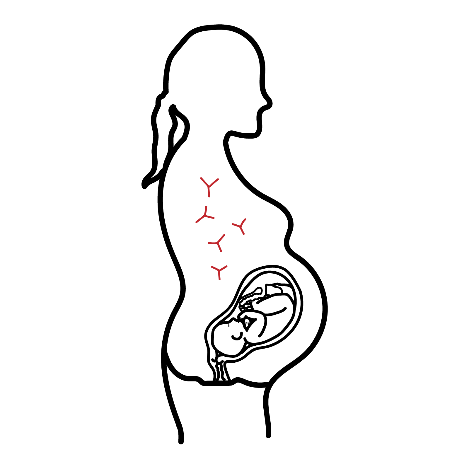
Rhesus haemolytic disease
Overview
Rhesus haemolytic disease, more accurately called RhD haemolytic disease of the fetus and newborn (HDFN), occurs when maternal IgG antibodies against the RhD antigen cross the placenta and destroy RhD-positive fetal red blood cells. It usually develops after an RhD-negative mother has previously been sensitised by pregnancy, fetomaternal haemorrhage or transfusion. Disease severity ranges from mild neonatal jaundice to severe fetal anaemia, hydrops fetalis, kernicterus or fetal death. Routine anti-D immunoglobulin prophylaxis has made severe RhD disease uncommon in countries with established prevention programs.
RhD incompatibility alone does not equal disease—the mother must first become alloimmunised.
Definition
RhD incompatibility: an RhD-negative mother carries an RhD-positive fetus.
Alloimmunisation: maternal formation of antibodies against foreign red-cell antigens.
Fetomaternal haemorrhage: passage of fetal red blood cells into the maternal circulation.
Hydrops fetalis: abnormal fetal fluid accumulation in at least two compartments, such as ascites, pleural effusion, pericardial effusion or skin oedema.
Direct antiglobulin test—DAT: detects maternal antibodies attached to neonatal red blood cells.
Aetiology & Risk Factors
RhD HDFN requires an RhD-negative mother, an RhD-positive fetus and maternal anti-D alloimmunisation.
Risk factors for sensitisation include:
- Previous pregnancy with an RhD-positive fetus
- Previous miscarriage, termination, ectopic or molar pregnancy
- Antepartum haemorrhage or placental abruption
- Abdominal trauma
- Amniocentesis, chorionic-villus sampling or cordocentesis
- External cephalic version
- Delivery of an RhD-positive infant
- Previous RhD-incompatible blood transfusion
- Failure, delay or inadequate dosing of anti-D immunoglobulin
- Large fetomaternal haemorrhage
The first recognised pregnancy can be affected when sensitisation occurred during an earlier unrecognised pregnancy, transfusion or sensitising event.
Pathophysiology
- An RhD-negative mother is exposed to RhD-positive fetal red blood cells.
- The initial immune response produces antibodies and immunological memory.
- On subsequent exposure, maternal IgG anti-D antibodies are produced.
- IgG crosses the placenta and binds to RhD-positive fetal red blood cells.
- Antibody-coated red cells are destroyed, producing fetal haemolytic anaemia.
- Severe anaemia causes increased cardiac output, tissue hypoxia and extramedullary haematopoiesis.
- Progressive disease may cause hepatosplenomegaly, liver dysfunction, heart failure and hydrops fetalis.
- Before birth, bilirubin is cleared through the placenta; after delivery, haemolysis can cause rapidly rising unconjugated bilirubin and neurological toxicity.
Clinical Manifestations
Fetal manifestations
- Fetal anaemia
- Increased fetal heart rate or reduced fetal movements
- Cardiomegaly and high-output cardiac failure
- Hepatosplenomegaly
- Placental thickening
- Polyhydramnios
- Ascites
- Pleural or pericardial effusions
- Generalised skin oedema
- Hydrops fetalis
- Intrauterine fetal death
Neonatal manifestations
- Pallor and anaemia
- Jaundice, often appearing within the first 24 hours
- Rapidly rising unconjugated bilirubin
- Hepatosplenomegaly
- Oedema or residual hydrops
- Respiratory distress
- Heart failure
- Poor feeding, lethargy or hypotonia
- Severe bilirubin toxicity:
- Acute bilirubin encephalopathy
- Kernicterus
- Late or persistent anaemia following birth.
Neonatal jaundice within the first 24 hours is pathological and requires urgent assessment.
Diagnosis & Investigations
Maternal screening
- ABO and RhD blood group
- Red-cell antibody screen at the first antenatal visit
- Repeat antibody screening at approximately 28 weeks
- Identify and quantify any clinically significant antibody.
- Distinguish passive anti-D following prophylaxis from true immune anti-D.
- Review previous pregnancies, transfusions and affected infants.
Determine fetal risk
- Paternal RhD testing may help assess whether the fetus could be RhD positive.
- Cell-free fetal DNA testing can determine fetal RHD genotype without an invasive procedure.
- If the fetus is confirmed RhD negative, it is not at risk from maternal anti-D.
Monitor an at-risk fetus
- Serial maternal anti-D antibody levels or titres.
- Serial ultrasound assessment for hydrops and fetal growth.
- Middle cerebral artery peak systolic velocity—MCA-PSV is the principal non-invasive test for fetal anaemia.
- MCA-PSV greater than 1.5 multiples of the median suggests significant fetal anaemia.
- Fetal blood sampling—cordocentesis—can directly measure fetal haemoglobin and allows immediate intrauterine transfusion when required.
Neonatal investigations
- Cord or neonatal ABO and RhD group
- Direct antiglobulin test
- Haemoglobin and haematocrit
- Reticulocyte count and blood film
- Total and direct bilirubin
- Serial bilirubin measurements
- Monitor glucose, acid–base status and cardiorespiratory function in severe disease.
Prevention
Anti-D immunoglobulin prevents sensitisation by clearing RhD-positive fetal cells before the maternal immune system develops lasting anti-D antibodies.
- It is given only to non-sensitised RhD-negative women.
- It does not treat established maternal alloimmunisation.
- In Australia, routine prophylaxis is:
- 625 IU at 28 weeks
- 625 IU at 34 weeks
- 625 IU following delivery of an RhD-positive infant, preferably within 72 hours.
- Give additional anti-D after recognised sensitising events.
- After 12+6 weeks, the Australian standard dose for a sensitising event is generally 625 IU.
- Following events after 20 weeks or after delivery, quantify fetomaternal haemorrhage to determine whether additional anti-D is required.
- Anti-D should be administered promptly and should not be delayed while awaiting fetomaternal-haemorrhage results.
Treatment
Antenatal treatment
- Refer sensitised pregnancies to a maternal–fetal medicine specialist.
- Serial MCA Doppler monitoring for fetal anaemia.
- Intrauterine transfusion with appropriately selected antigen-negative red blood cells when severe fetal anaemia is confirmed or strongly suspected.
- Repeat transfusions may be required until delivery.
- Corticosteroids for fetal lung maturation when preterm delivery is anticipated.
- Plan delivery according to fetal condition, gestation and timing of the last transfusion.
- A fetus at significant risk of anaemia is commonly delivered around 37–38 weeks if earlier delivery is not indicated.
Neonatal treatment
- Prompt neonatal assessment and serial haemoglobin and bilirubin monitoring.
- Maintain adequate feeding, hydration and temperature.
- Intensive phototherapy for significant unconjugated hyperbilirubinaemia, using gestation- and age-specific treatment thresholds.
- Compatible packed red-cell transfusion for symptomatic or severe anaemia.
- Exchange transfusion for severe, rapidly rising or treatment-refractory hyperbilirubinaemia or signs of acute bilirubin encephalopathy.
- Monitor for rebound hyperbilirubinaemia and delayed anaemia after discharge.
Complications & Prognosis
Fetal complications
- Severe anaemia
- High-output heart failure
- Hydrops fetalis
- Prematurity
- Intrauterine fetal death
- Complications of fetal blood sampling or intrauterine transfusion
Neonatal complications
- Severe unconjugated hyperbilirubinaemia
- Acute bilirubin encephalopathy
- Kernicterus, causing permanent neurological injury
- Anaemia requiring transfusion
- Heart failure and respiratory compromise
- Thrombocytopenia or neutropenia
- Late hyporegenerative anaemia
- Complications of exchange transfusion, including electrolyte disturbance, thrombosis, infection and cardiorespiratory instability.
- Prognosis is excellent when maternal sensitisation is prevented with appropriate anti-D prophylaxis.
- Mild neonatal disease usually resolves with monitoring and phototherapy.
- Severe fetal anaemia can often be successfully treated with specialist surveillance and intrauterine transfusion.
- Prognosis worsens with early-onset anaemia, hydrops fetalis, extreme prematurity or delayed treatment.
- Untreated severe hyperbilirubinaemia can cause permanent neurological disability or death.
- Maternal alloimmunisation is permanent, meaning subsequent antigen-positive pregnancies remain at risk and may be affected earlier or more severely.
References
- Kemper AR, Newman TB, Slaughter JL, et al. Clinical practice guideline revision: management of hyperbilirubinaemia in the newborn infant 35 or more weeks of gestation. Pediatrics. 2022;150(3):e2022058859. doi:10.1542/peds.2022-058859.
- National Blood Authority Australia. Guideline for the prophylactic use of Rh D immunoglobulin in pregnancy care. Canberra: National Blood Authority; 2024.
- Regan F, Shehata N, O’Brien K, et al. Guideline for the investigation and management of red cell antibodies in pregnancy: a British Society for Haematology guideline. Transfus Med. 2025;35(1):3–23. doi:10.1111/tme.13098.
- American College of Obstetricians and Gynecologists. ACOG Practice Bulletin No. 192: management of alloimmunization during pregnancy. Obstet Gynecol. 2018;131(3):e82–e90.
- Mari G, Norton ME, Stone J, et al. Society for Maternal-Fetal Medicine Clinical Guideline No. 8: the fetus at risk for anaemia—diagnosis and management. Am J Obstet Gynecol. 2015;212(6):697–710. doi:10.1016/j.ajog.2015.01.059.








Members only discussions coming soon…