

Neonatal Respiratory Distress Syndrome
Overview
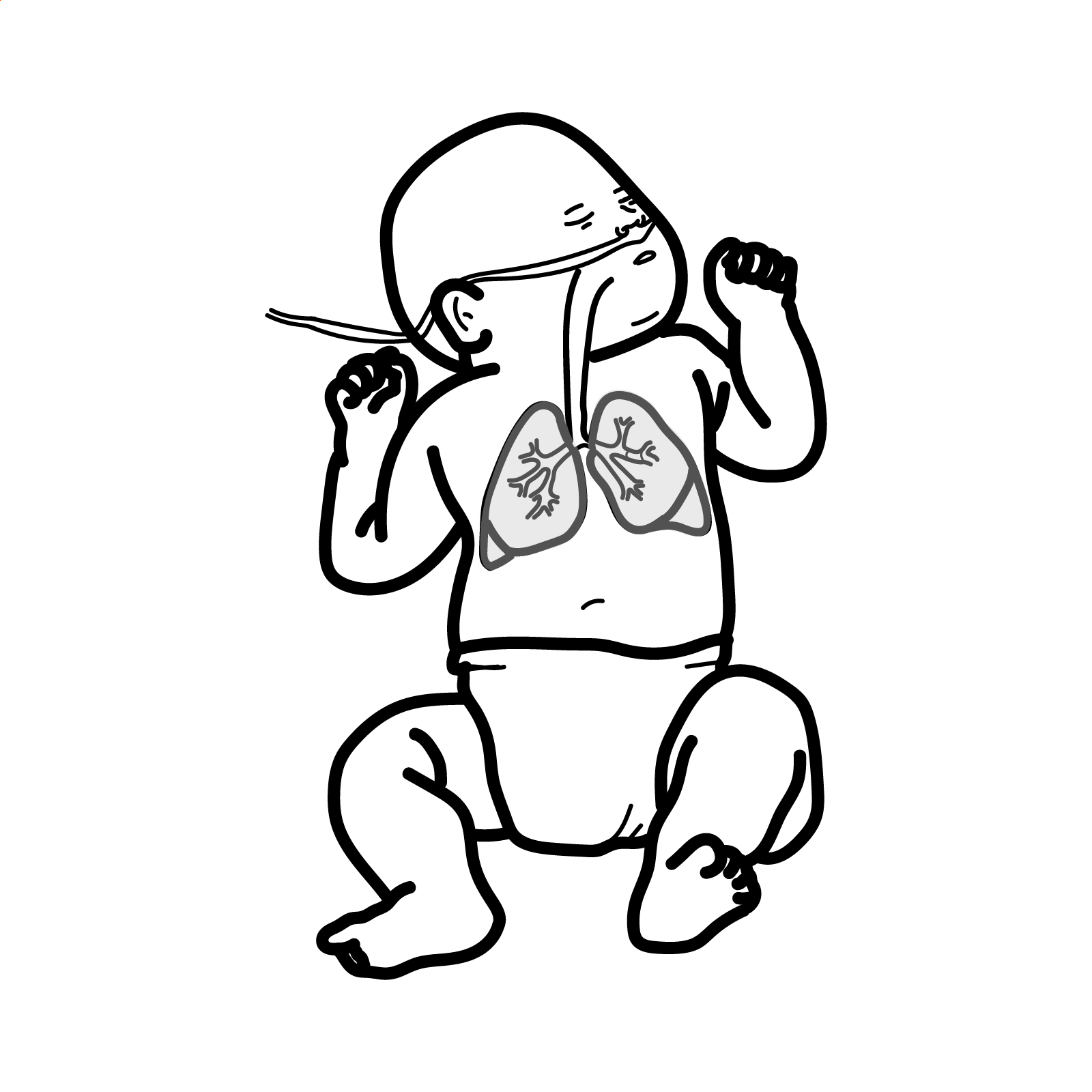
Neonatal respiratory distress syndrome (RDS), previously called hyaline membrane disease, is a disorder caused primarily by pulmonary surfactant deficiency and structural lung immaturity. It occurs mainly in premature infants and presents shortly after birth with tachypnoea, grunting, chest recession and increasing oxygen requirements. Severity increases as gestational age decreases.
Neonatal RDS is different from paediatric acute respiratory distress syndrome, which is an inflammatory lung injury occurring after insults such as pneumonia or sepsis.
>90% risk of RDS if fetus is born 28 weeks or less.
Definition
Surfactant: phospholipid–protein substance produced by type II pneumocytes that reduces alveolar surface tension.
Functional residual capacity: volume of air remaining in the lungs after passive expiration.
Continuous positive airway pressure—CPAP: non-invasive support that maintains positive airway pressure throughout the respiratory cycle.
Less-invasive surfactant administration—LISA: delivery of surfactant through a thin tracheal catheter while the infant continues breathing on CPAP.
Bronchopulmonary dysplasia—BPD: chronic lung disease of prematurity associated with prolonged oxygen or respiratory-support requirements.
What to notice
Detailed image description
Anatomy & Physiology
Transition from foetal life to neonatal life requires:
- Fetal lungs are filled with fluid from pulmonary secretions
- Most fluid moves out into amniotic fluid with breathing movements
- Labour onset causes release of catecholamines, fluid in lungs is largely squeezed out with passage of the chest through the birth canal
- With normal chest recoil, the infant’s lungs fill with air.
- Surfactant is released from type II pneumocytes to lower surface tension and establish residual lung volume
- Onset of regular breathing
- ↑pulmonary blood flow as a result of increased systemic vascular resistance and ↓pulmonary vascular resistance
Infants born by caesarean section are more likely to have retained lung fluid.
Pulmonary Surfactant
- Pulmonary surfactant is a complex mixture of lipids and proteins
- The main function of surfactant is to decrease surface tension of the alveoli allowing for maintenance of functional residual capacity preventing atelectasis and lung injury.
- The surfactant proteins also contribute to natural immunologic defences and assist with the spreading of surfactant throughout the alveoli and the recycling of surfactant between cells and the airspaces.
Aetiology & Risk Factors
Major risk factor
- Prematurity, particularly birth before 32 weeks
- Risk increases substantially with decreasing gestational age.
Other risk factors
- Absence of antenatal corticosteroid exposure
- Maternal diabetes
- Caesarean birth without labour
- Male sex
- Multiple pregnancy, particularly the second-born twin
- Perinatal hypoxia or asphyxia
- Hypothermia
- Previous sibling affected by neonatal RDS
- Genetic abnormalities affecting surfactant production or function
- Very rapid delivery before normal physiological transition has occurred
Maternal diabetes can delay fetal surfactant maturation through fetal hyperinsulinaemia. Antenatal corticosteroids accelerate lung maturation and reduce RDS, intraventricular haemorrhage and neonatal mortality when preterm birth is anticipated
Neonatal RDS is primarily caused by surfactant deficiency in premature lungs.
Pathophysiology
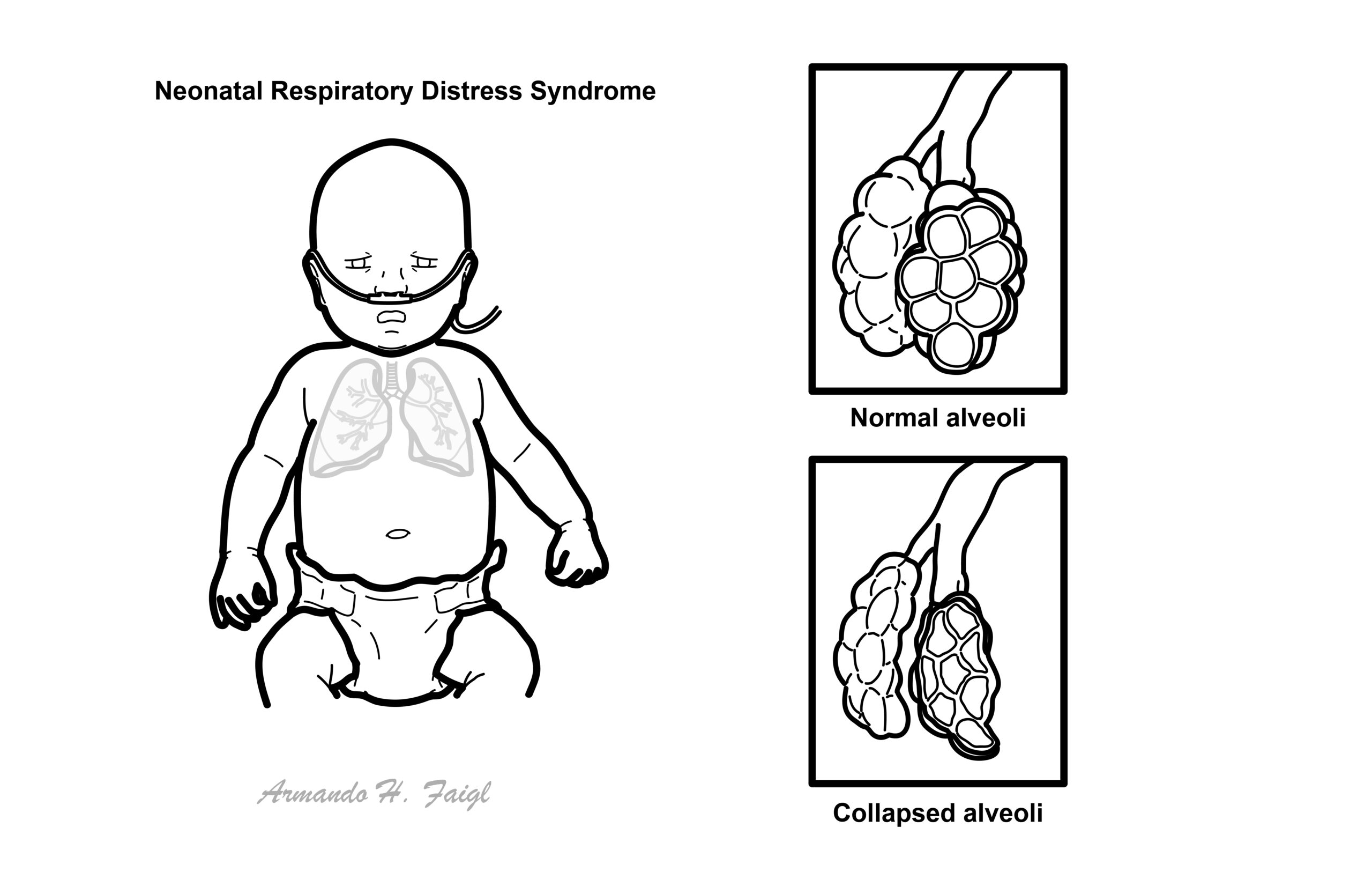
- The primary cause of RDS is deficiency of pulmonary surfactant, which is developmentally regulated.
- The fetal lung is filled with fluid and provides no respiratory function until birth
- Fetal alveolar development is occurring in utero. Week 20 of gestation, surfactant is slowly expressed.
- Pre-term delivery → immature lungs/surfactant → RDS
- Gene mutations for proteins in surfactant → surfactant deficiency and/or dysfunction → RDS
- Insufficient surfactant stores leads to:
- Decreased compliance of the lungs
- Inability of the infant to maintain air in the lung at the end of expiration resulting in a tiny area of collapsed lung (microatelectasis).
- ↑ effort on inhalation with microatelectasis → Ventilation and perfusion mismatch → Hypoxaemia, Tachypnoea, laboured breathing
- Lung inflammation and respiratory epithelial injury, which may result in pulmonary oedema and increased airway resistance
Surfactant is a surface-active lipoprotein complex comprised of a mixture of phospholipids (90%), proteins (10%), and a small portion of other neutral lipids. The main function of surfactant is to decrease surface tension of the alveoli allowing for maintenance of functional residual capacity preventing atelectasis and lung injury. The surfactant proteins also contribute to natural immunologic defenses and assist with the spreading of surfactant throughout the alveoli and the recycling of surfactant between cells and the airspaces.
Clinical Manifestation
Usually occur at or soon after birth (first 4-6 hours)
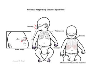
- Tachypnoea
- Laboured breathing
- Cyanosis
- Grunting
- Nasal flaring
- Intercostal and subcostal retraction
Tachypnoea, grunting, recession and rising oxygen requirements beginning shortly after birth are typical.
Diagnosis
Diagnosis is based on:
- Prematurity or another relevant risk factor
- Respiratory distress beginning shortly after birth
- Increasing oxygen or respiratory-support requirements
- Compatible chest imaging
- Exclusion of other causes of neonatal respiratory distress
Treatment should not be delayed until classical radiographic changes appear, because early CPAP and surfactant can prevent progression to severe radiographic disease.
- Pulse oximetry
- Blood gas
- Chest X-ray
- Classical findings include:
- Reduced lung volumes
- Diffuse fine reticulogranular or ground-glass appearance
- Air bronchograms
- Widespread atelectasis
- Classical findings include:
Imaging also helps identify air leaks, pulmonary haemorrhage, pneumonia or incorrect tube and line positioning. Lung ultrasound is increasingly used to assess lung aeration and support decisions about surfactant treatment.
- FBC
- Blood cultures and inflammatory markers if sepsis is suspected
- Blood glucose
- Electrolytes, calcium and renal function
- Haematocrit
- Echocardiography if congenital heart disease, patent ductus arteriosus or pulmonary hypertension is suspected
Treatment
- Resuscitation
- Oxygen and respiratory support
- Use the lowest FiO₂ required to maintain target oxygen saturation.
- Avoid excessive oxygen because of retinopathy of prematurity and lung injury.
- CPAP is the preferred initial respiratory support for spontaneously breathing premature infants.
- Surfactant therapy (decreases alveolar surface tension, improves lung compliance, and maintains functional residual capacity)
- +/- Intubation: mechanical ventilation
- +/- IV antibiotics
- Prophylactic caffeine is recommended for infants born before 32 weeks in current European guidance.
More info on Neonatal resuscitation
CPAP is first-line respiratory support for a spontaneously breathing premature infant.
Surfactant administration should be considered in any premature intubated infant with a presumed diagnosis of RDS.
| Indications for intubations |
| Failure of CPAP or non-invasive ventilation |
| Severe respiratory acidosis |
| Severe recurrent apnoea |
| Persistent hypoxaemia |
| Need for intubation during resuscitation |
| Exhaustion or poor respiratory effort |
Prevention
- Transfer pregnancies at high risk of very premature delivery to a tertiary perinatal centre where possible.
- Give a single course of antenatal corticosteroids when preterm birth before 34 weeks is anticipated and active neonatal care is planned.
- The greatest benefit occurs when birth follows more than 24 hours but less than approximately 7–10 days after treatment.
- Maternal magnesium sulfate before anticipated birth below 32 weeks reduces the risk of cerebral palsy but does not directly treat RDS.
Complications & Prognosis
- Dependent on general appearance at birth and severity of underlying lung disease. Long-term risks of chronic lung disease.
- Classically worsens for 2-3days, then diuresis occurs recovery over 3-4days. Subsequent improvement is coincident with increased production of endogenous surfactant with resolution of symptoms by one week of age.
- Prolonged ventilation
- Increased risk of bronchopulmonary dysplasia
- Sudden deterioration
- Pneumothorax
- Endotacheal tube blockage or displacement
- Mechanical failure with the ventilator
- Increase in the severity of the underlying lung disease
- Massive intraventricular haemorrhage
- Necrotizing enterocolitis
- Patent ductus arteriosus
If baby suddenly deteriorates despite initially improving think of the possible causes of sudden deterioration.
References
- Sweet DG, Carnielli VP, Greisen G, Hallman M, Klebermass-Schrehof K, Ozek E, et al. European consensus guidelines on the management of respiratory distress syndrome: 2025. Neonatology. 2026;1–26. doi:10.1159/000551062.
- Queensland Clinical Guidelines. Respiratory distress and continuous positive airway pressure. Brisbane: Queensland Health; 2024.
- Sweet DG, Carnielli VP, Greisen G, Hallman M, Klebermass-Schrehof K, Ozek E, et al. European consensus guidelines on the management of respiratory distress syndrome: 2022 update. Neonatology. 2023;120(1):3–23. doi:10.1159/000528914.
- Polin RA, Carlo WA; Committee on Fetus and Newborn. Surfactant replacement therapy for preterm and term neonates with respiratory distress. Pediatrics. 2014;133(1):156–163. doi:10.1542/peds.2013-3443.
- World Health Organization. WHO recommendations for care of the preterm or low-birth-weight infant. Geneva: World Health Organization; 2022.





Members only discussions coming soon…