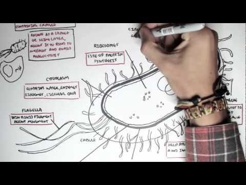
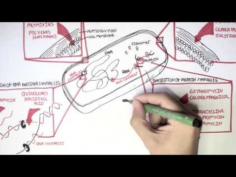
0:00 Hello, in this video, we're going to talk about bacteria and bacteria structure 0:10 . 0:10 Bacteria is what's called a unicellular prokaryotic organism. 0:14 So unicellular means each bacteria is a single living cell. 0:18 Prokaryotic means that they lack a nucleus and membrane-bound organelles. 0:24 Unlike viruses, bacteria are metabolically active and capable of independent 0:29 replication. 0:30 A useful analogy is to think of bacteria as really self-contained factories. 0:34 They can generate energy, their own energy, they can synthesize proteins, they 0:38 can replicate 0:38 their DNA, and they can divide without needing to enter another cell, like 0:44 viruses. 0:45 We will look at the different structures of the bacteria and use examples of 0:48 some bacteria 0:49 that cause disease in humans. 0:51 Now here I am drawing a bacteria, but the bacteria is a rod-shaped and it's 0:57 just meant 0:57 to represent a typical bacteria. 1:00 But again, all bacteria are different, we're just using this diagram to 1:04 illustrate all 1:06 the important structures that bacteria may have. 1:13 Let's begin by talking about the bacterial cell envelope. 1:18 The cell envelope refers to the layers that surround the bacterial cytoplasm. 1:23 It consists of two things, the cell membrane and in most bacteria, a cell wall. 1:29 The cell membrane is really a phospholipid bilayer that regulates transports, 1:36 nutrients 1:36 and waste. 1:38 It maintains the electrochemical gradient and plays a role in energy production 1:43 . 1:43 You can think of these as controlled security gate around the bacteria. 1:54 The cell wall lies outside the membrane and provides structural rigidity, 2:02 protection specifically 2:04 from osmotic lysis, so basically dying due to pressure. 2:12 It is composed of important structures called peptidoglycans, a rigid mesh made 2:18 of sugars 2:19 and amino acids that is really unique to bacteria. 2:24 This uniqueness explains why the cell wall is a major antibiotic target. 2:31 There are some clinical relevance, Streptococcus pyogenes is a bacteria that 2:36 relies on its 2:36 cell wall. 2:38 This particular bacteria is highly susceptible to penicillin because the cell 2:43 wall is. 2:44 Staphylococcus aureus uniqueness is that it can alter these structures called 2:49 penicillin 2:50 binding proteins and so they can become resistant to standard penicillin and 2:55 can develop what's 2:56 called methicillin resistance. 2:59 So this is MRSA. 3:04 Because we talked about the cell wall, this is a good segue to talk about gram 3:07 positive 3:08 versus gram negative bacteria. 3:11 Bacteria are commonly classified based on the cell wall architecture, which 3:16 determines 3:16 their appearance on the gram stain and their antibiotic susceptibility. 3:22 So really, bacteria can be divided into two broad groups. 3:27 Now gram positive bacteria typically have a thick peptidoglycan layer, no outer 3:33 membrane. 3:34 They retain this crystal violet stain and so appear purple. 3:39 Examples of a gram positive bacteria include Staphylococcus aureus and strep 3:43 pneumoniae. 3:44 These organisms are generally more susceptible to beta-lactam antibiotics, so 3:49 like penicillin. 3:52 Gram negative bacteria, on the other hand, have a thin peptidoglycan layer. 3:56 They have an outer membrane. 3:59 They do not retain crystal violet and so they appear pink on the gram stain. 4:06 Examples to remember include E. coli and Niceria meningitis. 4:12 The outer membrane of gram negative bacteria acts as an additional permeability 4:19 barrier, 4:19 making these organisms harder to treat. 4:24 Now the outer membrane, which we already spoke about, which is made up of a 4:30 phospholipid 4:31 bilayer, contains also these things called lipopolysaccharides, some bacteria. 4:37 The outer membrane specifically of the gram negative bacteria contains the lip 4:42 opolysaccharides 4:44 LPS. 4:45 So LPS is found in gram negative bacteria. 4:48 For different lipopolysaccharides, lipid A is an example and it's an endotoxin, 4:53 triggers 4:54 the innate immune activation. 4:56 There's the O antigen, which allows for antigenic variability and immune 5:02 evasion, so running 5:03 away or evading the immune system of the human, for example. 5:09 So E. coli or Klebsiella bacteria can cause septic shock due to lipopolysacchar 5:15 ide driven 5:16 cytokine release because lipopolysaccharides triggers a really strong immune 5:23 activation 5:24 in the human and so can cause quite a significant shock response. 5:29 And this explains why gram negative sepsis is often severe. 5:41 The cytoplasm of the bacteria contains enzymes, metabolites and genetic 5:47 material. 5:47 Because bacteria are prokaryotic organisms, they lack the nucleus and so the 5:53 DNA is located 5:54 in what's called the nucleoid region. 5:57 Now the genetic material, bacteria can either have a single, circular or double 6:03 -stranded 6:04 DNA chromosomes. 6:07 They could also have something called plasmids, which are really extra DNA. 6:13 Plasmid itself are small, they could be circular. 6:18 They are not essential for survival but can confer advantages such as the 6:24 actual genetic 6:25 material can have some form of gene for antibiotic resistance or a gene for 6:34 toxin production. 6:36 Ribosomes are really like a upgrade for certain bacteria. 6:40 Examples of this include the ESBL gene in E. coli, which really causes quite 6:47 significant 6:49 antibiotic resistance. 6:52 And carbapenin resistance are often found in plasmids, particularly in Klebsie 6:59 lla pneumonia. 7:01 Next we talk about ribosomes and protein synthesis. 7:04 The ribosomes are structures that help make proteins. 7:08 Proteins are important for bacterial survival. 7:13 Bacteria ribosomes are what's called 70 S ribosomes, they're composed of 30 S 7:18 subunits 7:18 and 50 S subunits. 7:20 They are structurally distinct from human 80 S ribosomes, allowing selective 7:25 antibiotic 7:26 targeting. 7:29 Ribosomes include macrolides, which inhibit the 50 subunit ribosomes, which are 7:34 very 7:35 effective for microplasmid pneumoniae, or amino glycosides, which target the 30 7:41 S subunits, 7:42 which are effective for gram-negative aerobes, for example. 7:48 Some bacteria possess additional external structures that enhance survival and 7:53 pathogenicity. 7:55 A capsule is a polysaccharide layer, external to the cell wall, that inhibits 8:00 phagocytosis 8:02 and increases virulence. 8:04 Examples of organisms that have this include streptococcus pneumoniae, which 8:09 has a capsule. 8:10 This allows it to cause invasive disease, but is also a very good vaccine 8:15 target. 8:16 Hemophilus influenza type B has also a capsule. 8:21 Legilla are found in some bacteria, and are these long, whip-like structures 8:25 responsible 8:25 for motility. 8:27 Salmonella uses a flagella, for example, to aid in gut invasion. 8:35 Pilli or fimbriae are short, hair-like projections found in some bacteria 8:40 involved in adherence 8:41 to host cells, or tissues, or attaching, as well as they are important for 8:46 horizontal 8:47 gene transfer. 8:48 An example is niceragonaria, which uses pilli to attach to the mucosal tissue, 8:56 and also 8:57 to allow it to really evade the immune system. 9:03 Finally, bacteria replicate through what's called binary fission, a rapid 9:11 process producing 9:14 two identical daughter cells. 9:17 Under optimal conditions, E. coli, for example, can divide every 20 minutes. 9:21 This rapid replication explains acute infection onset and rapid development of 9:26 antibiotic 9:27 resistance. 9:30 I hope that this overview of bacteria structure makes sense. 9:35 We talked about the bacterial cell envelope, which is made up of the cell 9:39 membrane and 9:39 cell wall. 9:40 But then we talked about the bacterial capsule, which is like an extra layer. 9:44 We talked about the flagella for motility, the pillow or the fimbriae for 9:49 attachment, 9:50 then the lipopolysaccharides on the membrane, which can be very toxic and 9:55 really triggered 9:56 immune response. 9:58 And very importantly, the cell wall, which allows us to differentiate between 10:02 gram-positive 10:03 and gram-negative bacteria. 10:04 Thank you for watching. 10:10 You