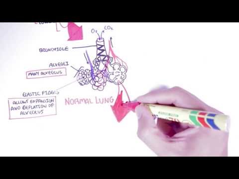
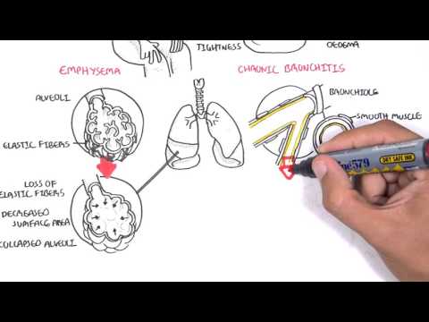
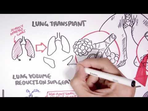
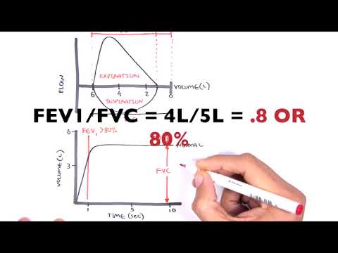
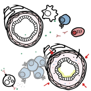
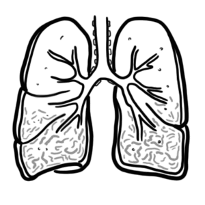
0:00 In this video we're going to look at the pathophysiology of emphysema. 0:05 Now emphysema is characterized by the destruction of the alveoli through the 0:10 breakdown of elastic 0:11 fibers by proteases secreted by immune cells. 0:16 So let's have a look at the pathophysiology. 0:19 So here we have a man and he has lungs with emphysema. 0:25 He is a heavy smoker which is the main cause of emphysema. 0:31 So let's zoom into his lungs. 0:33 We can see that his alveoli are affected, severely affected. 0:40 Here we see destruction of the alveoli. 0:43 It's walls and elastic fibers mainly by proteases which are chemicals secreted 0:50 by immune cells. 0:55 So let us begin first by looking at a normal alveoli and see how it progresses 1:05 to an alveoli 1:06 of emphysema. 1:10 So here we have the bronchioles, the alveoli, made up of many alveolus. 1:16 And here we have the elastic fibers which are found on the alveoli and on the 1:21 bronchioles. 1:22 The elastic fibers allow recoiling to occur during inhalation and exhalation of 1:33 gases. 1:33 So let us zoom into only one alveolus. 1:38 Here we can find, here's one alveolus, here we can find elastic fibers, epit 1:43 helial cells 1:44 and surfactant cells. 1:47 In the alveolus we can find alveolar macrophages that has a role in cleaning up 1:53 the alveolus 1:54 and protecting it during infections or against infections. 1:58 Here we have the blood supply to each of the alveoluses essentially and they 2:04 contain red 2:04 blood cells. 2:06 Essentially oxygen will be exchanged for carbon dioxide here if you remember. 2:13 Now what essentially happens is that normally the alveolus will secrete anti- 2:22 proteases which 2:24 will essentially protect it against protease activity. 2:29 So usually there's a balance between anti-protease and protease. 2:35 protease essentially cause destruction or damage and anti-protease will prevent 2:40 this 2:40 from occurring. 2:43 Emphysema is caused by the imbalance between anti-protease and protease 2:49 activity. 2:50 So if there's more protease activity there will be more damage, just remember 2:57 that. 2:58 So let's draw another diagram. 3:01 Now in most people the slow process of destruction of the alveoli, the elastic 3:08 fibres is initiated 3:09 through the inhalation of toxins such as cigarettes or from air pollution. 3:15 So cigarettes and air pollution contain oxidative toxins which if inhaled in 3:20 considerable amounts 3:22 will have devastating consequences. 3:25 These reactive oxidative toxins will essentially initiate an immune response 3:30 and inflammatory 3:32 response. 3:34 Remember that alveolar macrophages are normally found within the alveolus. 3:39 So exposure to these toxins from cigarettes will cause these macrophages to 3:44 begin secreting 3:45 many inflammatory mediators, inflammatory cytokines, mainly interleukin-6, 3:51 interleukin-8, interleukin-1, 3:54 TNF alpha and leukotrine B4. 3:59 Now what they will do is essentially these chemicals will enhance the immune 4:04 response. 4:05 For example interleukin-1 and TNF alpha can recruit neutrophils into the area, 4:11 a process 4:12 known as chemotaxis. 4:16 So here we can see more neutrophils coming into the area. 4:19 The neutrophils will actually begin secreting proteases, mainly elastase which 4:25 will begin 4:26 destroying the elastic fibres. 4:29 Not only this but the macrophage also secreats other chemicals such as metallop 4:35 roteases which 4:36 is another type of protease which causes damage to the tissues there. 4:41 So there's all these chemicals being secreted around this area from the immune 4:45 cells, the 4:46 neutrophils in the macrophages which will essentially aggravate the area 4:50 causing damage 4:50 to the surrounding tissue. 4:58 So again the neutrophils and macrophages are the main producers of proteases. 5:04 The main proteases remember is elastase and matrix metalloprotease. 5:10 So the elastase will cause destruction of the elastic fibres like so. 5:16 And the proteases will also damage the tissues, especially the metalloproteases 5:23 . 5:24 So this whole response is sort of continuing through the inhalation of toxins. 5:30 So the neutrophils are secreting elastase, the macrophages are still secreting 5:34 cytokines 5:35 which will recruit more and more immune cells. 5:39 As well we not only see neutrophils and macrophages, there will also be T lymph 5:45 ocytes coming into 5:46 the area, the T lymphocytes will also destroy tissue possibly through T cell 5:52 mediated apoptosis. 5:54 So the T cells will tell the tissues to basically kill itself. 6:01 After some time we also see collagen deposition, possible fibrosis. 6:08 Now from this diagram we can see that in an emphysema type situation we see a 6:14 lot of protease 6:16 activity. 6:18 And so we can see that there is an imbalance between protease activity and 6:22 antiprotease 6:23 activity, being protease having a much more substantial role. 6:32 And this, all this is the result of inhalation of toxins such as from cigarette 6:37 which will 6:38 create this sort of immune response. 6:43 But remember this diagram I am drawing up now is not, normally emphysema does 6:48 not occur 6:49 step by step as what I'm showing, so you got to keep this in mind. 6:56 Now in the lungs normally one of the main antiprotease in the lungs is alpha 7:01 antitripsin, 7:03 however some people suffer from alpha antitripsin deficiency, therefore they 7:09 are more susceptible 7:10 to get emphysema, because there is no antiprotease activity, I hope this makes 7:18 sense. 7:19 So as we can see through this diagram, the pathophysiology of emphysema is the 7:24 result 7:24 of the imbalance between protease and antiprotease activity with it being more 7:36 protease activity. 7:39 Now let's look at another thing called air trapping, which occurs a lot in em 7:45 physema. 7:46 Air trapping is essentially when we breathe air in and it becomes trapped and 7:51 it's very 7:52 difficult to breathe out, essentially exhale. 7:56 To understand the mechanism behind this we need to see the normal lungs first. 8:02 So here we have the normal alveolus that will expand during inhalation of air. 8:09 The elastic fibers will allow the alveolus to expand when we breathe in and 8:16 then during 8:17 exhalation the alveolus will essentially deflate, this is because of the recoil 8:23 of the elastic 8:24 fibers, big to small. 8:27 And so during exhalation air will flow out easily, hope that makes sense. 8:36 Now let's look at lungs with emphysema, so during inhalation the alveolus is 8:42 able to 8:43 expand with force, and this has to be with force because there is no elastic 8:49 fibers, 8:50 because they are destroyed remember. 8:53 Also, the loss of elasticity in the bronchioles will cause the bronchioles to 8:58 become more 8:59 narrower during inhalation. 9:02 So then during exhalation the narrowing of the bronchiol and the absence of a 9:08 recoil 9:08 system because of the destruction of the elastic fibers will cause air to be 9:14 trapped, making 9:15 it hard to breathe out. 9:21 So I hope that makes sense, so to summarize, in a person with emphysema air can 9:26 be trapped 9:27 within the alveoli. 9:29 The destruction of the elastic fibers will reduce recoil of airways, and so it 9:34 is difficult 9:35 to breathe out. 9:42 I hope you enjoyed this video on the pathophysiology of emphysema, thank you 9:45 for watching, the next 9:46 video will look at the treatments.