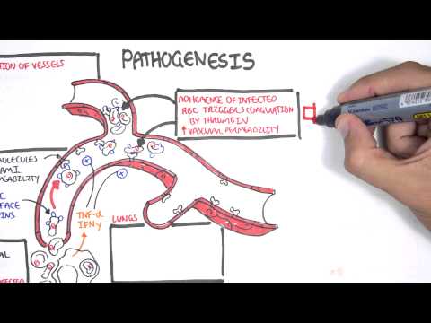
0:00 Pneumocystis urevechae causes pneumocystis pneumonia in immunocompromised 0:14 patients, so 0:15 basically patients who have a weak immune system. 0:20 These patients include people who have HIV, or those on long-term immunosupp 0:26 ressants, 0:27 such as prednisone. 0:30 Pneumocystis urevechae can cause mild to very severe life-threatening 0:34 respiratory infections. 0:36 Pneumocystis is actually a fungus that was first found to infect humans back in 0:40 World 0:41 War II in infants and children specifically who were malnourished and unwell. 0:47 At the time, Pneumocystis was thought to be a protozoan species because it had 0:52 many 0:52 forms that made it look like it was a protozoan under a microscope. 0:58 However, genomic sequences found that pneumocystis is much more closely related 1:03 to a fungus, 1:04 and so it was reclassified as a fungus and not a protozoan. 1:11 Pneumocystis urevechae is an interesting organism. 1:18 It loves the lungs. 1:19 They stay and live in the alveoli, and for the most part, do not cause any 1:23 problems in 1:24 people with a normal immune system. 1:28 There are three developmental stages of pneumocystis. 1:33 The trophic form, the sporeocyte, and the spores. 1:38 These stages all exist within the alveoli. 1:43 The trophic forms can replicate through binary fission into two near-identical 1:49 trophic forms, 1:51 and this is called asexual reproduction. 1:54 Alternatively, the trophic forms can undergo sexual reproduction, and here the 2:00 trophic 2:00 forms they mate, divide, and form many spores inside a closed wall. 2:07 This structure is called a sporosite. 2:11 A sporosite is a cell that contains many spores. 2:16 The sporosite will eventually rupture and release all the spores it contains. 2:22 Now these spores will then eventually become the trophic form of the organism 2:29 once again. 2:30 The trophic form of pneumocystis can then enter either the asexual or sexual 2:38 reproductive 2:38 cycle again. 2:42 Unlike most fungi, pneumocystis lacks ergosterol in its cell wall, therefore 2:48 commonly used 2:50 antifungal medications that actually target ergosterol synthesis such as the az 2:56 ole and 2:57 the amphotericin B, they do not work against pneumocystis. 3:02 The cell wall of pneumocystis contains beta-D-glucan, which is important 3:08 because beta-D-glucan 3:09 is a blood test that can be ordered. 3:13 Pneumocystis organisms carry a variety of surface glycoproteins, these proteins 3:19 on its 3:20 surface which are actually unique to different pneumocystis species. 3:26 So for example, there are many pneumocystis species, and each of these species 3:32 actually 3:33 infects only specific mammals. 3:36 So for humans, it's pneumocystis uravechae, which infects only humans. 3:43 And then you have pneumocystis carini, which only infects rats. 3:48 And then pneumocystis marina, which only infects mice. 3:54 Back in the day, pneumocystis carini was thought to be the one that infected 3:58 humans, but that's 3:59 not the case. 4:01 And so PCP is actually an abbreviation for pneumocystis pneumonia and not pneum 4:10 ocystis carini pneumonia. 4:18 So let's talk about the pathophysiology of pneumocystis pneumonia. 4:23 Well, pneumocystis is transmitted by the airborne route. 4:30 In healthy individuals, pneumocystis can colonize the lung and cause no 4:34 problems. 4:35 However, if a patient becomes immunocompromised, meaning they have a weak 4:40 immune system, pneumocystis 4:41 can lead to a terrible lung infection. 4:49 Once in the alveoli and the patient's immune system is down, the trophic form 4:54 of the fungus 4:54 attaches to the alveolar type 1 cells and undergoes proliferation. 5:01 Impaired humoral and T cell mediated immunity contribute to this uncontrolled 5:07 proliferation 5:08 of the fungus. 5:09 And the host immune response results in a production of inflammatory substances 5:14 , cytokines, such 5:15 as TNF alpha and interleukin 1, which contribute to the lung damage. 5:22 The principal histological finding in patients who have pneumocystis pneumonia 5:28 is a foamy 5:29 e-synophilic alveolar exudate. 5:32 There may be high-line membrane for patients, but there is also interstitial 5:37 fibrosis and 5:38 underlying edema as well. 5:44 The risk factor for developing pneumocystis pneumonia is therefore anything 5:48 that can cause 5:49 someone to have a weak immune system. 5:53 HIV was a common cause of pneumocystis pneumonia. 5:57 In fact, the rate of pneumocystis infection dramatically rose with the HIV 6:03 epidemic. 6:05 And this is because people who had HIV back in the time, their immune cells 6:11 were depleted. 6:12 And thanks to the discovery of effective antiretroviral treatment, the rates of 6:16 pneumocystis in patients 6:18 who have HIV have dropped significantly. 6:22 The most significant risk factors for pneumocystis pneumonia in patients 6:27 without HIV are really 6:29 those who take steroids and those with a defect in their cell-mediated immunity 6:37 . 6:38 Other risk factors include hematological malignancies, solid organ malignancies 6:42 , organ transplantation, 6:45 logical diseases, autoimmune diseases, and the use of other immunosuppressive 6:56 drugs. 6:57 The classic presentation of someone with pneumocystis pneumonia can vary, but 7:03 typically progressive 7:04 dyspnea, dry cough, fever, which is often mild, weight loss, and diffuse 7:11 bilateral, 7:12 interstitial infiltrates, ground glass changes, they call it on the x-ray. 7:19 Extra pulmonary manifestation of the fungus is rare because remember, these 7:22 guys, they 7:23 love the lungs. 7:29 Changes to order for anyone with suspected pneumocystis pneumonia is an ABG to 7:34 evaluate 7:35 severity of the hypoxemia. 7:38 Lactate dehydrogenase, LDH, can be ordered. 7:41 And this is actually a good clinical indicator of possible pneumocystis 7:46 pneumonia, specifically 7:48 in HIV patients. 7:51 As mentioned, beta-D glucan, which is the part of the cell wall of the fungus, 7:59 and so if 8:00 you check it in the blood, and if you don't find it, then it is helpful to rule 8:08 presence 8:09 of this fungus out. 8:12 Gold standard to diagnose pneumocystis pneumonia is actually identification of 8:16 the fungus through 8:18 respiratory secretions. 8:20 And this is either from induced sputum specimens or from bronco-alveolar lavage 8:28 , fluid from 8:29 the actual bronchus itself. 8:32 And once you obtain the respiratory specimens, there are many stains that can 8:35 be used to 8:36 help identify trophic forms or the cyst forms of the fungus. 8:41 Finally, PCR can also be used to really pin down the organism. 8:47 A CT chest is fundamental in suspected pneumonia cases, and CT classic findings 8:55 include bilateral 8:57 ground glass changes, such as in this image here. 9:02 You can see ground glass hazy type changes in both lobes of the lung. 9:09 Pyla, or mediasthenolymph adenopathy, is rare in pneumocystis pneumonia. 9:16 Finally, treatment. 9:22 Once the diagnosis of pneumocystis pneumonia is established through 9:26 identification of the 9:27 fungus through microscopy, or PCR, trimethoprim and sulfa methoxazole is used 9:34 first line. 9:36 This is typically known as backtrum. 9:38 There are alternative antimicrobials to use in those who cannot tolerate backtr 9:44 um. 9:44 And this includes klimdamycin and primaquin, dapsone or trimethoprim, and/or at 9:53 ovoquone. 9:54 Interestingly, steroids can be used as an adjunct for treatment in patients 10:00 with severe 10:01 pneumocystis pneumonia, as long as they don't have HIV. 10:05 In patients with HIV, corticosteroids as an adjunct or an addition for 10:11 treatment of severe 10:13 pneumocystis pneumonia is not recommended. 10:17 Prevention of pneumocystis pneumonia is very important. 10:20 And this is done also with trimethoprim and sulfa methoxazole as well. 10:25 Prophylaxis is given to those with a weak immune system, including if someone 10:31 is having 10:32 high dose steroids for a long duration. 10:36 And this also includes other immunosuppressants. 10:40 Prophylaxis is given to those with an immunodeficiency disorder, whether it's 10:44 acquired or congenital. 10:47 And also, prophylaxis is given to those who already had the infection in the 10:53 past. 10:54 And so this is secondary prophylaxis to prevent recurrence. 11:03 So in summary, pneumocystis pneumonia is caused by the organism pneumocystis u 11:08 revechi in immunocompromised 11:12 people, those with a weakened immune response. 11:15 Classically, patients present with a worsening cough, dysnia, and low-grade fe 11:21 vers. 11:22 On X-ray, you have increased markings bilaterally, ground-glass changes. 11:28 Treatment is bacterium. 11:30 So trimethoprim and sulfa methoxazole. 11:34 Finally, prophylaxis is important in those with a who are at risk as well. 11:40 Thank you for watching.