

Acute Urinary Retention
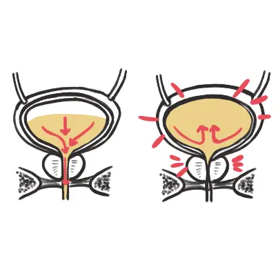
DEFINITION & OVERVIEW
Distinction from Chronic Retention: Acute retention is acutely painful with a tense, tender palpable bladder; Chronic retention is painless, with bladder volumes often > 1000–1500 mL and associated overflow incontinence and bilateral hydronephrosis.
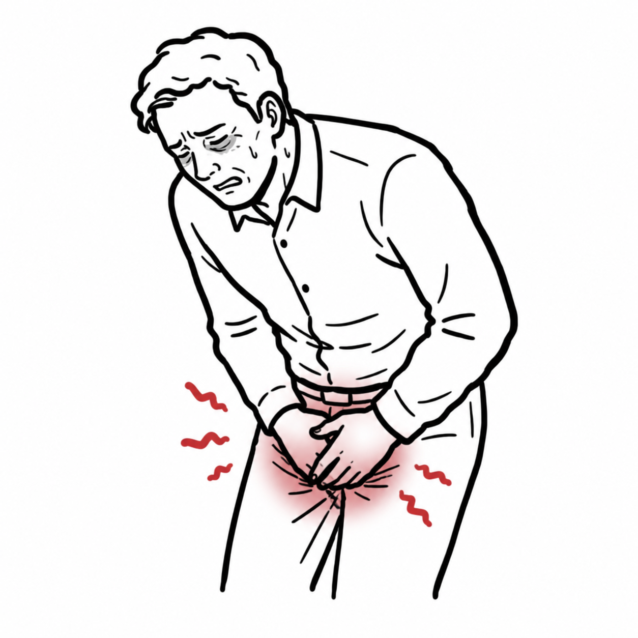
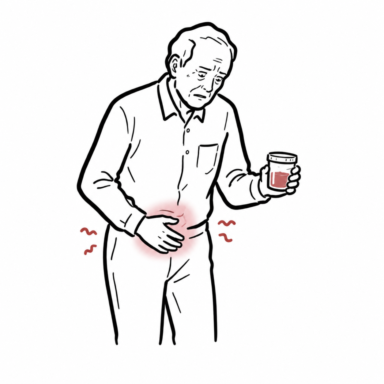
Definition: Acute Urinary Retention (AUR) is the sudden, painful inability to voluntarily void urine despite having a full urinary bladder.
Diagnostic Threshold: Confirmed on bedside ultrasound bladder scan or immediate post-catheterization drainage volume of > 300–500 mL of urine in an uncomfortable patient (often > 800–1000 mL).
Clinical Significance: The most common urological emergency. More than 90% of cases occur in men, primarily driven by underlying Benign Prostatic Hyperplasia (BPH).
Acute urinary retention is agonizingly painful. If a patient presents with painless urinary retention, a large palpable bladder, and urinary dribbling, they have Chronic Urinary Retention with Overflow Incontinence—screen immediately for bilateral hydronephrosis and postrenal acute kidney injury.
APPROACH
- Step 1: Emergency Triage & Red Flag Screening (Rule Out Neurological & Obstructive Crises)
- Spinal Cord / Cauda Equina Red Flags: Saddle anesthesia (loss of perianal sensation), bilateral lower limb radiculopathy/weakness, loss of anal sphincter tone -> Emergency MRI Whole Spine + Neurosurgery/Orthopaedic Spinal consult.
- Systemic Sepsis / Urosepsis: Fever, rigors, tachycardia, hypotension, delirium in elderly -> Blood cultures, broad-spectrum IV antibiotics (e.g., Ampicillin + Gentamicin or Ceftriaxone), and emergency drainage.
- Pelvic / Genital Trauma (Urethral Disruption): Blood at urethral meatus, high-riding prostate, perineal “butterfly” hematoma -> STRICTLY CONTRAINDICATED: Blind urethral catheterization. Perform Ascending Urethrogram (RUG) / Suprapubic catheter.
- Step 2: Immediate Diagnostic & Therapeutic Decompression
- Confirm distension via Bedside Ultrasound Bladder Scan (Volume > 300–500 mL).
- Attempt Urethral Catheterization:
- Standard: 14–16 Fr Foley catheter with copious water-soluble lubricant.
- Difficult/Enlarged Prostate: 16–18 Fr Tiemann / Coudé tip catheter (curved tip pointed anteriorly at 12 o’clock).
- Macroscopic Hematuria / Clots: 20–24 Fr 3-way catheter + manual clot washout.
- If Urethral Catheter Fails: Immediate Urology consult for flexible cystoscopic-assisted catheterization or Suprapubic Catheter (SPC) insertion.
- Step 3: Post-Decompression Clinical Monitoring
- Measure and record the immediate residual volume drained at 10–15 minutes.
- Monitor for Post-Obstructive Diuresis (POD) (Urine output > 200 mL/hr for >= 2 consecutive hours) and transient decompression hematuria.
- Check Serum Creatinine and Electrolytes (K+, Na+) to assess for postrenal AKI.
In acute pelvic trauma, blood at the urethral meatus indicates a urethral tear. Never force a urethral Foley catheter; doing so can convert a partial urethral tear into a complete transection.
DIFFERENTIAL DIAGNOSIS
- Outflow Obstruction (Mechanical / Structural – Most Common):
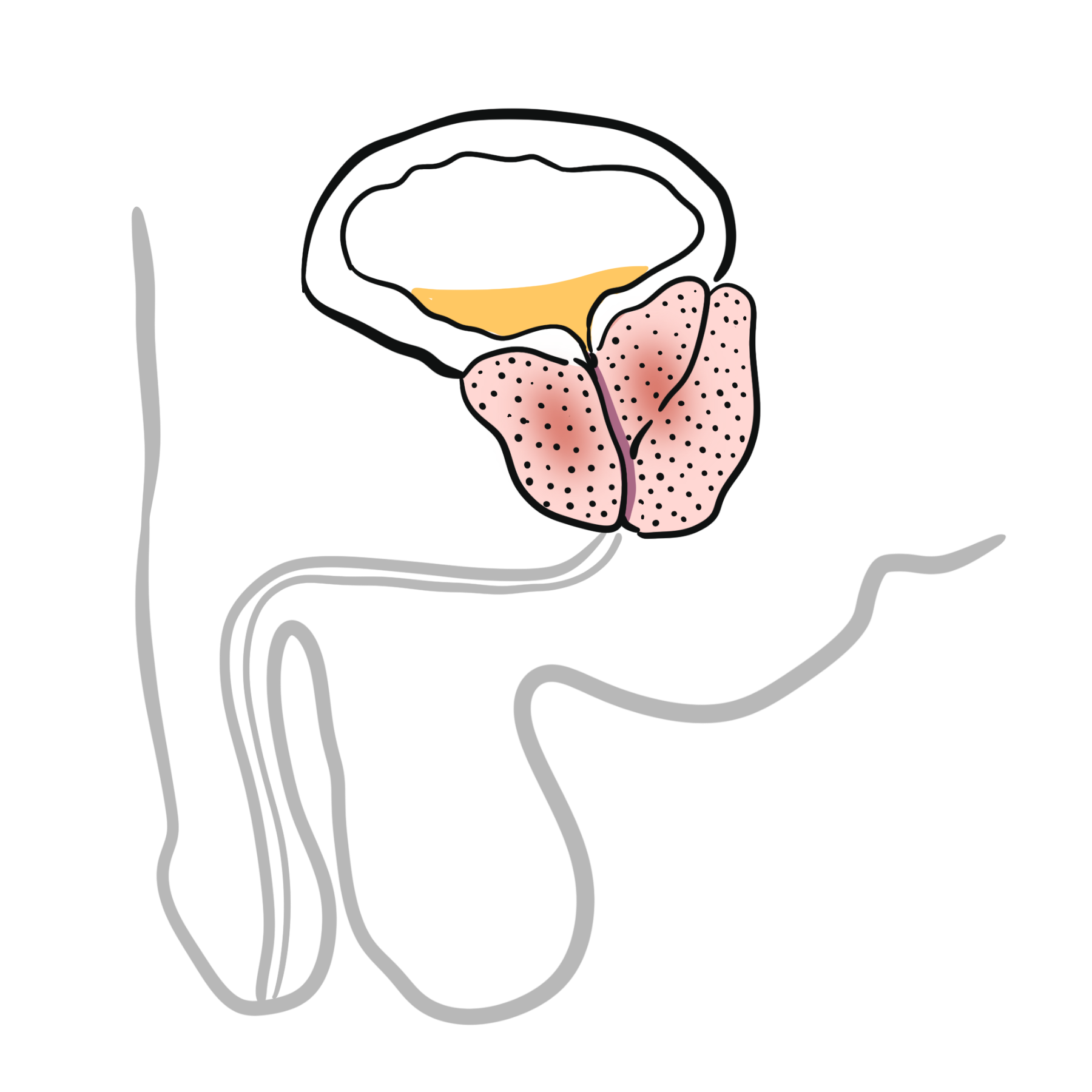
- Benign Prostatic Hyperplasia (BPH – ~70% of Male Cases):
- Distinguishing features: Age > 50 years; preceding lower urinary tract symptoms (LUTS: weak stream, hesitancy, nocturia, frequency); symmetrically enlarged, smooth, non-tender prostate on DRE.
- Prostate Adenocarcinoma:
- Distinguishing features: Hard, nodular, asymmetrical prostate on DRE; elevated age-adjusted PSA, constitutional symptoms, bone pain.
- Urethral Stricture Disease:
- Distinguishing features: Younger men with history of urethral trauma (straddle injury), previous instrumentation/catheterization, or gonococcal urethritis.
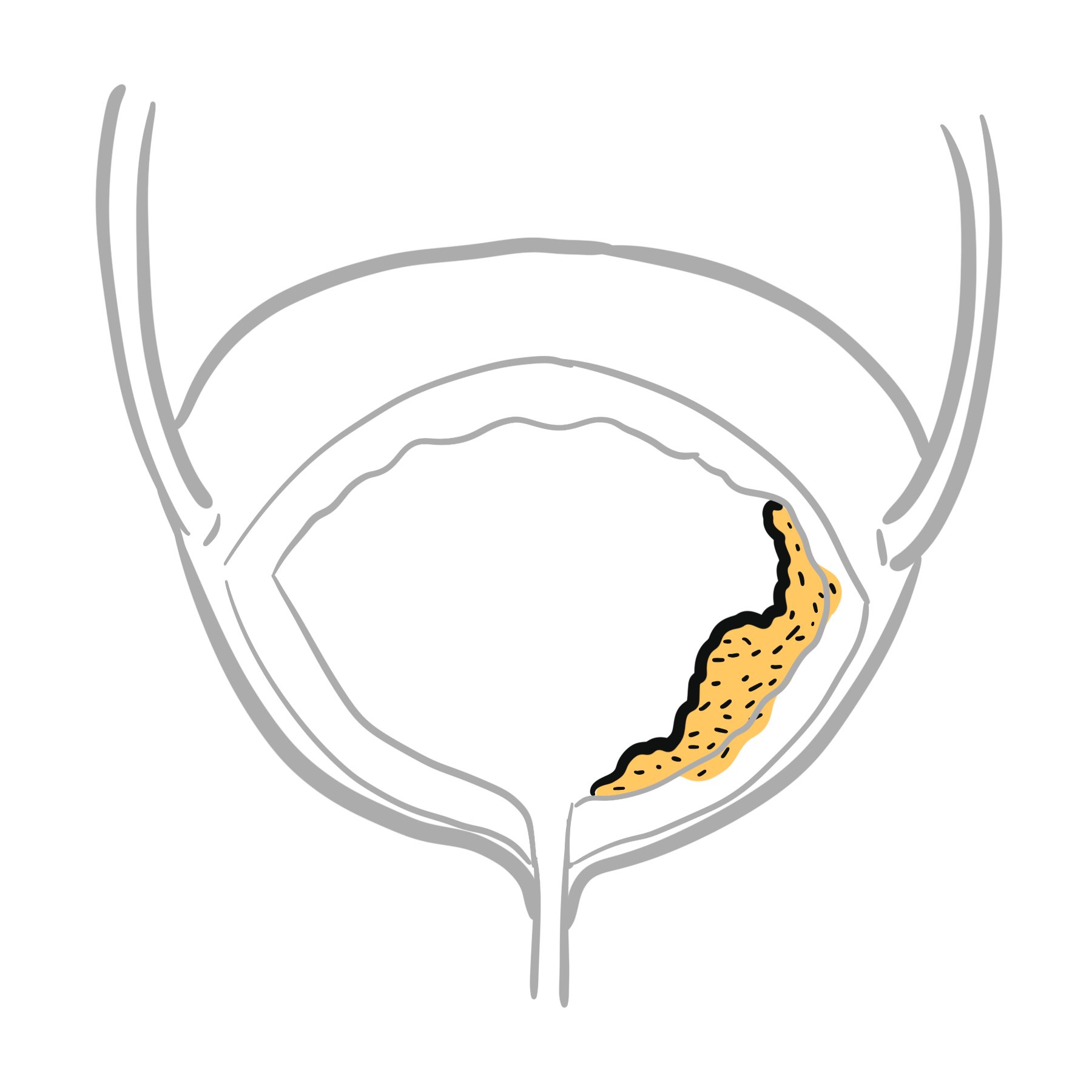
- Clot Retention (Hemorrhagic Cystitis, Bladder/Prostate Tumor):
- Distinguishing features: Gross hematuria preceding retention; catheter aspirates organized dark red clots.
- Incarcerated Gravid Uterus (Females, Weeks 12–16 of Pregnancy):
- Distinguishing features: Retroverted uterus becomes trapped in the pelvic hollow beneath the sacral promontory, compressing the urethra.
- Benign Prostatic Hyperplasia (BPH – ~70% of Male Cases):
- Infectious & Inflammatory Causes:
- Acute Bacterial Prostatitis:
- Distinguishing features: High fever, pelvic/perineal pain, dysuria, pyuria; exquisitely tender, hot, boggy prostate on gentle DRE (avoid vigorous prostatic massage due to bacteremia risk).
- Severe Cystitis / Urethritis / Genital Herpes (HSV):
- Distinguishing features: Severe dysuria and reflex detrusor inhibition due to vulvar/urethral ulceration.
- Acute Bacterial Prostatitis:
- Neurological & Pharmacological Causes:
- Cauda Equina Syndrome / Acute Cord Compression:
- Distinguishing features: Painless or paradoxical retention with overflow, bilateral sciatica, motor weakness, saddle anesthesia, absent bulbocavernosus reflex.
- Medication-Induced Urinary Retention (Very Common Trigger):
- Offending Agents: Anticholinergics (Atropine, Oxybutynin), Antihistamines, Tricyclic Antidepressants (Amitriptyline), Sympathomimetics (Pseudoephedrine), Opioids (inhibit detrusor contractility), Calcium Channel Blockers.
- Cauda Equina Syndrome / Acute Cord Compression:
In acute bacterial prostatitis, do NOT perform vigorous prostatic massage or repeat rough rectal examinations—this can precipitate bacteremia and uroseptic shock.
INVESTIGATIONS
- First-Line / Bedside Diagnostics:
- Bedside Ultrasound Bladder Scan: Immediate non-invasive quantification of urinary volume (confirms retention vs. anuria from renal failure/shock).
- Digital Rectal Examination (DRE): Evaluates prostate size/contour/tenderness, anal sphincter tone, and fecal impaction.
- Focused Neurological Exam: Perianal sensation (S2–S4), lower extremity motor strength, deep tendon reflexes.
- Urinalysis & Mid-Stream Urine Culture: Check for leukocyte esterase, nitrites, microscopic hematuria, and bacterial pathogens.
- Serum Biochemistry: Urea, Creatinine, Electrolytes (evaluates postrenal AKI and hyperkalemia).
- Targeted / Specialized Workup:
- Renal Tract Ultrasound: Evaluates for bilateral hydronephrosis, renal cortical thinning, bladder diverticula, or calculi.
- MRI Whole Spine: Mandatory if saddle anesthesia, motor deficits, or spinal cord compression is suspected.
- Serum Prostate-Specific Antigen (PSA): Delay formal PSA testing for >= 2–4 weeks post-AUR and catheterization, as acute distension, catheter trauma, and urinary infection cause transient marked false elevations in PSA.
- Flexible Cystourethroscopy / Ascending Urethrogram: Evaluates urethral strictures, bladder neck contractures, or bladder masses once the acute phase has settled.
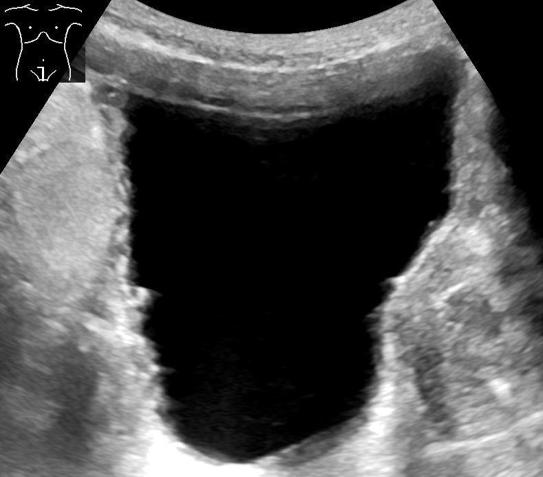
Ultrasound showing a distended, trabeculated urinary bladder. Image: Mikael Häggström, CC0, via Wikimedia Commons.
What to notice
Detailed image description
Source: Wikimedia Commons asset page. Creator: Mikael Häggström. Licence: CC0 1.0 Universal. Imported without modification for the Acute Urinary Retention article.
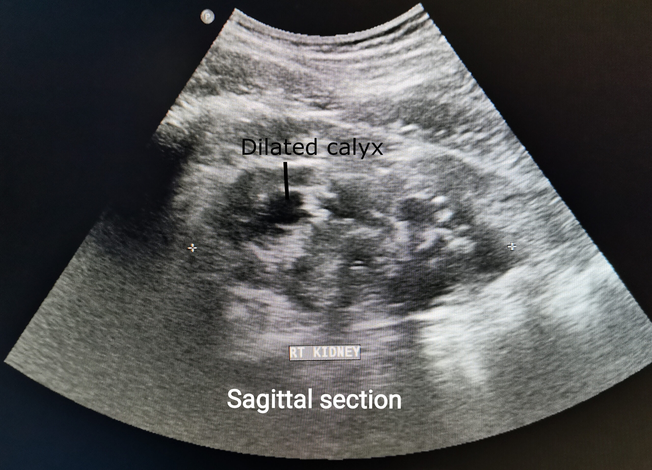
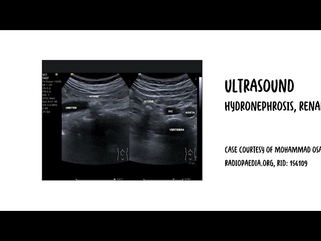
Ultrasound of the right kidney showing moderate hydronephrosis with dilated renal calyces. Image: Cerevisae/Wikimedia Commons, CC BY-SA 4.0.
What to notice
Detailed image description
Source: https://commons.wikimedia.org/wiki/File:Ultrasound_of_right_kidney_moderate_hydronephrosis.jpg. Creator: Cerevisae. Licence: Creative Commons Attribution-ShareAlike 4.0 International (https://creativecommons.org/licenses/by-sa/4.0/). No modifications.
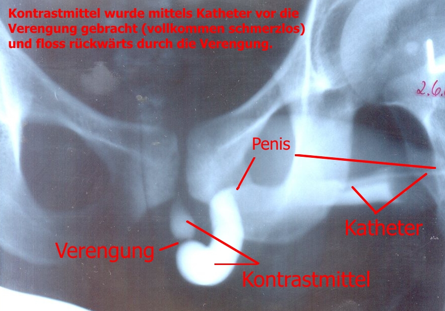
Retrograde urethrogram demonstrating a male urethral stricture. Image: STofffuchs, via Wikimedia Commons, CC BY 3.0.
What to notice
Detailed image description
Source: https://commons.wikimedia.org/wiki/File:Urethra_stricture.jpg Creator: STofffuchs Licence: Creative Commons Attribution 3.0 Unported: https://creativecommons.org/licenses/by/3.0/ Modifications: None.
Never measure serum PSA during an episode of acute urinary retention or immediately after catheterization. Acute bladder distension, UTI, and catheter instrumentation cause massive false-positive spikes in PSA levels.
CRITICAL MANAGEMENT
- Immediate Bladder Decompression:
- Complete, continuous emptying via Foley catheter (gradual clamping is outdated and unsupported by evidence; drain the bladder completely in one go).
- Document precise residual volume drained at insertion.
- Management of Post-Obstructive Diuresis (POD):
- Definition: Urine output > 200 mL/hr for >= 2 consecutive hours or > 3–4 L/24h post-decompression.
- Monitoring: Strict fluid balance chart (hourly urine output), daily weights, and serial electrolytes/creatinine.
- Fluid Replacement: If patient is unstable or developing electrolyte derangements, replace 50–75% of hourly urine output with IV isotonic crystalloids (e.g., 0.45% or 0.9% Normal Saline + KCl) to prevent severe hypovolemia while avoiding perpetuation of diuresis.
- Trial Without Catheter (TWOC) Protocol for BPH:
- Start Alpha-1 Blocker: Initiate Tamsulosin 0.4 mg PO daily (or Alfuzosin/Silodosin) immediately upon catheterization.
- Duration: Maintain catheter for 3–7 days while on alpha-blocker therapy before attempting a TWOC (significantly increases TWOC success rate from ~30% to > 60%).
- TWOC Procedure: Remove catheter early in the morning; patient must pass adequate volume (> 200 mL) with a post-void residual < 100–200 mL on ultrasound scan. If TWOC fails -> re-catheterize and refer for elective surgical resection (TURP).
When preparing a male patient with BPH-induced urinary retention for a Trial Without Catheter (TWOC), prescribe an Alpha-1 blocker (e.g., Tamsulosin) for at least 3 days prior to catheter removal to more than double the likelihood of successful spontaneous voiding.
REFERENCES
- Serlin DC, Heidelbaugh JJ, Stoffel JT. Urinary retention in adults: evaluation and initial management. Am Fam Physician. 2018;98(8):496–503. PMID:30277739.
- National Institute for Health and Care Excellence. Lower urinary tract symptoms in men: management [Internet]. London: NICE; 2010 [updated 2015 Jun 3; cited 2026 Aug 31]. Available from: https://www.nice.org.uk/guidance/cg97
- European Association of Urology. EAU guidelines on the management of non-neurogenic male lower urinary tract symptoms [Internet]. Arnhem: European Association of Urology; 2026 [cited 2026 Aug 31]. Available from: https://uroweb.org/guidelines/management-of-non-neurogenic-male-luts
- American Urological Association. Urotrauma guideline [Internet]. Linthicum (MD): American Urological Association; [cited 2026 Aug 31]. Available from: https://www.auanet.org/guidelines-and-quality/guidelines/urotrauma-guideline
- Fisher E, Subramonian K, Omar MI. The role of alpha blockers prior to removal of urethral catheter for acute urinary retention in men. Cochrane Database Syst Rev. 2014;2014(6):CD006744. doi:10.1002/14651858.CD006744.pub3
- Halbgewachs C, Domes T. Postobstructive diuresis: pay close attention to urinary retention. Can Fam Physician. 2015;61(2):137–142. PMID:25821871.




Members only discussions coming soon…