

Blount’s Disease
Overview
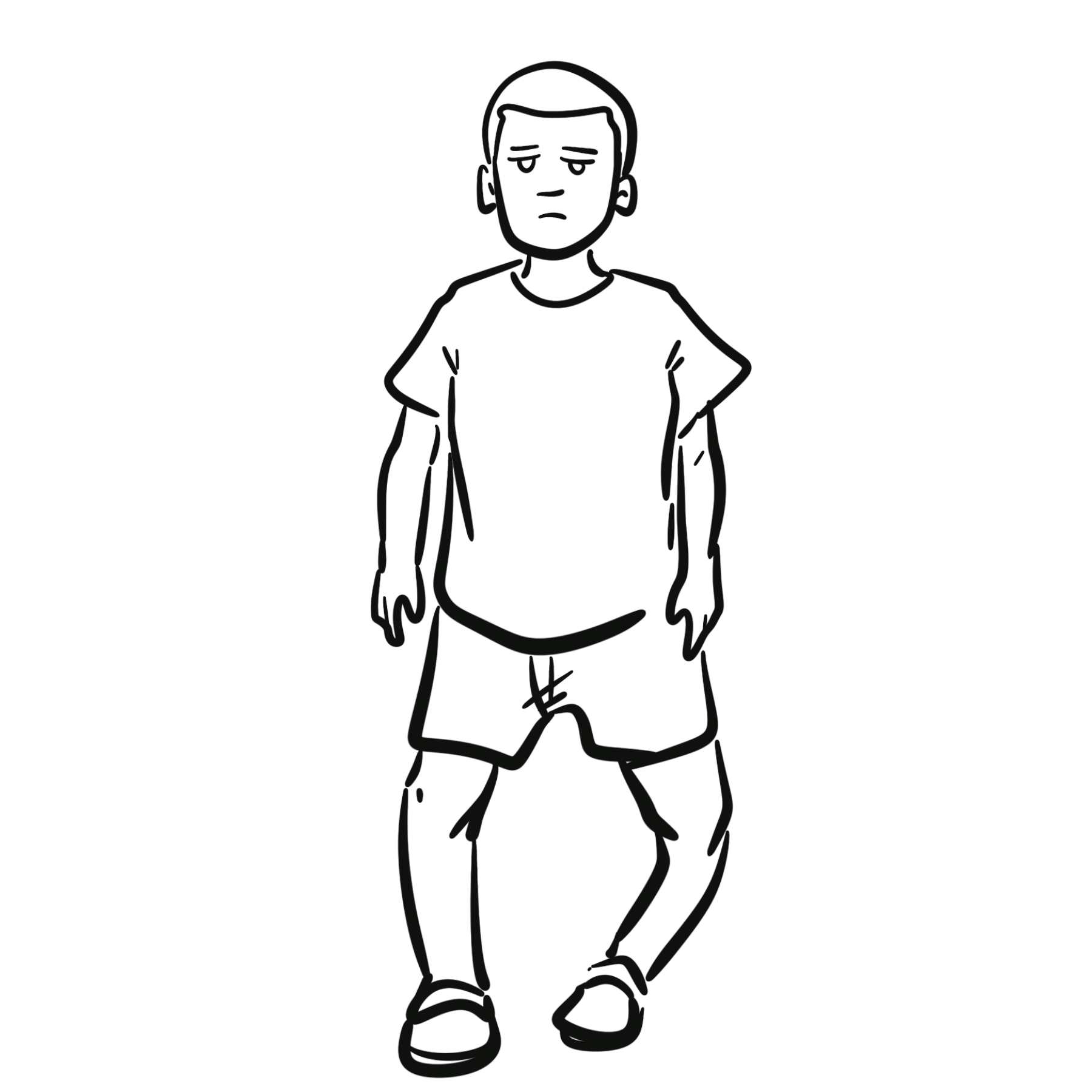
Blount’s disease, also called tibia vara, is a developmental growth disorder of the posteromedial proximal tibial physis. Asymmetric growth produces progressive genu varum centred below the knee, commonly with procurvatum, internal tibial torsion and relative shortening. It differs from physiological bowing because the deformity persists or progresses rather than following normal spontaneous correction.¹,²
Early-onset disease usually becomes apparent during the first years of walking and is often bilateral. Late-onset disease presents in older children or adolescents, is more often unilateral and may cause activity-related medial knee pain. Delayed recognition allows progressive physeal injury, joint-line obliquity, instability, limb-length discrepancy and premature medial-compartment osteoarthritis.²–⁴
Definition
- Blount’s disease
- Progressive varus deformity caused by disordered growth of the posteromedial proximal tibial physis.
- Genu varum
- Coronal-plane alignment in which the knees remain apart when the ankles are together.
- Tibia vara
- Varus angulation centred in the tibia; commonly used as a synonym for Blount’s disease.
- Lateral thrust
- Abrupt lateral movement of the knee during weight-bearing, suggesting dynamic instability.
Anatomy & Physiology
The proximal tibial physis contributes substantially to tibial length and alignment. Normally, growth is balanced across its medial and lateral portions, maintaining a near-horizontal knee joint line and directing the mechanical axis through the central knee.
Normal lower-limb alignment changes with age. Infants commonly have physiological genu varum; the legs usually move towards neutral by about 18–24 months and then into physiological valgus during early childhood. Interpretation therefore requires the child’s age, symmetry, site of angulation and change over time—not the appearance of bowing alone.¹
Aetiology & Risk Factors
Aetiology
The aetiology is multifactorial and incompletely understood. Excessive compressive loading across the posteromedial proximal tibial physis suppresses endochondral growth, but mechanical loading alone does not explain unilateral disease or severe disease in children without obesity. Biological and genetic susceptibility are therefore likely contributors.¹,²
Risk Factors
- overweight or obesity, particularly in late-onset disease
- greater mechanical loading across an already varus proximal tibia
- early walking, although the strength and consistency of this association are uncertain
- family history or genetic susceptibility
- populations in which Blount’s disease is more prevalent; ethnicity should not be used to exclude the diagnosis
Obesity increases medial physeal loading and is associated with disease progression and treatment failure. However, Blount’s disease also occurs in children of normal weight. Weight alone neither confirms nor excludes the diagnosis.
Pathophysiology
- Varus alignment concentrates compressive force on the posteromedial proximal tibial physis.
- Growth and endochondral ossification become relatively inhibited medially while lateral growth continues.
- The proximal tibia develops progressive varus, increased posterior slope (procurvatum), internal rotation and shortening.
- The medial metaphysis develops beaking, sclerosis and irregularity; the medial epiphysis and physis may become depressed and fragmented.
- Dynamic lateral thrust and ligamentous laxity increase abnormal joint loading.
- Advanced disease may form a medial physeal bar, causing fixed growth arrest, joint incongruity and recurrent deformity after simple correction.
Because the deformity is three-dimensional, correction of the coronal varus alone may leave clinically important internal tibial torsion, procurvatum or limb-length discrepancy. Treatment planning must identify every component of the deformity.
Clinical Manifestations
Early-onset disease
- progressive bowing that becomes more apparent after walking begins
- bilateral deformity more often than in late-onset disease
- abrupt varus centred at the proximal tibia rather than a gentle curve through the whole limb
- intoeing from internal tibial torsion
- palpable medial proximal tibial prominence or metaphyseal beak
- lateral thrust during stance in more advanced disease
- relative shortening when unilateral
- usually little or no pain in toddlers
Late-onset disease
- progressive unilateral or asymmetric genu varum
- activity-related medial knee pain
- limp, reduced endurance or difficulty with sport
- proximal tibial varus with internal torsion and increased posterior tibial slope
- possible distal femoral varus, distal tibial valgus or planovalgus foot compensation
- lateral thrust or knee instability in severe deformity
A lateral thrust is not simply cosmetic bowing. It indicates dynamic coronal-plane instability and should prompt specialist assessment.
Diagnosis & Investigations
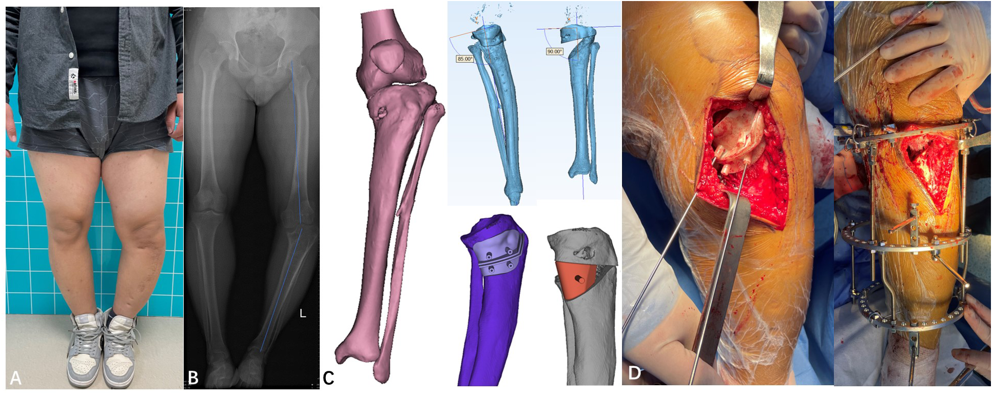
Diagnosis is based on the pattern and progression of deformity together with weight-bearing radiographs. No blood test diagnoses Blount’s disease. The central task is to distinguish pathological, progressive proximal tibial varus from physiological bowing and other causes of genu varum.¹–³
Clinical assessment
- age at onset and whether the bowing is worsening, stable or improving
- walking age, growth pattern, weight trajectory and family history
- pain, limp, functional limitation and previous trauma or infection
- standing alignment, symmetry and the level at which the limb angulates
- gait assessment for intoeing and lateral thrust
- hip and knee range of motion, ligamentous laxity and rotational profile
- leg lengths and examination for skeletal dysplasia or metabolic bone disease
First-Line Imaging
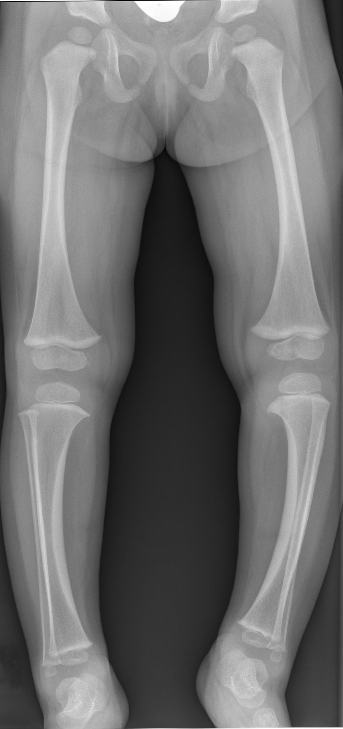
Obtain bilateral standing anteroposterior long-leg radiographs from the hips to the ankles once the child can stand reliably.
A lateral knee or tibial radiograph is useful for assessing posterior tibial slope and procurvatum.¹,³,⁴
Radiographic features supporting Blount’s disease include:
- medial metaphyseal beaking
- widening or irregularity of the medial physis
- delayed or irregular medial epiphyseal ossification
- posteromedial slope or depression
- proximal tibial varus
- medial physeal-bar formation in advanced disease
Metaphyseal–Diaphyseal Angle
The Levine–Drennan metaphyseal–diaphyseal angle helps assess the likelihood of progression in young children with bowing.
- Less than 10°: Physiological bowing is strongly favoured.
- 11–16°: Indeterminate; clinical and radiographic follow-up is required.
- Greater than 16°: High risk of progression and Blount’s disease.
These thresholds support but do not replace assessment of the child’s age, symmetry, progression and characteristic radiographic abnormalities.¹
A single borderline radiograph does not settle the diagnosis. In a young child with an angle of 11–16°, progression on correctly positioned serial weight-bearing radiographs is more informative than indiscriminate additional testing.
Selective Investigations
Further investigations should be requested only when they are likely to alter diagnosis or management.
MRI
MRI may be considered in delayed, advanced or recurrent early-onset disease when assessment of the following will affect surgical planning:
- physeal viability
- suspected physeal-bar formation
- medial tibial plateau depression
- unossified epiphyseal cartilage
- meniscal abnormalities
- articular surface morphology
Bone Age
Bone-age assessment may be useful in late-onset disease when remaining growth determines whether guided growth remains feasible.
CT Rotational Study
CT is rarely required. It may be considered when clinically important tibial torsion cannot be adequately defined for operative planning.
Blood Tests
Blood tests are not routinely required.
Calcium, phosphate, alkaline phosphatase, vitamin D, renal function or other targeted tests should be requested only when the presentation raises concern for rickets, renal osteodystrophy or another metabolic bone disorder.
Important Differential Diagnoses
| Condition | Features favouring the diagnosis | Features against Blount’s disease |
|---|
| Physiological genu varum | Symmetrical, gentle bowing at an appropriate age that improves over time | No focal medial metaphyseal beak or progressive proximal tibial varus |
| Rickets | Widened wrists, growth disturbance, metaphyseal cupping or fraying and biochemical abnormalities | Systemic skeletal abnormalities rather than isolated posteromedial proximal tibial disease |
| Skeletal dysplasia | Disproportionate stature, widespread skeletal abnormalities or relevant family history | Multifocal skeletal disease rather than isolated mechanical physeal change |
| Post-traumatic or post-infective physeal arrest | Relevant history with focal physeal-bar formation or asymmetrical growth arrest | Does not follow the characteristic developmental pattern |
| Focal fibrocartilaginous dysplasia | Usually unilateral focal tibial bowing with a characteristic cortical lesion | Different anatomical site and radiographic morphology |
Classification
Classification by age at onset
- Early-onset or infantile Blount’s disease: develops in early childhood, commonly before 4 years; often bilateral and more likely to develop medial plateau depression and physeal arrest.
- Late-onset Blount’s disease: develops in later childhood or adolescence; more often unilateral and commonly associated with obesity, knee pain and deformity involving the distal femur as well as the tibia.
Langenskiöld radiographic stages
The six-stage Langenskiöld system describes the radiographic evolution of early-onset disease. It is most useful as a shared description of morphology and natural history rather than as a stand-alone treatment algorithm.³
- Stage I: medial metaphyseal beaking.
- Stage II: saucer-shaped defect of the medial metaphysis.
- Stage III: the saucer deepens into a step.
- Stage IV: the epiphysis bends down over the medial beak.
- Stage V: double-epiphysis appearance with severe posteromedial depression.
- Stage VI: medial physeal bony bar and fixed growth arrest.
Langenskiöld staging was designed for early-onset disease. Late-onset disease is better described according to the anatomical source and magnitude of deformity, remaining growth, torsion, limb-length discrepancy and mechanical-axis deviation.
Treatment
Management should be led by paediatric orthopaedics. The aim is to restore a stable, plantigrade limb with a near-normal mechanical axis and joint orientation while preserving growth and minimising recurrence. Treatment depends on age, radiographic stage, progression, deformity magnitude, instability, obesity, physeal-bar formation and remaining growth.²–⁴
Observation and risk-factor management
- Observe only when physiological bowing remains likely or the diagnosis is indeterminate, with planned clinical and radiographic review rather than open-ended reassurance.
- Support healthy weight management without delaying orthopaedic correction of progressive deformity.
- Physiotherapy may improve strength and function but cannot reverse pathological physeal growth disturbance.
Bracing
A knee-ankle-foot orthosis may be considered for selected children younger than about 3 years with mild early-onset disease, particularly unilateral Langenskiöld stage I or II disease. Bilateral involvement, obesity, instability, more advanced stage and delayed treatment predict poorer response. Failure to show correction should prompt timely surgical reassessment.³
Guided growth
Lateral proximal tibial hemiepiphysiodesis can gradually correct mild-to-moderate deformity when sufficient growth remains. It does not correct established internal tibial torsion or an intra-articular medial plateau deformity and may fail or rebound, particularly with severe deformity, high body mass index or limited remaining growth.³,⁴
Corrective osteotomy and reconstruction
- Proximal tibial osteotomy provides acute or gradual correction of varus; technique is selected according to deformity complexity and surgical expertise.
- Multiplanar osteotomy or circular external fixation can address varus, procurvatum, internal torsion and shortening together.
- Earlier correction of progressive early-onset disease—ideally before fixed medial physeal arrest—reduces recurrence risk compared with late correction.
- Advanced disease may require physeal-bar management, medial tibial plateau elevation, epiphysiodesis or staged limb-length procedures in addition to realignment.
- Late-onset disease may require concurrent correction of distal femoral varus, distal tibial valgus or limb-length discrepancy.
A straight-looking tibia is not the only treatment endpoint. If the mechanical axis, knee joint line, rotation or limb length remains abnormal, pathological loading and functional impairment may persist despite apparent cosmetic correction.
Complications & Prognosis
Complications
- progressive multiplanar deformity and gait deterioration
- lateral knee instability and thrust
- medial tibial plateau depression and joint incongruity
- physeal bar formation and recurrent varus
- limb-length discrepancy
- meniscal and cartilage overload
- premature medial-compartment knee osteoarthritis
- treatment complications including implant failure, undercorrection, overcorrection, recurrence, delayed union, non-union, neurovascular injury and compartment syndrome
Prognosis
Prognosis is best when progressive disease is recognised early and alignment is corrected before irreversible medial physeal arrest or substantial joint-surface deformity develops. Higher age at correction, obesity, advanced Langenskiöld stage, severe medial physeal slope, instability and previous recurrence are associated with more complex treatment and a greater risk of recurrent deformity.²–⁴
Children require follow-up through growth because rebound or recurrent varus may occur after bracing, guided growth or osteotomy. Untreated or residual deformity increases the risk of pain, functional limitation and premature knee osteoarthritis in adolescence or adulthood.²,⁴
Correction is not the end of surveillance. Blount’s disease can recur as growth continues, so lower-limb alignment and limb length must be followed until skeletal maturity.
References
- Dakshina Murthy TS, Taqi M, De Leucio A. Blount disease. In: StatPearls [Internet]. Treasure Island (FL): StatPearls Publishing; 2026 [updated 2024 Jan 9; cited 2026 Aug 20]. Available from: https://www.ncbi.nlm.nih.gov/books/NBK560923/
- Maré PH, Thompson DM. Infantile Blount’s disease. SA Orthop J. 2020;19(3). doi:10.17159/2309-8309/2020/v19n3a8
- Pediatric Orthopaedic Society of North America. Blount’s disease – infantile [Internet]. Oakbrook Terrace (IL): POSNA [cited 2026 Aug 20]. Available from: https://posna.org/physician-education/study-guide/blount-s-disease-infantile
- Pediatric Orthopaedic Society of North America. Adolescent Blount disease [Internet]. Oakbrook Terrace (IL): POSNA [cited 2026 Aug 20]. Available from: https://posna.org/physician-education/study-guide/adolescent-blount-disease




Members only discussions coming soon…