

Cardiac Sarcoidosis
Overview
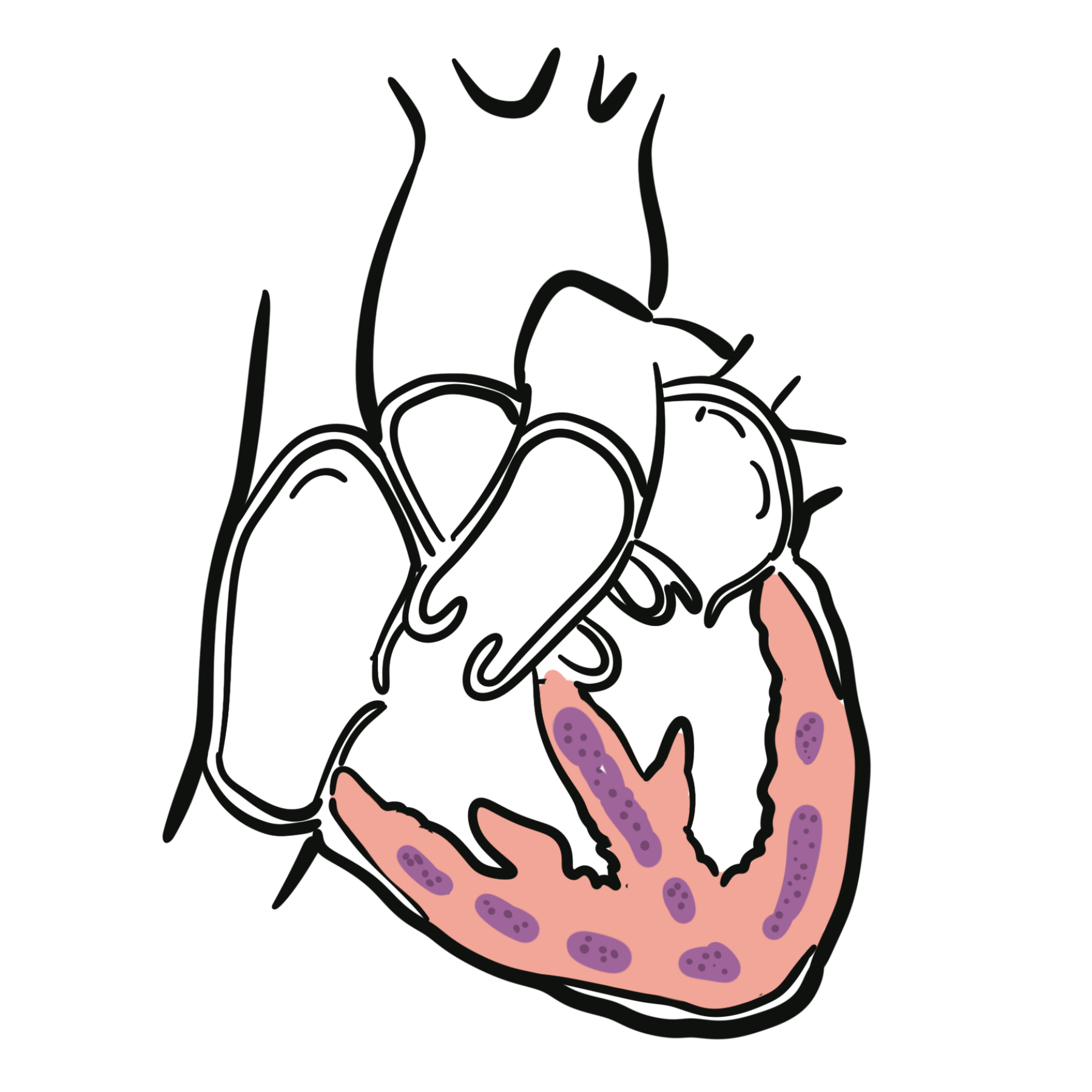
Cardiac sarcoidosis is an inflammatory heart disease caused by myocardial infiltration with non-necrotising granulomas. Inflammation may subsequently resolve, recur, or progress to permanent fibrosis. The disease is often patchy and may affect the conduction system, ventricular myocardium, papillary muscles, valves, pericardium, or coronary microcirculation.1
Cardiac involvement may occur as part of multisystem sarcoidosis or, less commonly, without clinically apparent disease elsewhere—termed isolated cardiac sarcoidosis. Its presentation ranges from asymptomatic abnormalities on cardiac imaging to high-grade atrioventricular block, ventricular arrhythmias, heart failure, or sudden cardiac death.1–3
Diagnosis is challenging because there is no single sufficiently sensitive reference test. A probability-based diagnosis is usually made by integrating the clinical presentation, electrocardiography, cardiac imaging, assessment for extracardiac sarcoidosis, and—where feasible—histological evidence.1,2
Definition
- Cardiac sarcoidosis
- Granulomatous inflammation involving the heart, with or without sarcoidosis in other organs.
- Isolated cardiac sarcoidosis
- Cardiac sarcoidosis without clinically demonstrable extracardiac involvement.
- Late gadolinium enhancement (LGE)
- A cardiac MRI finding that usually represents myocardial injury or fibrosis; it is not specific to sarcoidosis.
- Myocardial FDG uptake
- Uptake of ¹⁸F-fluorodeoxyglucose on PET that may indicate metabolically active myocardial inflammation when physiological uptake has been adequately suppressed.
Anatomy & Physiology
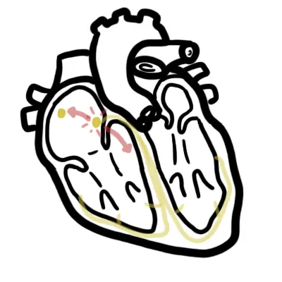
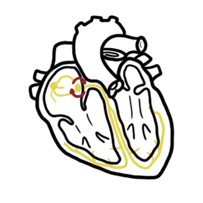
The cardiac conduction system travels through the atrioventricular node, His bundle, and bundle branches within the basal interventricular septum. Sarcoid inflammation involving this region can therefore cause bundle branch block or advanced atrioventricular block.
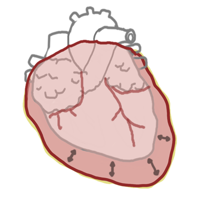
Granulomatous inflammation may involve either ventricle, although the basal septum, left ventricular free wall, papillary muscles, and right ventricular insertion regions are frequently affected. Disease is characteristically patchy rather than confined to a single coronary territory.
The clinical consequences depend on both the location and phase of disease:
- Inflammation can produce oedema, myocardial injury, conduction disturbance, and enhanced ventricular automaticity.
- Healing and fibrosis create fixed conduction block and re-entry circuits that predispose to ventricular tachycardia.
- Extensive myocardial involvement causes systolic or diastolic ventricular dysfunction.
- Right ventricular involvement may produce right-sided heart failure or an arrhythmogenic cardiomyopathy-like phenotype.
Aetiology & Risk Factors
Aetiology
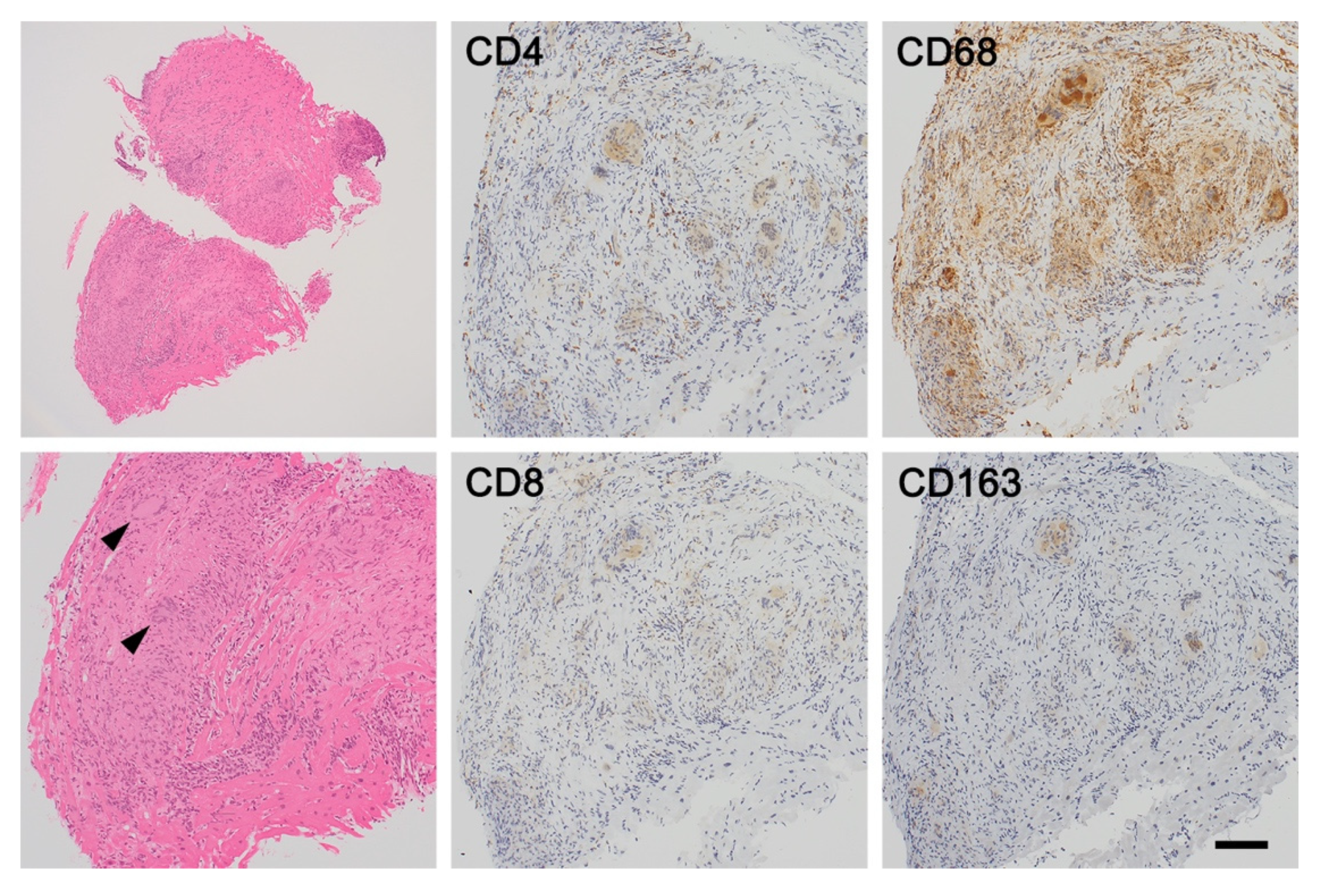
The cause of sarcoidosis is incompletely understood. The prevailing model involves an exaggerated cell-mediated immune response to one or more unidentified antigens in a genetically susceptible person. Activated T lymphocytes and macrophages aggregate to form non-necrotising granulomas. Persistent inflammation may lead to myocardial scarring and remodelling.1,4
Potential environmental, occupational, microbial, and genetic contributors have been investigated, but no single trigger accounts for all cases.
Risk Factors
Factors associated with sarcoidosis or a greater likelihood of cardiac involvement include:
- Established extracardiac sarcoidosis, particularly when accompanied by cardiac symptoms or an abnormal ECG
- Family history or genetic susceptibility to sarcoidosis
- Ancestry associated with a higher incidence or more severe phenotype of systemic sarcoidosis
- Middle adulthood, although cardiac sarcoidosis can occur across a broad age range
- Multisystem or longstanding inflammatory disease
Traditional cardiovascular risk factors do not cause cardiac sarcoidosis, although coexisting coronary artery disease, hypertension, or valvular disease may complicate its recognition and management.
Cardiac disease may be the first or only clinically apparent manifestation of sarcoidosis. The absence of pulmonary symptoms, lymphadenopathy, or a previous sarcoidosis diagnosis therefore does not exclude it.
Pathophysiology
The pathological process usually evolves through overlapping inflammatory and fibrotic phases:
- An antigen-driven immune response activates T lymphocytes and macrophages.
- Non-necrotising granulomas form within affected myocardium.
- Active inflammation causes oedema, myocyte injury, conduction disturbance, and electrical instability.
- Resolution may occur, but recurrent or persistent inflammation promotes replacement fibrosis.
- Patchy scar creates regions of slow conduction that support re-entrant ventricular arrhythmias.
- Extensive myocardial injury and remodelling lead to ventricular dysfunction and heart failure.
Active inflammation and established scar may coexist. This distinction is clinically important because immunosuppression primarily targets inflammation, whereas fixed scar may continue to cause atrioventricular block or ventricular tachycardia even after inflammatory activity improves.1,4
Why might ventricular tachycardia continue despite apparently successful treatment of inflammation?
The inflammatory trigger may have subsided, but the resulting myocardial scar can remain as a permanent re-entry substrate. Management may therefore require both immunosuppression and arrhythmia-directed treatment.
Clinical Manifestations
Clinical presentation depends on the location, burden, and activity of myocardial involvement.
Conduction Disease
Inflammation or fibrosis of the basal septum may cause:
- First-degree atrioventricular block
- Bundle branch block
- Mobitz II second-degree atrioventricular block
- Complete heart block
- Sinus-node dysfunction, less commonly
Unexplained high-grade atrioventricular block in a patient younger than approximately 60 years is an important clinical red flag.1
Arrhythmias
Ventricular arrhythmias may result from active inflammation, established scar, or both. Presentations include:
- Frequent premature ventricular complexes
- Non-sustained ventricular tachycardia
- Sustained monomorphic or polymorphic ventricular tachycardia
- Ventricular fibrillation
- Palpitations, presyncope, syncope, or sudden cardiac arrest
Atrial fibrillation and atrial tachycardia may occur, particularly with atrial enlargement or advanced ventricular disease.
Heart Failure
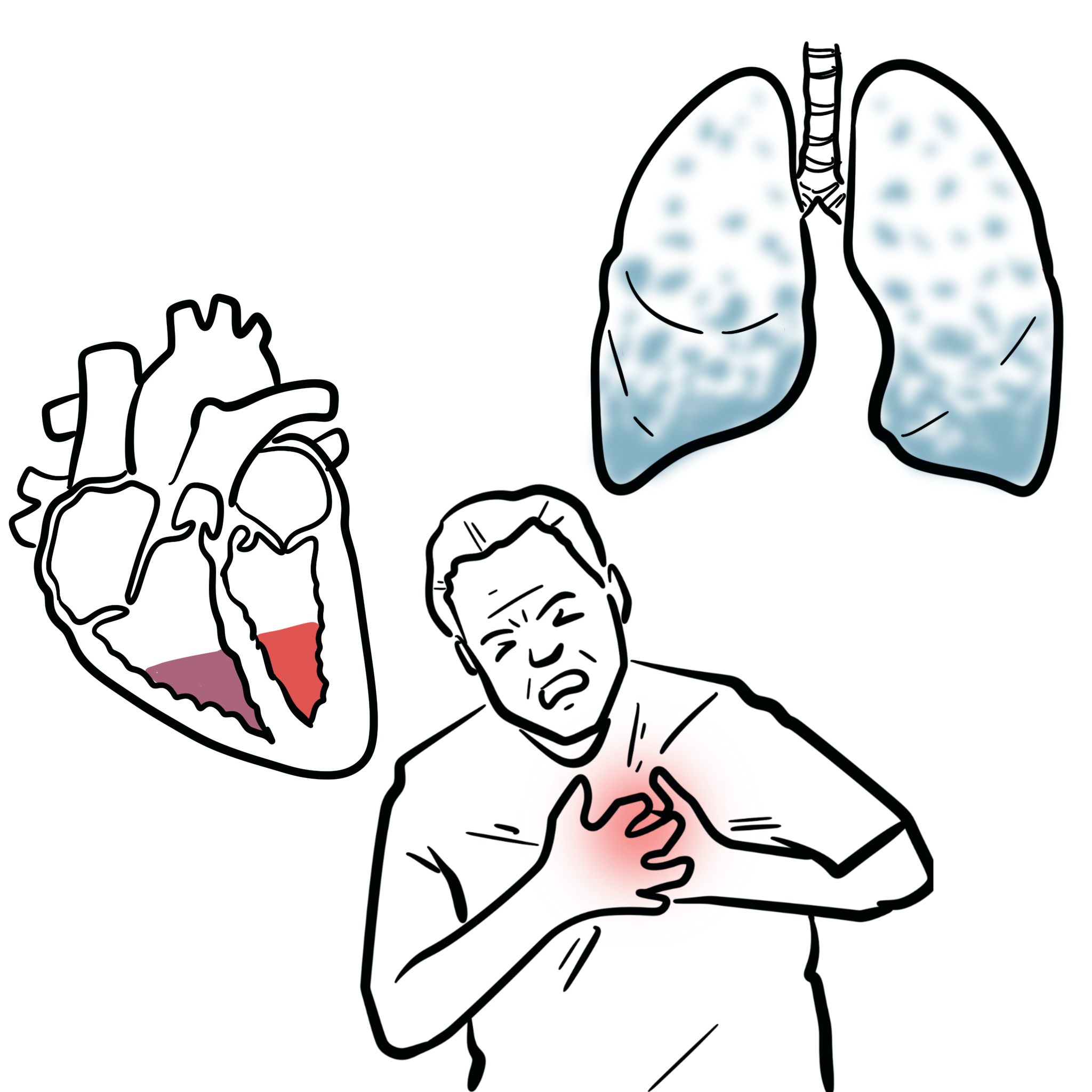
- Exertional dyspnoea and fatigue
- Orthopnoea or paroxysmal nocturnal dyspnoea
- Peripheral oedema or raised jugular venous pressure
- Left, right, or biventricular systolic dysfunction
- Regional wall-motion abnormalities or ventricular aneurysm
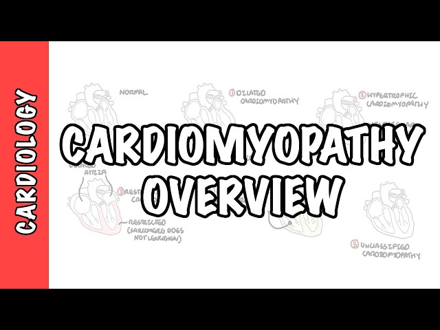
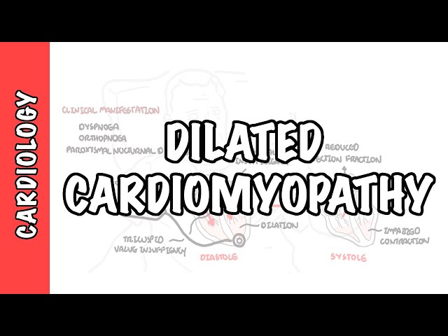
Cardiac sarcoidosis can resemble dilated cardiomyopathy, myocarditis, ischaemic cardiomyopathy, or arrhythmogenic right ventricular cardiomyopathy.
Other Manifestations
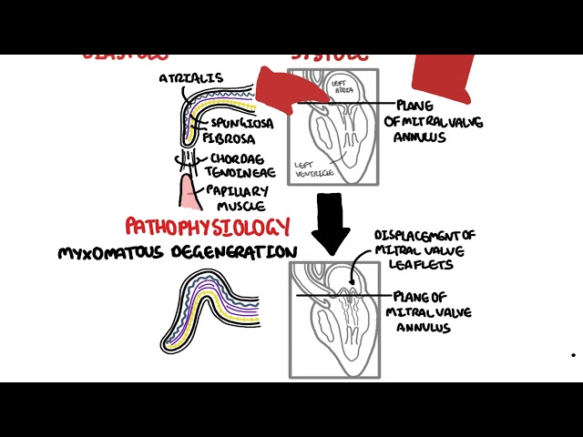
- Mitral regurgitation due to papillary muscle involvement or ventricular remodelling
- Pericardial effusion or pericarditis
- Chest discomfort caused by inflammation or arrhythmia
- Intracardiac thrombus in severely dysfunctional or aneurysmal ventricles
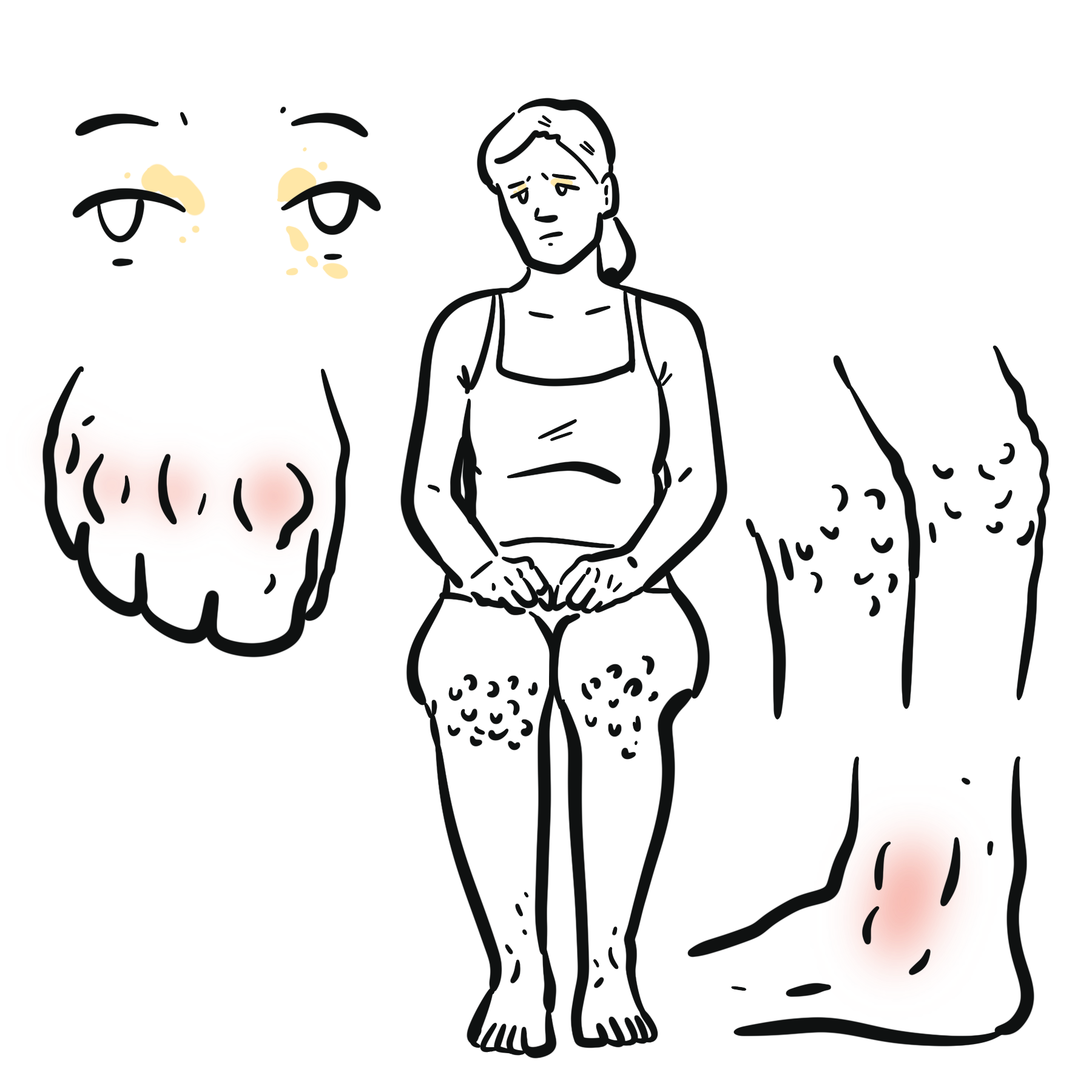
- Systemic features such as cough, uveitis, skin lesions, lymphadenopathy, or neurological symptoms
Consider cardiac sarcoidosis in a younger or middle-aged adult with otherwise unexplained high-grade atrioventricular block, sustained ventricular arrhythmia, regional ventricular aneurysm, basal septal thinning, or non-ischaemic cardiomyopathy—especially when extracardiac sarcoidosis is present.
Diagnosis & Investigations
Diagnosis is based on concordant clinical, imaging, and pathological evidence. Investigations should answer four practical questions:
- Is there evidence of cardiac involvement?
- Is myocardial inflammation currently active?
- Is there a safer extracardiac site for biopsy?
- What is the risk of heart failure or sudden cardiac death?
Initial Cardiac Assessment
Electrocardiography and rhythm monitoring
- A 12-lead ECG should be obtained when cardiac sarcoidosis is suspected.
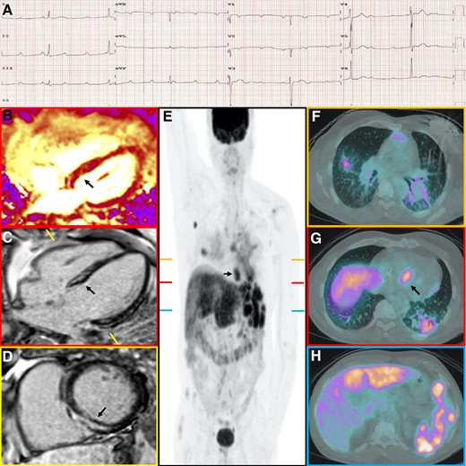
- Relevant abnormalities include atrioventricular block, bundle branch block, fragmented QRS complexes, pathological Q waves, ventricular ectopy, ventricular tachycardia, and atrial arrhythmias.
- Transthoracic echocardiography assesses ventricular function, haemodynamics, and alternative structural diagnoses.
- Echocardiography may be normal despite clinically important cardiac sarcoidosis and cannot establish or exclude the diagnosis by itself.1,3
A patient with pulmonary sarcoidosis has palpitations and presyncope, but their left ventricular ejection fraction is normal. Is cardiac sarcoidosis excluded?
No. Patchy inflammation or scar can cause conduction disease and ventricular arrhythmias before conventional echocardiographic function becomes abnormal. Rhythm monitoring and advanced imaging may still be required.
Advanced Cardiac Imaging
Cardiac magnetic resonance imaging
- Preferred advanced structural investigation when available and not contraindicated.
- Late gadolinium enhancement is often patchy or multifocal, mid-myocardial or subepicardial, located in the basal septum or lateral wall, distributed outside a single coronary territory, and may involve either ventricle.
¹⁸F-FDG PET with myocardial perfusion imaging
- Identifying active myocardial inflammation
- PET can also identify extracardiac disease and a more accessible biopsy site.
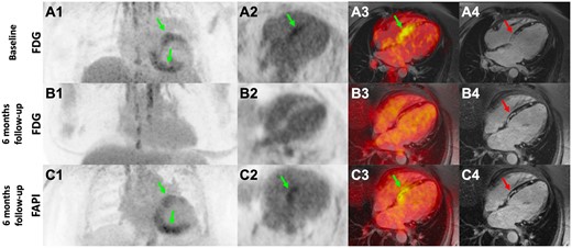
Cardiac MRI and FDG-PET provide complementary information: MRI is best established for myocardial injury, scar, ventricular structure, and prognosis; FDG-PET is best suited to metabolically active inflammation and selected treatment monitoring. Neither test should be interpreted in isolation.
Assessment for Extracardiac Sarcoidosis and Tissue Diagnosis
- Chest imaging should assess for pulmonary disease and mediastinal or hilar lymphadenopathy
- Whole-body PET or targeted imaging may identify lymph nodes, lung, skin, or other organs suitable for biopsy.
- When tissue confirmation is required, biopsy of an accessible extracardiac lesion is usually preferred because it is safer and often has a higher diagnostic yield than endomyocardial biopsy.
- Endomyocardial biopsy has limited sensitivity because myocardial involvement is patchy.
Histology showing non-necrotising granulomas supports sarcoidosis only after infections, foreign-body reactions, drug reactions, and other granulomatous diseases have been considered.
Targeted Laboratory Assessment
- Cardiac troponin may indicate ongoing myocardial injury.
- BNP or NT-proBNP helps assess haemodynamic stress and heart-failure severity.
- Full blood count, renal function, liver profile, calcium, and relevant infection screening provide baseline information before immunosuppression.
- Serum angiotensin-converting enzyme has limited sensitivity and specificity and should not be used to rule cardiac sarcoidosis in or out.3
Treatment
Management should be individualised through a multidisciplinary team that may include cardiology, cardiac electrophysiology, cardiac imaging, respiratory medicine, rheumatology, and other organ specialists.
Treatment has four complementary goals:
- Suppress clinically significant active inflammation.
- Prevent sudden cardiac death and control arrhythmias.
- Treat conduction disease and heart failure.
- Monitor for progression, relapse, and treatment toxicity.
Immunosuppression
- Corticosteroids are the usual first-line therapy for clinically significant active cardiac sarcoidosis.
- A steroid-sparing agent may be introduced to reduce corticosteroid exposure or treat persistent disease.
- Methotrexate is commonly used; alternatives include azathioprine and mycophenolate mofetil.
- Refractory disease may require specialist consideration of a tumour necrosis factor inhibitor.
Response is assessed using symptoms, ECG or device data, ventricular function, biomarkers where informative, and selective repeat FDG-PET. Suppression of PET activity does not eliminate risk arising from established scar.
Management of Conduction Disease
Symptomatic bradycardia or advanced atrioventricular block requires pacing according to standard indications. Although conduction may improve with immunosuppression, recovery is unpredictable and permanent pacing should not be deferred when clinically indicated.
When cardiac sarcoidosis causes an indication for permanent pacing, consider whether an implantable cardioverter-defibrillator is more appropriate than a pacemaker alone. The same patient may also be at risk of ventricular tachyarrhythmia and sudden cardiac death.
Ventricular Arrhythmia and Sudden-Death Prevention
An implantable cardioverter-defibrillator is indicated for survivors of cardiac arrest, patients with sustained ventricular tachycardia, and patients with severely reduced left ventricular systolic function despite appropriate treatment.1,6
It should also be considered in selected patients with extensive myocardial scar, unexplained suspected arrhythmic syncope, a permanent pacing indication, moderate ventricular dysfunction, or inducible sustained ventricular tachycardia. Risk assessment should not rely on left ventricular ejection fraction alone.
Active inflammation is treated with immunosuppression when appropriate. Antiarrhythmic medication may be required. Catheter ablation can reduce recurrent ventricular tachycardia burden, although multiple scar-related circuits and ongoing inflammation can limit long-term success.
Heart Failure Management
Patients with ventricular dysfunction should receive guideline-directed heart-failure therapy unless contraindicated, with diuretics for congestion and cardiac resynchronisation therapy according to standard criteria. Selected patients with refractory advanced heart failure or uncontrollable ventricular arrhythmias may require assessment for mechanical circulatory support or cardiac transplantation.
Complications & Prognosis
Complications
- Complete atrioventricular block
- Sustained ventricular tachycardia or ventricular fibrillation
- Sudden cardiac death
- Left, right, or biventricular heart failure
- Ventricular aneurysm
- Functional mitral or tricuspid regurgitation
- Intracardiac thrombus and systemic embolism
- Recurrent implantable cardioverter-defibrillator therapies
- Adverse effects of long-term immunosuppression
Prognosis
Clinical outcome is highly variable. Some patients remain stable with limited disease, whereas others develop progressive heart failure or life-threatening arrhythmias.
Adverse prognostic markers include reduced left or right ventricular ejection fraction, extensive LGE or myocardial scar, sustained ventricular arrhythmia, high-grade atrioventricular block, persistent inflammatory activity, advanced heart failure, and extensive right ventricular involvement.
Early recognition, treatment of active inflammation, appropriate device therapy, and guideline-directed heart-failure management can improve outcomes. However, ventricular arrhythmia and sudden death may occur even when left ventricular ejection fraction is preserved.
Why is a normal or near-normal ejection fraction insufficient reassurance?
Ejection fraction measures global pump function, but cardiac sarcoidosis is patchy. A relatively small strategically located scar can disrupt the conduction system or sustain ventricular tachycardia without causing major global systolic impairment.
References
- Cheng RK, Kittleson MM, Beavers CJ, Birnie DH, Blankstein R, Bravo PE, et al. Diagnosis and management of cardiac sarcoidosis: a scientific statement from the American Heart Association. Circulation. 2024;149(21):e1197–e1216. doi:10.1161/CIR.0000000000001240.
- Birnie DH, Sauer WH, Bogun F, Cooper JM, Culver DA, Duvernoy CS, et al. HRS expert consensus statement on the diagnosis and management of arrhythmias associated with cardiac sarcoidosis. Heart Rhythm. 2014;11(7):1305–1323. doi:10.1016/j.hrthm.2014.03.043.
- Terasaki F, Azuma A, Anzai T, Ishizaka N, Ishida Y, Isobe M, et al. JCS 2016 guideline on diagnosis and treatment of cardiac sarcoidosis—digest version. Circ J. 2019;83(11):2329–2388. doi:10.1253/circj.CJ-19-0508.
- Aftab A, et al. Cardiac sarcoidosis: diagnosis and management. Front Cardiovasc Med. 2024;11:1394075. doi:10.3389/fcvm.2024.1394075.
- Chareonthaitawee P, Beanlands RS, Chen W, Dorbala S, Miller EJ, Murthy VL, et al. Joint SNMMI–ASNC expert consensus document on the role of ¹⁸F-FDG PET/CT in cardiac sarcoid detection and therapy monitoring. J Nucl Med. 2017;58(8):1341–1353. doi:10.2967/jnumed.117.196287.
- Al-Khatib SM, Stevenson WG, Ackerman MJ, et al. 2017 AHA/ACC/HRS guideline for management of patients with ventricular arrhythmias and the prevention of sudden cardiac death. Circulation. 2018;138(13):e272–e391. doi:10.1161/CIR.0000000000000549.


Members only discussions coming soon…