

Buerger’s Disease
Overview
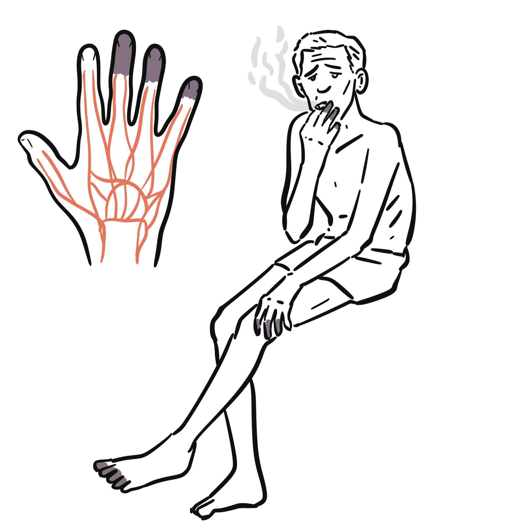
Buerger’s disease, also called thromboangiitis obliterans, is a non-atherosclerotic inflammatory and thrombotic disorder affecting small- and medium-sized arteries and veins, predominantly in the distal limbs. It is strongly associated with tobacco exposure and causes progressive digital ischaemia, superficial thrombophlebitis, painful ulcers and gangrene.1–3
The disease classically affects younger people who smoke and have few conventional atherosclerotic risk factors. Although historically described predominantly in men, Buerger’s disease also occurs in women. Its prevalence is higher in regions where tobacco use is common, including parts of the Middle East, South and East Asia, and the Mediterranean.1,2
Diagnosis is clinical and requires:
- Compatible distal limb ischaemia.
- Current or recent tobacco exposure.
- Characteristic distal vascular abnormalities.
- Careful exclusion of atherosclerosis, embolic disease, connective tissue disease, other vasculitides and thrombophilia.
Definition
- Buerger’s disease
- A tobacco-associated, segmental, non-atherosclerotic inflammatory and thrombotic disease affecting small- and medium-sized arteries and veins of the extremities.
- Thromboangiitis obliterans
- The pathological name for Buerger’s disease, describing inflammation and thrombosis of affected vessels with progressive luminal occlusion.
- Critical limb-threatening ischaemia
- Chronic limb ischaemia associated with ischaemic rest pain, non-healing ulceration or gangrene.
Anatomy & Physiology
The distal circulation of the hands and feet depends on small muscular arteries, digital arteries and an extensive network of collateral vessels. These vessels provide oxygenated blood to tissues with limited tolerance of prolonged ischaemia, particularly the fingertips, toes and nail beds.
Buerger’s disease most often affects:
- Distal radial and ulnar arteries.
- Palmar and digital arteries.
- Distal anterior tibial, posterior tibial and peroneal arteries.
- Plantar and digital arteries.
- Adjacent superficial veins.
- Small nerves travelling within the affected neurovascular bundles.
Because the disease is predominantly distal, brachial and femoral pulses may remain palpable despite severe hand or foot ischaemia. Distal collateral vessels develop around occluded arterial segments and may produce the characteristic “corkscrew” appearance on angiography.1–3
Normal proximal pulses do not exclude severe Buerger’s disease. A patient may have palpable radial or pedal pulses while the digital arteries are critically compromised.
Aetiology & Risk Factors
Aetiology
The exact cause is uncertain, but tobacco exposure is central to disease initiation and persistence. Tobacco constituents may trigger endothelial dysfunction, heightened vascular inflammation, abnormal vasoconstriction and thrombosis in genetically susceptible individuals.1–3
Unlike atherosclerosis, the primary abnormality is not lipid deposition within an arterial plaque. The inflammatory process is concentrated within an occlusive thrombus, while the internal elastic lamina and overall architecture of the vessel wall are relatively preserved, particularly during the acute phase.1,2
Immune dysregulation, altered endothelial responses and increased cellular sensitivity to tobacco components have been proposed. No single genetic variant or circulating biomarker establishes the diagnosis.
Risk Factors
The major risk factors and clinical associations are:
- Cigarette smoking.
- Use of cigars, pipes or smokeless tobacco.
- Ongoing nicotine exposure.
- Cannabis smoking, which may produce or contribute to a similar distal arteritis.
- Younger age at the onset of limb ischaemia.
- Male sex, although the sex difference is becoming less pronounced.
- Residence in regions with high tobacco consumption and increased disease prevalence.
Conventional atherosclerotic risk factors such as longstanding diabetes, marked hyperlipidaemia and older age make isolated Buerger’s disease less likely and should prompt careful consideration of alternative or concurrent vascular disease.
The disease is named after Leo Buerger, who described the pathological findings in amputated limbs in 1908. Felix von Winiwarter had reported a similar condition several decades earlier.
Pathophysiology
Buerger’s disease is a segmental inflammatory thrombosis involving arteries and veins. The pathological appearance evolves through three broad phases:
- Acute phase: An inflammatory thrombus occludes the vessel lumen. Neutrophils, microabscesses and multinucleated giant cells may be present, while the vessel wall and internal elastic lamina remain relatively preserved.
- Subacute phase: The thrombus begins to organise, with persistent inflammation and recanalisation.
- Chronic phase: The thrombus becomes fibrotic and organised, producing permanent vascular occlusion. The vessel may become difficult to distinguish from other chronic occlusive disorders.
Multiple distal arterial segments may be affected, with relatively normal vessels between diseased areas. Adjacent superficial veins can develop migratory thrombophlebitis, and involvement of nearby nerves contributes to disproportionate pain and sensory symptoms.1–3
Progressive arterial obstruction reduces tissue perfusion. Initially, this produces cold sensitivity and exertional pain in the foot, calf, hand or forearm. Further reduction in perfusion causes rest pain, particularly in the digits. Persistent ischaemia results in non-healing ulcers, infection and gangrene.
Collateral vessels form around the occluded arterial segments. These small, tortuous collaterals can produce a “corkscrew” appearance on angiography. This finding supports the diagnosis in the correct clinical context but is not specific to Buerger’s disease.2,3
“Corkscrew collaterals” are supportive, not diagnostic by themselves. Similar collateral patterns can occur in systemic sclerosis, connective tissue disease, vasculitis and other chronic distal occlusive disorders.
Clinical Manifestations
Buerger’s disease usually presents with symptoms of distal extremity ischaemia. More than one limb may be affected, and upper-limb involvement is an important clue.
Early Manifestations
- Cold sensitivity of the hands or feet.
- Raynaud phenomenon.
- Digital paraesthesia, numbness or burning pain.
- Claudication involving the foot arch, calf, hand or forearm.
- Reduced exercise tolerance of an affected limb.
- Migratory superficial thrombophlebitis.
Foot-arch or instep claudication in a younger person who smokes is particularly suggestive of distal arterial disease. Upper-extremity claudication is unusual in atherosclerosis and should increase suspicion for Buerger’s disease or another non-atherosclerotic vascular disorder.
Progressive Ischaemia
As perfusion deteriorates, manifestations include:
- Persistent digital rest pain.
- Pain that worsens with elevation and improves when the limb is dependent.
- Cool, pale or cyanotic fingers and toes.
- Delayed capillary refill.
- Reduced or absent distal pulses.
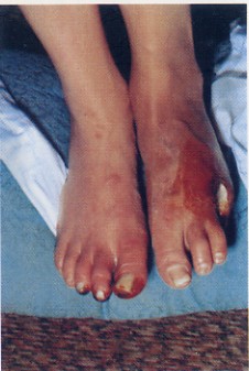
- Painful, sharply demarcated digital ulcers.
- Nail and skin trophic changes.
- Digital gangrene.
- Secondary soft-tissue infection or osteomyelitis.
The Allen test may demonstrate impaired arterial supply to the hand. An abnormal test in a young person with limb ischaemia supports distal arterial occlusion but does not establish the cause.
Superficial Thrombophlebitis
Migratory superficial thrombophlebitis may precede arterial symptoms. It presents as tender, erythematous, cord-like superficial veins that appear in different locations over time.
Visceral, coronary, cerebral and renal arterial involvement is rare. Symptoms suggesting these territories should prompt reconsideration of the diagnosis and investigation for another vasculitis, embolic disorder or atherosclerotic disease.
The characteristic clinical combination is a younger person with substantial tobacco exposure, distal upper- or lower-limb ischaemia, migratory superficial thrombophlebitis or Raynaud phenomenon, and few conventional atherosclerotic risk factors.
Diagnosis
There is no single diagnostic blood test for Buerger’s disease. Diagnosis depends on recognising a compatible clinical pattern, demonstrating distal vascular occlusion and excluding more common or treatable mimics.1–3
Non-Invasive Vascular Assessment
Ankle–brachial index
The ankle–brachial index may identify lower-limb arterial obstruction, but it can remain normal when disease is confined to arteries distal to the ankle. A normal ankle–brachial index therefore does not exclude Buerger’s disease.
Toe pressures and digital waveforms
Toe–brachial pressure, digital systolic pressure, pulse-volume recordings and photoplethysmography are more useful when disease is predominantly distal. They can:
- Objectively document digital hypoperfusion.
- Assess the severity of ischaemia.
- Provide a baseline for monitoring.
- Help evaluate the likelihood of wound healing.
Duplex ultrasonography
Duplex ultrasound can demonstrate distal arterial occlusion and assess proximal vessels for atherosclerotic disease, aneurysm or another source of embolisation. Its ability to visualise very small digital arteries is limited.
Angiography
CT angiography, MR angiography or catheter angiography may be used when the diagnosis remains uncertain, severe ischaemia requires anatomical definition or revascularisation is being considered.
Characteristic findings include:
- Segmental occlusion of distal small- and medium-sized arteries.
- Involvement of multiple limbs.
- Relative sparing of proximal arteries.
- Absence of significant calcified atherosclerosis.
- Tortuous “corkscrew” collateral vessels around occluded segments.
- Abrupt distal arterial cut-offs.
- Poor distal runoff.2,3
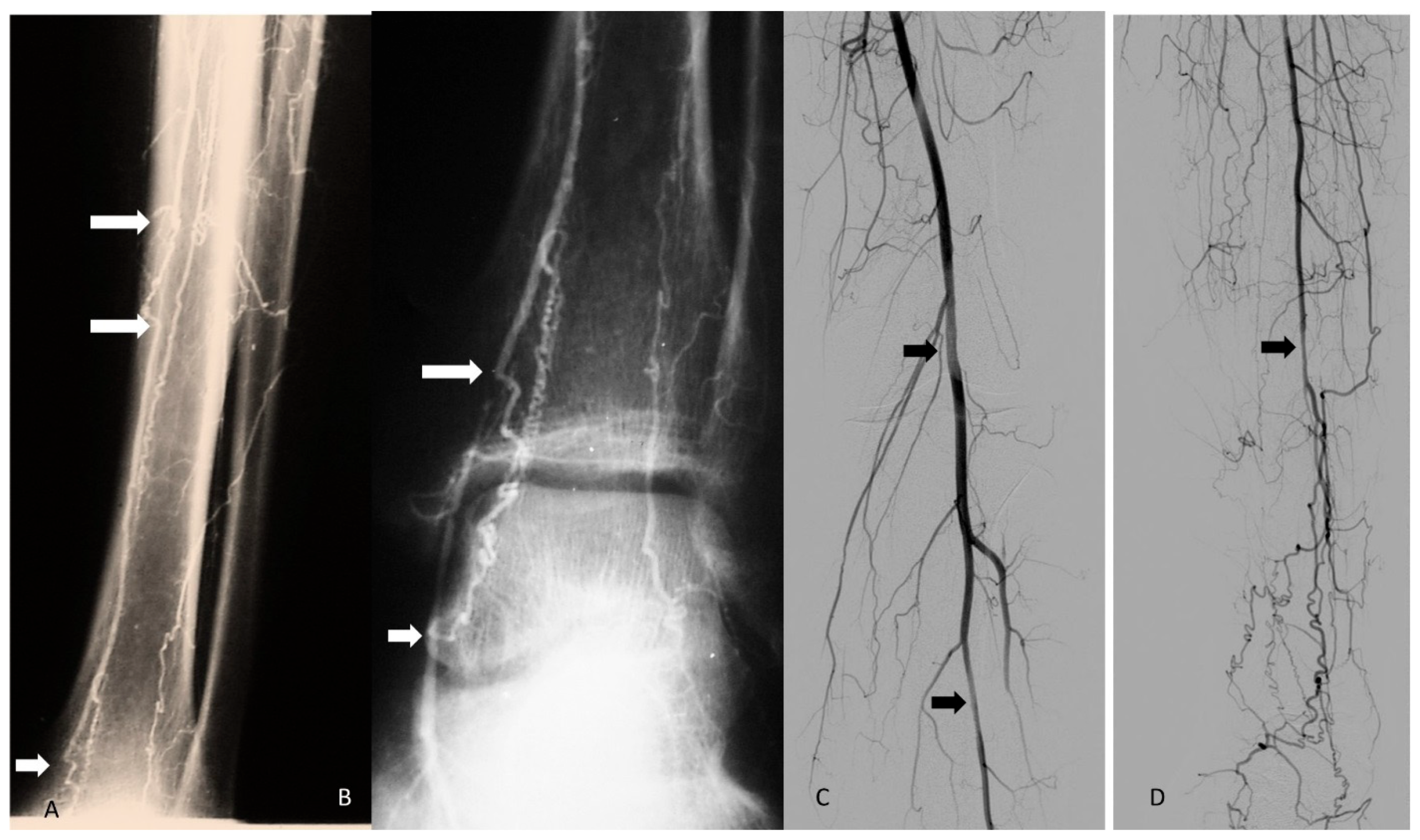
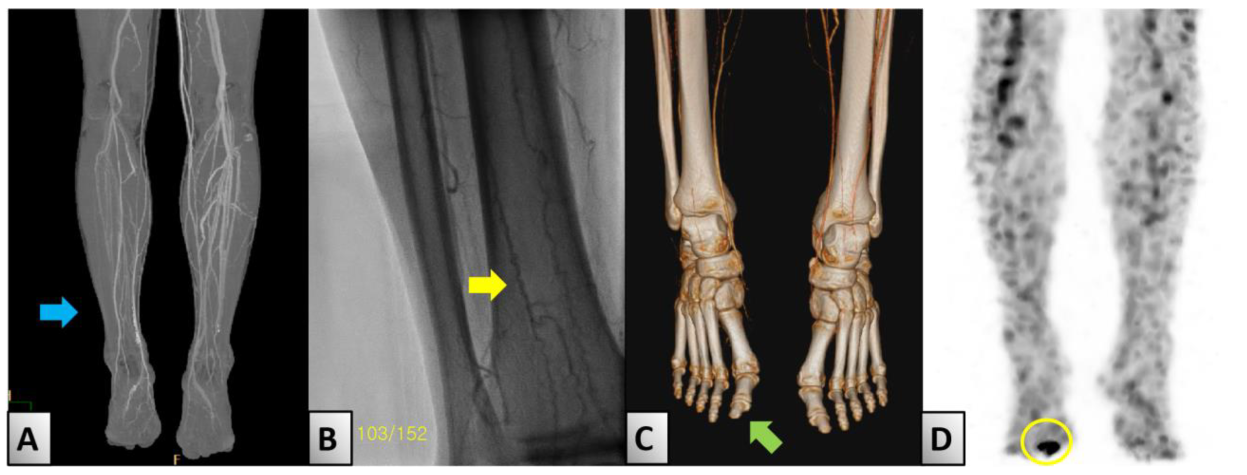
Catheter angiography provides the greatest detail of small distal vessels but is invasive. Angiographic findings must be interpreted with the clinical presentation because no individual pattern is entirely specific.
Targeted Exclusion of Important Mimics
Investigations should be directed toward plausible alternatives rather than ordered as an indiscriminate vasculitis panel.
Useful tests commonly include:
- Full blood count.
- Electrolytes, renal function and liver function.
- Fasting glucose or HbA1c.
- Lipid profile.
- C-reactive protein and erythrocyte sedimentation rate.
- Urinalysis.
- ANA with disease-specific antibodies when connective tissue disease is suspected.
- Antiphospholipid antibodies when thrombosis or pregnancy morbidity raises suspicion.
- Thrombophilia testing when the presentation, age or family history makes an inherited or acquired hypercoagulable state plausible.
Inflammatory markers are often normal in Buerger’s disease. Markedly elevated inflammatory markers, renal abnormalities, cytopenias or systemic manifestations should prompt investigation for infection, systemic vasculitis or connective tissue disease.
Echocardiography and rhythm assessment are appropriate when an embolic source is plausible. Blood cultures should be obtained if infective endocarditis or septic embolisation is suspected.
Histopathology
Biopsy is not routinely required. It may be considered when the presentation is atypical or another vasculitis cannot be excluded.
Do not diagnose Buerger’s disease solely because a young person who smokes has digital ischaemia. First exclude embolism, connective tissue disease, antiphospholipid syndrome, other vasculitides, atherosclerosis and drug-associated vasculopathy.
Treatment
Complete Tobacco and Nicotine Abstinence
Complete cessation of tobacco exposure is the most important intervention and the only measure consistently shown to prevent disease progression and reduce the risk of amputation.1,2,4
“Cutting down” is not treatment. The therapeutic target is complete and permanent abstinence from tobacco and nicotine exposure.
Limb Protection and Wound Care
Patients should be advised to:
- Keep affected limbs warm without applying direct heat.
- Avoid cold exposure and vasoconstrictive medicines where possible.
- Use well-fitting protective footwear.
- Inspect the feet and hands daily.
- Avoid burns, cuts and repetitive trauma.
- Obtain early podiatry and wound-care review.
- Treat cellulitis, abscess or osteomyelitis promptly.
- Maintain appropriate analgesia and nutrition.
Debridement should be undertaken cautiously because poorly perfused tissue may not heal. Dry, non-infected gangrene may be managed expectantly while tissue viability declares itself, whereas infected or wet gangrene requires urgent surgical assessment.
Pharmacological Treatment
Intravenous iloprost
Intravenous iloprost may improve rest pain and promote ulcer healing in severe ischaemia. Evidence suggests it is more effective than aspirin for these outcomes, although evidence that it prevents major amputation or changes long-term disease progression is limited.4
Antiplatelet therapy may be used for associated arterial disease or other cardiovascular indications. It does not replace tobacco cessation, and evidence that aspirin alone alters the course of Buerger’s disease is weak.
Vasodilator therapy
A calcium-channel blocker may help coexisting Raynaud phenomenon or vasospasm. Vasodilators are less likely to benefit a digit supplied through a completely occluded artery and may worsen perfusion pressure if they cause systemic hypotension.
Routine long-term anticoagulation, corticosteroids and immunosuppressive therapy are not established treatments for uncomplicated Buerger’s disease.
Revascularisation
Surgical or endovascular revascularisation is frequently difficult because disease is distal, segmental and associated with poor target vessels. It may be considered when a suitable distal target exists and limb salvage is otherwise threatened.
Continued tobacco exposure substantially reduces the likelihood of durable benefit.
Sympathectomy
Lumbar or cervical sympathectomy may reduce vasospasm and provide temporary pain relief in selected patients. It does not remove the underlying arterial obstruction, and evidence for durable ulcer healing or prevention of amputation is limited.
Spinal Cord Stimulation and Other Limb-Salvage Approaches
Spinal cord stimulation may be considered in highly selected patients with refractory ischaemic pain who are not candidates for revascularisation. Evidence remains limited.
Therapeutic angiogenesis and cell-based treatments are investigational. Available studies are small and at high risk of bias, and these therapies should not be presented as established alternatives to tobacco cessation or standard vascular care.5
Amputation
Amputation may be required for:
- Irreversible gangrene.
- Uncontrolled infection.
- Non-healing ulceration with severe tissue destruction.
- Refractory ischaemic pain.
- A non-viable limb or digit.
The most distal level likely to heal should be selected. Continued smoking increases the risk of recurrent disease and further amputation in the same or another limb.
Complications & Prognosis
Complications
- Chronic ischaemic rest pain.
- Recurrent digital ulceration.
- Cellulitis and soft-tissue infection.
- Osteomyelitis.
- Digital or limb gangrene.
- Minor or major amputation.
- Chronic neuropathic pain.
- Functional impairment and loss of employment.
- Depression, anxiety and reduced quality of life.
- Recurrent disease in other limbs with continued tobacco exposure.
Prognosis
Prognosis is strongly determined by tobacco abstinence. Complete cessation can arrest progression and substantially reduce the risk of amputation. Continued smoking is associated with recurrent ischaemia, tissue loss and repeated amputations.1,2
Established necrosis does not reverse immediately after smoking cessation, and some patients still require wound care, prostacyclin therapy or surgery for complications already present. However, abstinence remains essential even in advanced disease because it protects remaining viable tissue and other limbs.
Patients require ongoing follow-up to assess:
- Maintenance of tobacco and nicotine abstinence.
- New rest pain or tissue loss.
- Ulcer healing.
- Infection.
- Functional status.
- Need for vascular, pain, podiatry or rehabilitation services.
References
- Olin JW. Thromboangiitis obliterans (Buerger’s disease). N Engl J Med. 2000;343(12):864–869. doi:10.1056/NEJM200009213431207
- Piazza G, Creager MA. Thromboangiitis obliterans. Circulation. 2010;121(16):1858–1861. doi:10.1161/CIRCULATIONAHA.110.942383
- Fazeli B, Poredos P, Liew A, et al.; VAS International Buerger Working Group. The angiography pattern of Buerger’s disease: challenges and recommendations. J Clin Med. 2025;14(14):4841. doi:10.3390/jcm14144841
- Cacione DG, Moreno DH, Nakano LCU. Pharmacological treatment for Buerger’s disease. Cochrane Database Syst Rev. 2020;5:CD011033. doi:10.1002/14651858.CD011033.pub4
- Cacione DG, do Carmo Novaes F, Moreno DH. Stem cell therapy for treatment of thromboangiitis obliterans (Buerger’s disease). Cochrane Database Syst Rev. 2018;10:CD012794. doi:10.1002/14651858.CD012794.pub2
- Vijayakumar A, Tiwari R, Kumar Prabhuswamy V. Thromboangiitis obliterans (Buerger’s disease)—current practices. Int J Inflam. 2013;2013:156905. doi:10.1155/2013/156905
- Nam HJ, Kim SY, Byeon JY, Choi HJ. Extremely rare case of successful treatment of foot ulcer associated with Evans’ syndrome and Buerger’s disease. Medicina (Kaunas). 2024;60(7):1147. doi:10.3390/medicina60071147






Members only discussions coming soon…