

Ankle Sprain (Lateral Ligament Injury)
Overview
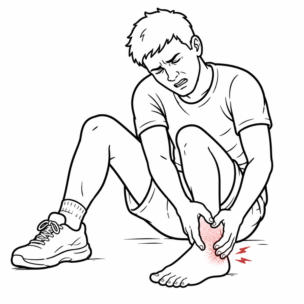
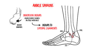
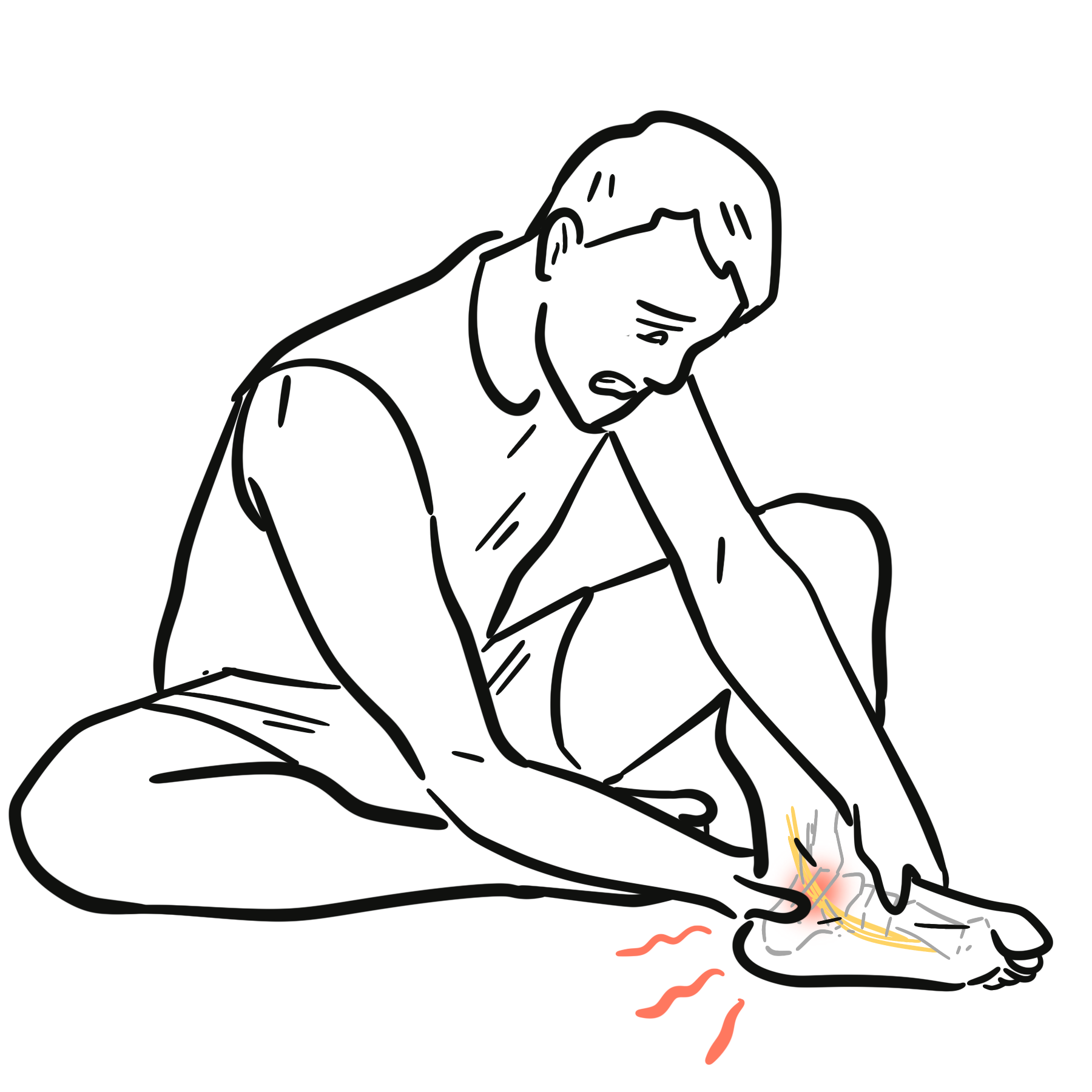
A lateral ankle sprain is an injury to one or more ligaments on the lateral side of the ankle, usually caused by excessive inversion with the foot plantarflexed. The anterior talofibular ligament is injured most commonly, followed by the calcaneofibular ligament and, in severe injuries, the posterior talofibular ligament.1
Most lateral ankle sprains can be managed non-operatively with early protected loading and structured rehabilitation. Inadequate rehabilitation increases the risk of recurrent sprain, persistent symptoms and chronic ankle instability.1
Fracture, syndesmotic injury, osteochondral injury, tendon injury and neurovascular compromise must be considered before diagnosing an uncomplicated lateral ligament sprain.
Definition
- Lateral ankle sprain
- Traumatic injury to the lateral ligament complex of the ankle, usually caused by an inversion and plantarflexion mechanism.
- Anterior talofibular ligament
- The ligament extending from the lateral malleolus to the talus; it is the lateral ligament most frequently injured during an ankle sprain.
- Calcaneofibular ligament
- The ligament extending from the lateral malleolus to the calcaneus; injury suggests a more substantial lateral ligament sprain.
- Chronic ankle instability
- Persistent perceived or mechanical ankle instability, recurrent sprains or episodes of giving way after an initial ankle sprain.
- Syndesmotic ankle sprain
- Injury to the distal tibiofibular ligament complex, commonly called a high ankle sprain.
Anatomy & Physiology
The ankle mortise is formed by the distal tibia and fibula surrounding the talar dome. It permits predominantly dorsiflexion and plantarflexion while transmitting load between the leg and foot.
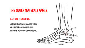
The lateral ligament complex comprises:
- Anterior talofibular ligament: resists anterior translation of the talus and inversion, particularly in plantarflexion.
- Calcaneofibular ligament: resists inversion, particularly when the ankle is neutral or dorsiflexed.
- Posterior talofibular ligament: the strongest lateral ligament; it is usually injured only in severe trauma or dislocation.
Dynamic stability is provided by the peroneal muscles, neuromuscular control, proprioception and coordinated lower-limb movement. Injury can therefore produce functional instability even after the ligament has structurally healed.
Aetiology & Risk Factors
Aetiology
The usual mechanism is rapid inversion of a plantarflexed foot, producing sequential loading of the anterior talofibular and calcaneofibular ligaments.
Common situations include:
- Landing on another player’s foot.
- Sudden cutting or change of direction.
- Running on an uneven surface.
- Missing a step or kerb.
- A direct force that drives the ankle into inversion.
A forceful external-rotation or dorsiflexion mechanism should raise suspicion for syndesmotic injury rather than an isolated lateral ankle sprain.
Risk Factors
- Previous ankle sprain.
- Incomplete rehabilitation.
- Impaired balance or proprioception.
- Reduced ankle dorsiflexion.
- Reduced ankle or lower-limb strength.
- Participation in court, field or jumping sports.
- Failure to use indicated bracing or taping after a previous sprain.
- Poor sport-specific conditioning.
- Fatigue and reduced neuromuscular control.
Pathophysiology
Excessive inversion and plantarflexion initially load the anterior talofibular ligament. Greater force can extend the injury to the calcaneofibular ligament and, less commonly, the posterior talofibular ligament.
Ligament disruption causes bleeding, oedema and an inflammatory response. Pain and swelling inhibit muscle activation and alter gait. Damage to ligament mechanoreceptors can impair proprioception and sensorimotor control.
During healing, collagen is deposited and remodelled along lines of mechanical stress. Appropriate progressive loading encourages functional recovery, whereas excessive early loading can aggravate injury and prolonged immobilisation can contribute to stiffness, weakness and impaired balance.
Persistent deficits in strength, dorsiflexion, proprioception or movement control can produce recurrent giving way and chronic ankle instability.1
A ligament may have healed sufficiently to permit routine walking while sensorimotor control and sport-specific function remain impaired. This helps explain why returning to sport based only on pain reduction or elapsed time can result in recurrent sprain.
Classification
Clinical grading provides a broad description of severity but may be difficult to determine accurately immediately after injury.
| Grade | Typical structural injury | Common clinical findings |
|---|---|---|
| Grade I | Ligament stretch or minor fibre disruption | Mild tenderness and swelling; little functional loss; no clear mechanical instability |
| Grade II | Partial ligament tear | Moderate pain, swelling and bruising; painful or restricted weight-bearing; possible mild-to-moderate laxity |
| Grade III | Complete ligament rupture | Marked swelling, bruising and functional loss; substantial laxity may be present; associated injury must be excluded |
Pain and guarding can limit the accuracy of stress testing during the acute phase. Reassessment after several days may better define ligament tenderness, laxity and functional impairment.
Clinical Manifestations
Typical features include:
- A history of inversion injury.
- Lateral ankle pain.
- Swelling around the lateral malleolus.
- Bruising that may track into the foot.
- Difficulty weight-bearing.
- Reduced ankle range of motion.
- Tenderness over the anterior talofibular or calcaneofibular ligament.
- A sensation of instability or giving way.
Clinical Examination
Palpate:
- Anterior talofibular ligament.
- Calcaneofibular ligament.
- Posterior talofibular ligament.
- Distal tibiofibular syndesmosis.
- Medial deltoid ligament.
- Peroneal tendons.
- Achilles tendon.
- Relevant bony landmarks.
Special tests include:
- Anterior drawer test: assesses anterior talofibular ligament laxity.
- Talar tilt test: assesses calcaneofibular ligament and lateral complex laxity.
- Squeeze and external-rotation tests: may support suspected syndesmotic injury.
Stress tests should be interpreted alongside the mechanism, tenderness pattern, functional impairment and the uninjured ankle.
Findings Suggesting Another or Additional Injury
- Focal bony tenderness.
- Gross deformity.
- Inability to bear weight.
- Pain proximal to the ankle joint.
- Medial ankle tenderness.
- Pain over the distal tibiofibular syndesmosis.
- Tenderness at the base of the fifth metatarsal or navicular.
- Achilles tendon discontinuity or a positive calf-squeeze test.
- Peroneal tendon subluxation or focal tendon tenderness.
- Locking, catching or persistent joint-line pain.
- Disproportionate pain, paraesthesia, pallor or reduced pulses.
Diagnosis
A lateral ankle sprain is diagnosed clinically after excluding fracture and other important injuries. Assessment should include the mechanism, ability to bear weight, location of maximal tenderness, swelling, bruising, range of motion, neurovascular status and examination of the entire ankle and foot.
Ottawa Ankle and Foot Rules
An ankle radiograph is indicated when there is pain in the malleolar zone and any of the following:
- Bone tenderness along the posterior edge or tip of the lateral malleolus.
- Bone tenderness along the posterior edge or tip of the medial malleolus.
- Inability to bear weight both immediately after the injury and during assessment for four steps.
A foot radiograph is indicated when there is pain in the midfoot zone and any of the following:
- Bone tenderness at the base of the fifth metatarsal.
- Bone tenderness over the navicular.
- Inability to bear weight both immediately after the injury and during assessment for four steps.
When the Ottawa rules are negative and can be reliably applied, routine radiography is generally unnecessary. When the rules are positive, a standard ankle or foot radiographic series is appropriate.2
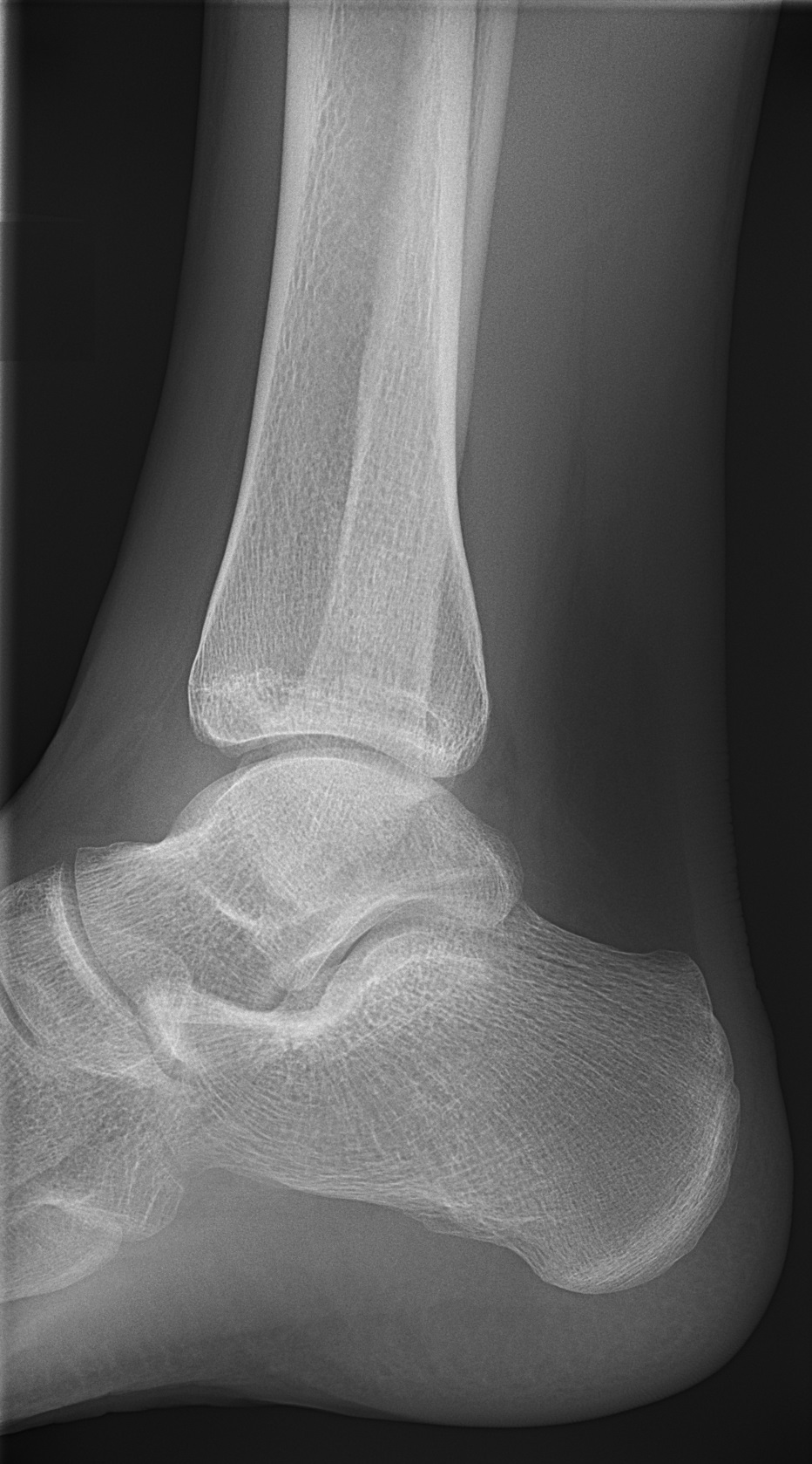
Lateral ankle radiograph showing no fracture after persistent pain following an ankle sprain. Image: Mikael Häggström, via Wikimedia Commons. CC0 1.0.
What to notice
Detailed image description
Source: https://commons.wikimedia.org/wiki/File:X-ray_of_normal_ankle_-_lateral.jpg. Creator: Mikael Häggström. Licence: CC0 1.0 Universal Public Domain Dedication. No modifications.
The Ottawa rules use focal bony tenderness and weight-bearing ability to guide radiography. Tenderness confined to the soft tissues anterior or inferior to the lateral malleolus supports ligament injury but does not replace a complete examination.
Additional Imaging
- Ultrasound: may demonstrate ligament or tendon injury but is operator-dependent and is not routinely required.
- MRI: consider for persistent pain, recurrent instability, suspected syndesmotic injury, osteochondral lesion, occult fracture or significant tendon injury.
- CT: useful when an occult or complex fracture is suspected or for operative planning.
Urgent specialist assessment is required for fracture-dislocation, neurovascular compromise, open injury, tendon rupture, severe syndesmotic injury or suspected compartment syndrome.
Treatment
Acute Management
Initial treatment aims to protect the injured tissues, control symptoms and restore safe movement:
- Protect the ankle from further injury.
- Use compression and elevation to help manage swelling.
- Apply ice for short-term analgesia if helpful, while avoiding direct skin injury.
- Provide simple analgesia or a short course of a non-steroidal anti-inflammatory drug when appropriate.
- Use an external support, such as a brace or taping.
- Begin progressive weight-bearing according to symptoms.
- Use crutches temporarily when a normal or near-normal gait is not possible.
A short period of immobilisation may be appropriate for a severe sprain, but prolonged immobilisation should be avoided. For more severe injuries, immobilisation is generally limited to approximately 10 days before progressive rehabilitation.1
Rehabilitation
A structured rehabilitation programme should include:
- Pain-limited active ankle movement.
- Restoration of dorsiflexion and normal gait.
- Progressive calf and peroneal strengthening.
- Balance and proprioceptive training.
- Dynamic postural-control exercises.
- Hopping, landing and change-of-direction drills.
- Sport-specific conditioning.
- Graduated return to full training and competition.
Manual therapy may provide short-term improvements in pain and dorsiflexion when combined with exercise. It should not replace active rehabilitation.1
Early protected weight-bearing and exercise-based rehabilitation are preferred to prolonged rest. Pain relief alone does not indicate restoration of strength, balance or readiness for sport.
Prevention of Recurrence
- Complete neuromuscular and balance rehabilitation.
- Continue home balance exercises after symptoms improve.
- Use bracing or taping during high-risk sport when indicated.
- Restore ankle dorsiflexion and lower-limb strength.
- Address landing, cutting and sport-specific movement control.
- Avoid returning solely because swelling has decreased.
Surgery is not routinely required after an acute lateral ankle sprain. It may be considered for persistent mechanical instability and recurrent sprains despite comprehensive rehabilitation.
Return to Activity & Sport
Return should be based on recovery across multiple domains rather than a fixed number of days. The PAASS framework recommends assessing:3
- Pain: during sport and over the previous 24 hours.
- Ankle impairments: range of motion, strength, endurance and power.
- Athlete perception: confidence, perceived stability and psychological readiness.
- Sensorimotor control: proprioception and dynamic balance.
- Sport and functional performance: hopping, jumping, agility, sport-specific drills and ability to complete a full training session.
The athlete should demonstrate a progressive return from walking and running to multidirectional and sport-specific tasks without meaningful pain, instability or loss of movement quality.
Complications & Prognosis
Complications
- Recurrent lateral ankle sprain.
- Chronic ankle instability.
- Persistent pain, swelling or stiffness.
- Reduced dorsiflexion.
- Impaired proprioception and balance.
- Peroneal tendon injury or subluxation.
- Syndesmotic injury.
- Osteochondral lesion of the talus.
- Occult fracture.
- Anterior or anterolateral ankle impingement.
- Post-traumatic osteoarthritis.
- Reduced confidence and avoidance of sport.
Prognosis
Most uncomplicated lateral ankle sprains improve with non-operative treatment, but recovery time varies with injury severity, associated injury, rehabilitation quality and functional demands.1
Poor prognostic features include:
- Previous recurrent sprains.
- Greater initial pain and inability to bear weight.
- Marked swelling or restricted range of motion.
- Syndesmotic or osteochondral injury.
- Persistent balance, strength or movement-control deficits.
- Premature return to sport.
- Failure to complete rehabilitation.
Persistent pain, swelling, locking, recurrent giving way or inability to progress rehabilitation should prompt reassessment for an alternative or associated diagnosis.
References
- Martin RL, Davenport TE, Fraser JJ, Sawdon-Bea J, Carcia CR, Carroll LA, et al. Ankle stability and movement coordination impairments: lateral ankle ligament sprains revision 2021. J Orthop Sports Phys Ther. 2021;51(4):CPG1–CPG80. doi:10.2519/jospt.2021.0302.
- Expert Panel on Musculoskeletal Imaging. ACR Appropriateness Criteria® acute trauma to the ankle. J Am Coll Radiol. 2020;17(11S):S355–S366. doi:10.1016/j.jacr.2020.09.014.
- Smith MD, Vicenzino B, Bahr R, Bandholm T, Cooke R, Mendonça LM, et al. Return to sport decisions after an acute lateral ankle sprain injury: introducing the PAASS framework—an international multidisciplinary consensus. Br J Sports Med. 2021;55(22):1270–1276. doi:10.1136/bjsports-2021-104087.






Members only discussions coming soon…