

Osteosarcoma
Overview
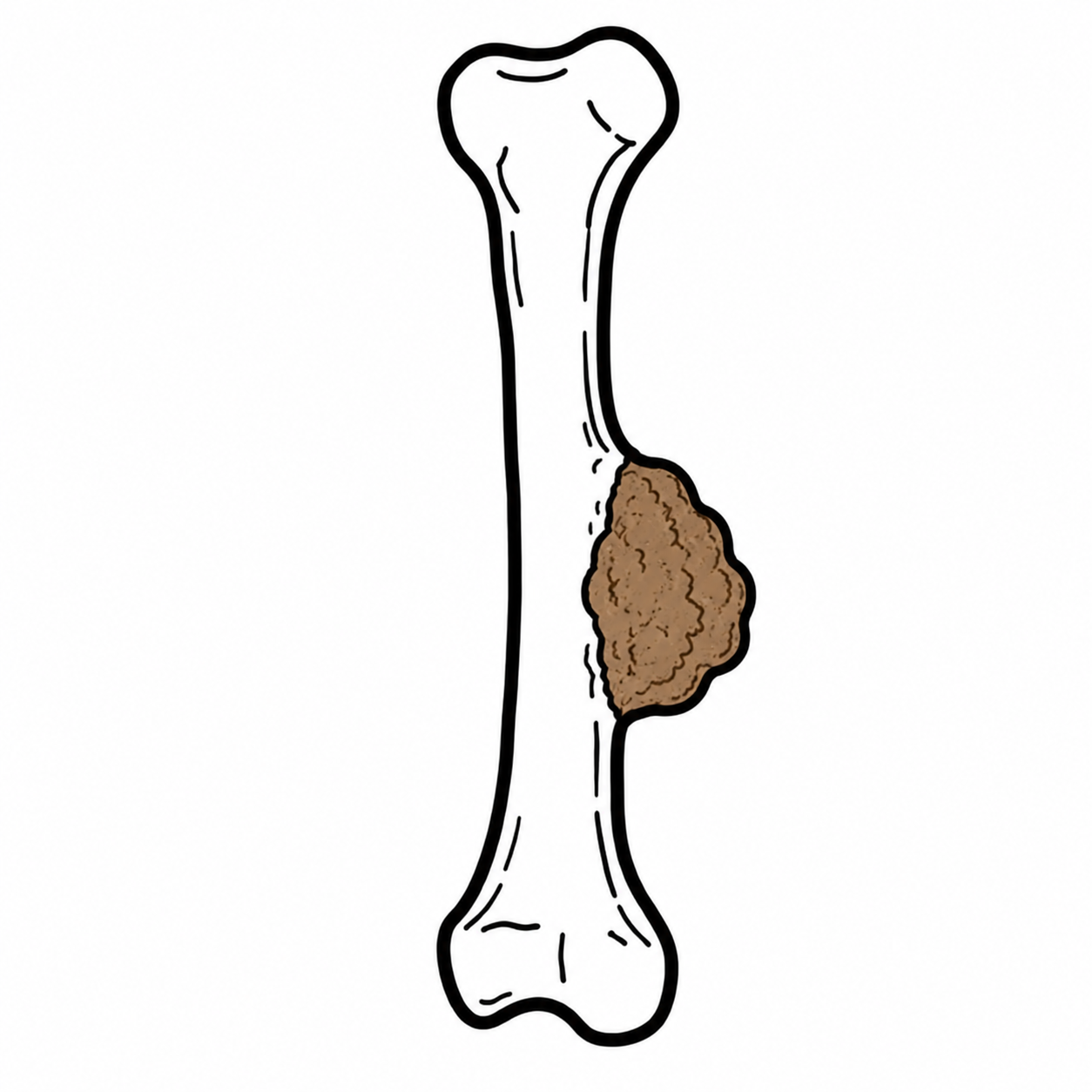
Osteosarcoma is an aggressive primary malignant bone tumour characterised by malignant cells directly producing osteoid or immature bone. It occurs mainly in adolescents and young adults, particularly during periods of rapid bone growth.
Incidence in people aged 0–19 years is approximately 5.4 cases per million per year. Common sites:
- Distal femur
- Proximal tibia
- Proximal humerus
More than half of childhood and adolescent tumours arise in the long bones around the knee.
Definition
Osteosarcoma: malignant bone tumour in which tumour cells directly produce osteoid.
Osteoid: unmineralised bone matrix produced by osteoblasts; its malignant production confirms the diagnosis.
Skip lesion: separate tumour focus within the same bone, discontinuous from the primary tumour.
Aetiology & Risk Factors
- Most cases are sporadic, with no identifiable cause.
- Risk factors include:
- Previous radiotherapy
- Paget disease of bone, particularly in older adults
- Hereditary retinoblastoma—RB1
- Li–Fraumeni syndrome—TP53
- Rothmund–Thomson syndrome—RECQL4
- Werner syndrome
Clinical Manifestations
- Progressive localised bone pain, often present for weeks.
- Pain may:
- Be worse at night
- Increase with activity
- Initially be attributed to a sporting injury
- Local swelling or palpable mass.
- Reduced movement of an adjacent joint.
- Limp or reduced limb function.
- Pathological fracture with minor or no trauma.
- Constitutional symptoms such as fever and weight loss are less common than in Ewing sarcoma.
Persistent focal bone pain—particularly pain lasting weeks, worsening at night or associated with swelling—should not automatically be attributed to sport or trauma.
Diagnosis
Initial investigations
- Plain X-ray of the affected bone is the initial imaging investigation.
- MRI of the entire affected bone and surrounding soft tissues:
- Defines local tumour extent.
- Assesses neurovascular structures and adjacent joint involvement.
- Detects skip lesions within the same bone.
Definitive diagnosis
- Biopsy
- Diagnosis requires a core-needle or open surgical biopsy.
- Histology demonstrates malignant tumour cells producing osteoid
- Staging once diagnosis confirmed
- CT chest: evaluates pulmonary metastases.
- PET-CT/PET-MRI or bone scan: evaluates bone and other metastatic disease.
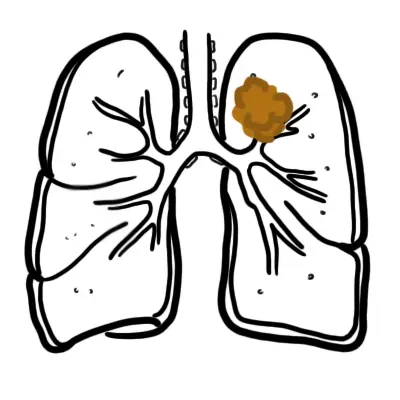
- Approximately 20% of patients have detectable metastases at presentation; the lungs are the most common site.
Do not biopsy a suspected bone sarcoma before specialist referral; the biopsy track must be incorporated into definitive resection.
Always stage the lungs with CT, as they are the most common site of metastasis.
Other investigations
- ALP and LDH are not diagnostic but elevated levels may be associated with higher tumour burden and poorer prognosis.
- Baseline cardiac assessment and hearing tests are commonly required before potentially cardiotoxic or ototoxic chemotherapy.
Treatment
Treatment should occur in a specialist sarcoma centre using a multidisciplinary team.
Localised high-grade osteosarcoma
- Neoadjuvant chemotherapy before surgery.
- Common regimen in children and younger adults:
- MAP: high-dose methotrexate, doxorubicin and cisplatin.
- Wide surgical resection with clear margins:
- Limb-sparing surgery is possible in more than 80% of extremity tumours.
- Amputation may be required when clear margins cannot otherwise be achieved or major neurovascular structures are involved.
- Postoperative chemotherapy is then continued.
- Surgery alone is inadequate because microscopic metastatic disease is commonly present even when imaging is negative.
Metastatic disease
- Systemic multi-agent chemotherapy.
- Surgical removal of the primary tumour.
- Complete resection of all detectable metastases where possible, particularly lung metastases.
- Patients with resectable lung-only metastases may still achieve long-term survival.
Radiotherapy
- Osteosarcoma is generally managed primarily with surgery and chemotherapy.
- Radiotherapy may be considered when:
- The tumour is unresectable
- Surgical margins are positive or inadequate
- Local recurrence cannot be resected
- Palliation of painful metastatic disease is required.
Complications & Prognosis
- Pulmonary metastases
- Bone metastases
- Local tumour recurrence
- Pathological fracture
- Chronic pain
- Loss of limb function
- Neurovascular compression
- Reduced mobility and disability
- Treatment related
- Surgery:
- Infection
- Delayed wound healing
- Prosthetic loosening or failure
- Limb-length discrepancy in growing children
- Fracture or need for revision surgery
- Amputation and long-term rehabilitation
- Chemotherapy related
Prognosis
- Patients with localised osteosarcoma treated with chemotherapy and complete surgical resection have approximately 60–70% five-year overall survival.
- Prognosis is substantially poorer with metastatic disease; Australian childhood data reported approximately 36% five-year survival for advanced osteosarcoma.
- Survival is poor when metastatic or recurrent disease cannot be completely surgically removed.
References
- National Cancer Institute. Osteosarcoma and undifferentiated pleomorphic sarcoma of bone treatment (PDQ): health professional version [Internet]. Bethesda: National Cancer Institute; updated 2024 Dec 2.
- Strauss SJ, Frezza AM, Abecassis N, Bajpai J, Bauer S, Biagini R, et al. Bone sarcomas: ESMO–EURACAN–GENTURIS–ERN PaedCan Clinical Practice Guideline for diagnosis, treatment and follow-up. Ann Oncol. 2021;32(12):1520–1536. doi:10.1016/j.annonc.2021.08.1995.
- Meltzer PS, Helman LJ. New horizons in the treatment of osteosarcoma. N Engl J Med. 2021;385(22):2066–2076. doi:10.1056/NEJMra2103423.
- Gill J, Gorlick R. Osteosarcoma: a review of diagnosis, management, and treatment strategies. Clin Adv Hematol Oncol. 2021;19(8):498–507.
- Cancer Australia. Bone tumours [Internet]. Canberra: Australian Government; updated 2025 Jul 22.
















Members only discussions coming soon…