Abdominal trauma can result in injuries that are intraperitoneal and retroperitoneal
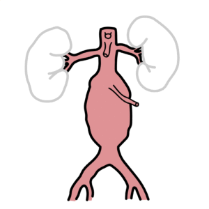
Retroperitoneal organs
Kidneys
GU tract
Duodenum
Pancreas.
Intraperitoneal injuries occur to the solid and hollow organs and the diaphragm.
Abdominal trauma is divided into blunt and penetrating mechanisms. Blunt abdominal trauma involves a crushing force that causes disruption of solid viscera (spleen or liver) or hollow viscera (intestine). These injuries are most common after falls or MVCs. Penetrating abdominal trauma occurs most commonly due to stab wounds and gunshot wounds that enter the intraperitoneal cavity.
Blunt Thoracic Injuries
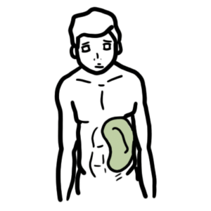
Splenic laceration
Liver laceration
Pancreatic injury
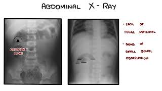
Bowel perforation
Duodenal haematoma
Diaphragmatic injury
Retropertioneal haemotoma
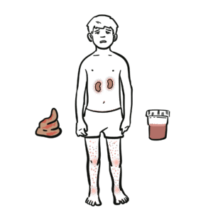
GU injury
Penetrating Injuries
Any organ
PAMAD
Preparation
Assessment – Primary Survey
Management
Assessment – Secondary Survey
Dislocation
Preparation
Staff
Equipment
Assessment – Primary Survey – ABCDE
Airway – Patent and Protected
Foreign bodies
Facial, mandibular, or tracheal/laryngeal fractures
Flail Chest – Paradoxical movement of chest wall segment
Consider diaphragm issues (injury at C3, C4, C5)
Watch for respiratory insufficiency
In the absence of major airway obstruction and flail chest, the presence of paradoxical breathing is considered highly suggestive of cervical spine injury
Management
High flow oxygen
Side note Ventilation may be reduced for a number of reasons, (1) diaphragm fatigue, (2) Progressively ascending spinal cord damage from primary damage or secondary ascending spinal cord oedema encroaching on C3-C5, (3) These same segments may be involved from primary injury and diaphragm may be partially paralysed and (4) Consequence of co-existing chest trauma.
Spinal immobilisation until spinal cord or unstable vertebral injury has been excluded on physical examination and investigations
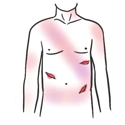
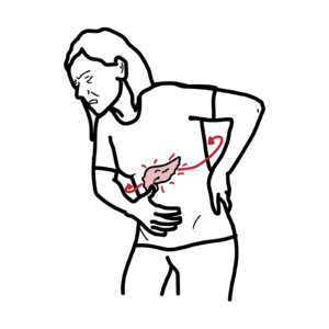
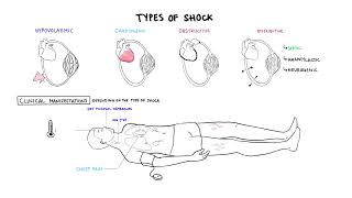
Kehr’s sign Pain in the shoulder that is not associated with tenderness or pain with shoulder ROM suggests that blood is present under the diaphragm, causing referred pain to the shoulder. This commonly occurs from a splenic or liver laceration.
Exposure/Environment
Increased risk of hyperthermia due to peripheral vasodilation


Discussion