Aortic Stenosis

Overview

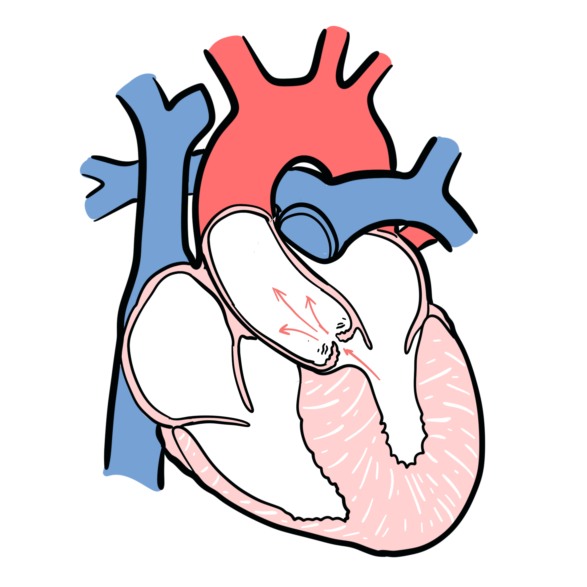
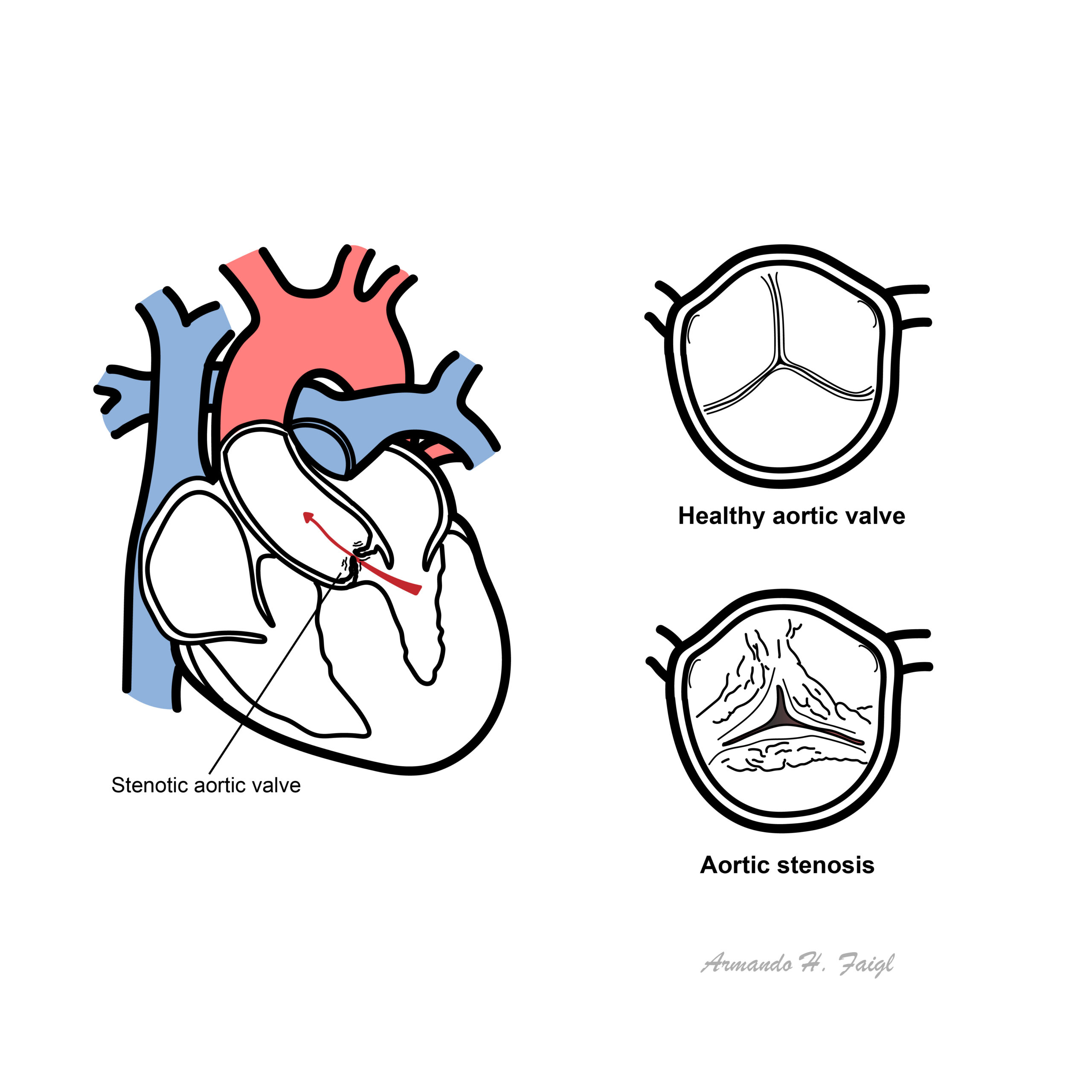
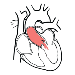
  • Aortic valve stenosis is characterised by obstruction of left ventricular outflow, resulting in inadequate cardiac output, decreased exercise capacity, heart failure, and death from cardiovascular causes.
  • Mild to moderate aortic stenosis is usually a symptomatic, but occasionally can be found on routine examination
  • Locations of stenosis - Valvular (common), supravalvular and subvalvular
  • Aortic Stenosis produces a "Mid Systolic Murmur", which is a murmur heard between the first heart sound (S1 - AV valve closure) and ends before the second heart sound (S2 - Aortic and Pulmonary valve closure)
  • Most common valvular heart disease in western countries and prevalence increase with increasing age
Overview

Aortic stenosis is narrowing of the aortic valve resulting in decreased blood flow from the heart (decrease cardiac output)

Cardiac Valves

The heart has 4 valves:

  • Aortic valve
  • Pulmonary valve
  • Tricuspid valve
  • Mitral valve (bicuspid valve)

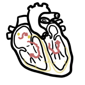
The Mitral and tricuspid valves are atrioventricular valves meaning they allow blood to move from the atrium to the ventricles of the heart. This occurs with ventricular diastole.

The aortic and pulmonary valve are tricuspid valves which when open allow blood to move to the aorta and pulmonary system respectively. This occurs during ventricular systole when the heart contracts.

Heart Valves

Surface anatomy of the heart valves

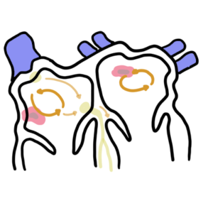
All of the heart valves except the mitral valve are usually tricuspid. However, there can be congenital bicuspid valves which can predispose one to valvular disease later on. The heart valves can be heard most prominent in the following regions of the chest

  • Aortic valve - Right 2nd intercostal space parasternal
  • Pulmonary valve - Left 2nd intercostal space parasternal
  • Tricuspid valve - Left 4th intercostal space parasternal
  • Mitral valve - Left 5 intercostal space mid-clavicular (below the nipple)

Risk Factors

  • Age >60
  • Congenitally unicuspid, bicuspid valve
  • Rheumatic heart disease
  • Chronic Kidney Disease

Signs and Symptoms

Sands

Aortic Stenosis classic triad of signs and symptoms are dizziness/syncope, dyspnoea, angina on exertion.


Cardiovascular Examination

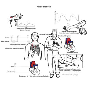
  • Slow rate of rise in carotid pulse (parvus and tardus)
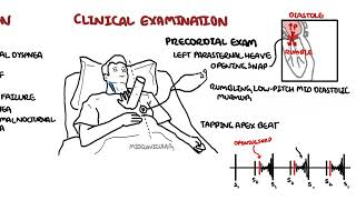
  • Auscultation - Aortic Valve (right second intercostal space parasternal)
    • Ejection systolic murmur - Mid to late peak intensity of the murmur generally begins after S1 and ends before S2 (mid systolic murmur)
    • Reduced intensity of S2, closing of the aortic valve
    • Atrial contraction, S4,  maybe heard
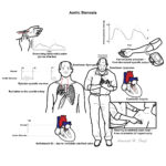
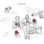
  • Auscultation - Apex (Left fifth intercostal space mid-clavicular)
    • Murmur may radiate to apex of heart
    • High frequency and louder murmur (Holosystolic murmur) can be confused with mitral regurgitation (Gallavardin phenomenon)

ExaminationCardiac Examination findings include parvus and tardus of the carotid artery and mid-systolic ejection murmur heard over the left second intercostal space parasternal. Murmur can radiate to the carotids.

Signs of severity

  1. Parvus et tardus
  2. Aortic thrill
  3. Length, harshness and lateness of the peak of the systolic murmur
  4. Atrial contraction, S4
  5. Parodical splitting of the second heart sound (delayed left ventricular ejection and aortic valve closure)
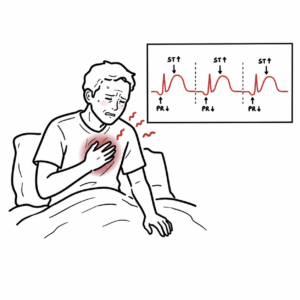
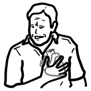
  6. Left ventricular failure
Think 
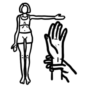
Squatting increases venous return and accentuate the murmur

Side note - Clinical Auscultation
Murmur Heart Sound
Mitral Stenosis High pitched early-diastolic murmur Loud S1
Mitral Regurgitation Pansystolic murmur radiates to the axilla Soft S1 loud S2
Aortic stenosis Ejection systolic "crescendo decresendo" murmur radiating to the carotids Soft S2
Aortic regurgitation early diastolic murmur Soft S2

Differential Diagnosis

Differential Diagnosis include other causes of mid-systolic murmurs

  • Mitral valve dysfunction
  • Hypertrophic cardiomyopathy
  • Aortic sclerosis
  • Atrial septal defect
  • Pulmonary stenosis

Investigations

  • Transthoracic echocardiogram - elevated aortic pressure
  • ECG - left ventricular hypertrophy and absent Q wave
  • X-ray - valve calcification
  • Cardiac MRI
  • Cardiac catheterisation - allows for direct measurement of the pressure gradient
investigation

Cardiac catheterization involves feeding a catheter through the femoral or radial artery towards the ascending aorta. This allows for visualization of the aortic valve. Tranthoracic echocardiogram allows non-invasive visualisation of the heart valve and is gold standard for diagnosis.

ECG

ECG changes in Aortic stenosis can include signs of left ventricular hypertyophy, absent Q wave and left ventricular strain (t-wave inversion)

Side Note:
ECG LVH

Diagnosis

  • Echocardiography is the primary test in diagnosis and evaluation of aortic stenosis
  • 50% stenosis of aortic valve

Aetiology

  • A congenitally abnormal valve (bicuspid or rarely unicuspid) with superimposed calcification
  • Calcific disease of a trileaflet valve
    • Age related
    • Typically in patients >65years
  • Rheumatic valve disease
  • Congenital aortic stenosis (rare)
    • congenital stenosis - abnormal valve is present from birth
    • acquired stenosis - abonrmal calve beomes stenotic overtime
Three major causes of Aortic Stenosis

Three major causes of Aortic Stenosis

Pathophysiology

The natural history of AS begins with a prolonged asymptomatic period.

Pathology

  • Calcified, stiff aortic valve leaflets
  • Calcification and dilation of  the ascending aorta
  • Left ventricular hypertrophy
  • Possible left atrial enlargement
  • Mitral annular calcification -> mitral regurgitation

Managment

Indications for surgery
Symptomatic
Critical obstruction based on catheterisatin
  • Long term anticoagulant (following valve replacement)
  • Long term antibiotic prophylaxis
  • Balloon valvuloplasty (for inoperable patients)
    • 80% re-stenosis in 1 year
    • ~14% develop mild/moderate reguritation
  • Transcatheter aortic valve replacement (for inoperable patients)
  • Surgical aortic valve replacement
    • mechanical
    • bioprosthetic
Management of Aortic Stenosis depends on operable and inoperable patients. For symptomatic oeprable patients, valve replacement is advised. For young patients, mechanical valve is suggested because of durability.

Management of Aortic Stenosis depends on operable and inoperable patients. For symptomatic operable patients, valve replacement/repair is advised. For young patients, mechanical valve is suggested because of durability.


Emergency (unstable)

  • Surgical aortic valve replacement
  • Transcatheter aortic valve replacement

Complication and Prognosis

Complication

  • Sudden Cardiac Death
  • Arrythmias
  • Endocarditis
  • Bleeding tendency
  • Systemic Embolism

Prognosis Mortality in patients with AS dramatically increases after the development of cardiac symptoms. The rate of death is 50% at 2 years for patients with symptomatic disease unless aortic valve replacement is performed promptly.

References

Jarcho, JA 2014, "Aortic-Valve Stenosis - From Patients at Risk to Severe Valve Obstruction", The New England Journal of Medicine, vol. 371, no. 8, pp. 744-56.
UpToDate
Best Practice
Aortic stenosis

Summary of Aortic Stenosis Aortic Stenosis is narrowing of the aortic valve resulting in increased work of heart. Mid-systolic ejection murmur is heard on auscultation. Exertional dizziness/syncope, dyspnea and angina are classic features. Electrocardiography is gold standard for diagnosis and treatment involves surgical valve repair, placement and medication.

Video: Aortic Stenosis


Contents
cross