

Arrythmias
Overview
An arrythmia refers to any disturbance in cardiac electrical activity that is not normal sinus rhythm with normal atrioventricular conduction. This may include changes in rate, rhythm or site of origin of the electrical signal for contraction.
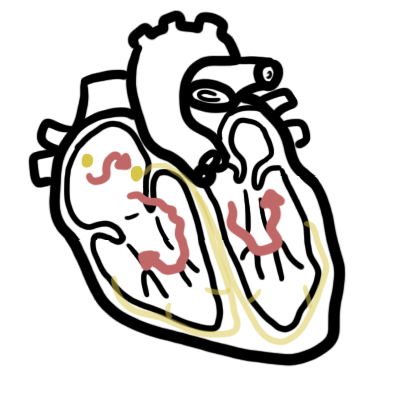
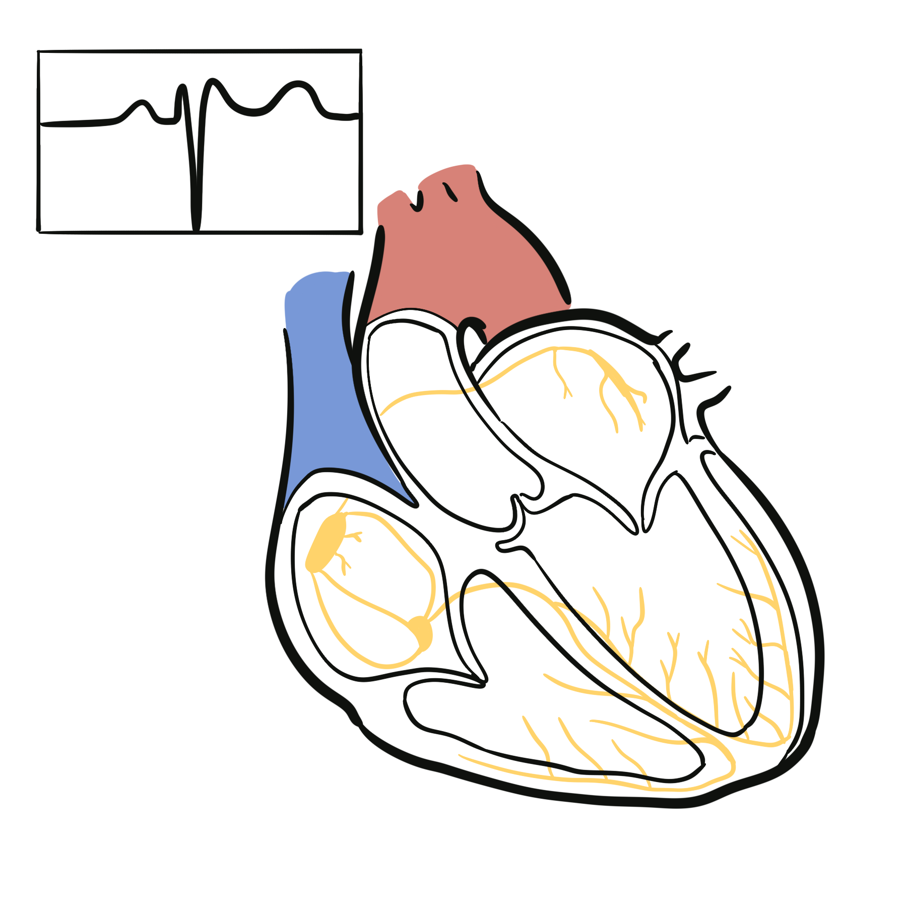
Normal conduction of the heart
For the heart to pump normally:
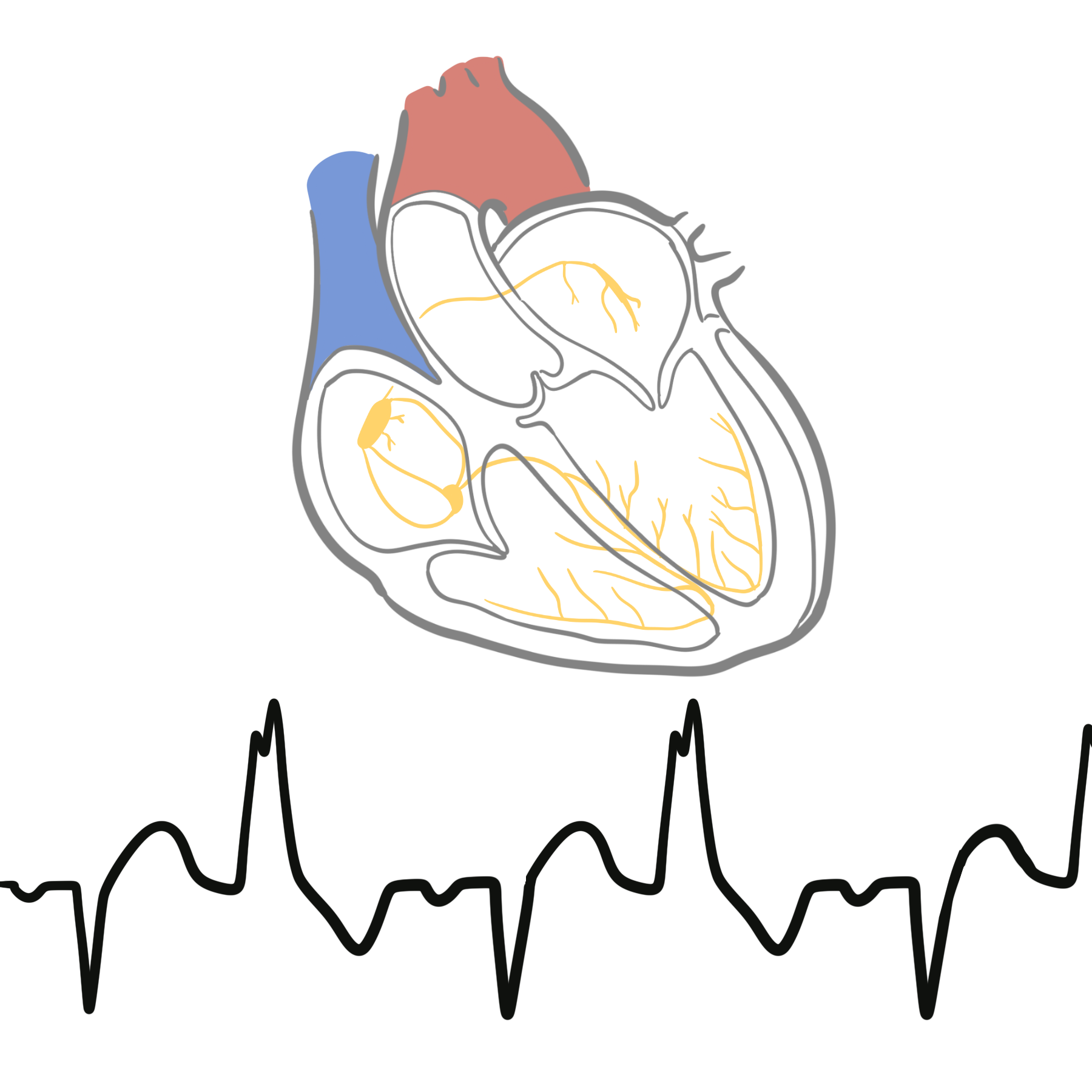
- SA node initiates an action potential spreading the current in to the right atrium (RA) and left atrium (LA) via Bachmann’s bundle
- SA node → AV node
- AV node delays conduction to ensure ventricles contract AFTER blood is emptied into the ventricles from the atria
- AV node → Bundle of His
- Bundle of His → Purkinje Fibres
- Purkinje fibres → cardiomyocytes (contractile cells)
Classification of Arrythmias
Arrhythmias are classified clinically and electrophysiologically across three primary axes:
- Heart rate and ECG morphology (the clinical/bedside framework)
- Anatomical site of origin
- Cellular electrophysiological mechanism.
Clinical & ECG Morphologic Classification
The primary bedside approach categorizes rhythms first by rate (<60 bpm vs. >100 bpm), then by QRS duration, and finally by rhythm regularity.
Arrhythmia
│
├── Bradyarrhythmias (< 60 bpm)
│   ├── Sinus Node Dysfunction (SND)
│   └── Atrioventricular (AV) Blocks
│
└── Tachyarrhythmias (> 100 bpm)
    ├── Narrow QRS Complex (≤ 120 ms) ── Supraventricular
    │   ├── Regular
    │   └── Irregular
    └── Wide QRS Complex (> 120 ms) ──── Ventricular or Aberrant SVT
        ├── Regular
        └── Irregular
Tachyarrhythmias (> 100 bpm)
Narrow-Complex Tachycardias (QRS ≤ 120 ms)
Conducted via the native His-Purkinje system; always supraventricular in origin (SVT).
| Rhythm | Regularity | Characteristic P-Wave / Atrial Activity | Common Mechanism |
| Sinus Tachycardia | Regular | Normal upright P waves in II, III, aVF | Physiological / enhanced automaticity |
| AVNRT | Regular | Pseudo-r’ in V1, pseudo-S in inferior leads (or buried) | Dual AV nodal pathway micro-reentry |
| AVRT (Orthodromic) | Regular | Inverted P waves in inferior leads, RP interval > 70 ms | Macro-reentry via accessory pathway |
| Atrial Tachycardia | Regular | Ectopic P wave morphology distinct from sinus P | Enhanced automaticity or triggered activity |
| Atrial Flutter | Regular (variable block) | Typical “sawtooth” flutter waves (~300 bpm, leads II, III, aVF) | Macro-reentry around tricuspid annulus (cavotricuspid isthmus-dependent) |
| Atrial Fibrillation | Irregularly irregular | Disorganized baseline fibrillatory waves (400–600 bpm), no distinct P waves | Multiple chaotic wavelets / pulmonary vein micro-reentry |
| Multifocal Atrial Tachycardia (MAT) | Irregular | Distinct P-wave morphologies with isoelectric baseline | Multiple ectopic firing foci (often seen in COPD) |
Wide-Complex Tachycardias (QRS > 120 ms)
Assumed to be Ventricular Tachycardia (VT) until proven otherwise (~80% of cases). The remaining ~20% represent SVT with aberrant conduction (bundle branch block) or antidromic AVRT.
| Morphology | Rhythm | Key Distinguishing Features |
| Monomorphic Regular | Monomorphic VT | AV dissociation, capture/fusion beats, extreme right-axis deviation (“northwest axis”), positive/negative precordial concordance, Joseph/Brugada criteria |
| SVT with Aberrancy | Typical RBBB or LBBB pattern with pre-existing or rate-dependent bundle branch block | |
| Antidromic AVRT | Anterograde conduction down accessory pathway; wide QRS with delta waves | |
| Polymorphic / Irregular | Polymorphic VT | Continuously changing QRS axis and morphology (normal baseline QTc) |
| Torsades de Pointes | Twisting of QRS axes around the isoelectric line; occurs in the setting of prolonged baseline QTc | |
| AF with WPW (Pre-excited AF) | Rapid, irregularly irregular wide complexes with varying QRS morphology and axis; high risk of degeneration to VF | |
| Ventricular Fibrillation (VF) | Chaotic, completely disorganized electrical deflection without discrete QRS, ST, or T waves |
Bradyarrhythmias (< 60 bpm)
Bradyarrhythmias are classified anatomically based on where conduction fails along the conduction hierarchy:
Sinus Node Dysfunction (Sick Sinus Syndrome)
- Sinus Bradycardia: Regular rhythm <60 bpm driven by normal SA node depolarisation.
- Sinus Arrest / Pause: Transient failure of SA node impulse generation (pause not an exact multiple of the baseline P-P interval).
- Sinoatrial Exit Block: Impulse is generated by the SA node but fails to exit into surrounding atrial myocardium.
- Tachy-Brady Syndrome: Alternating paroxysms of SVTs (commonly atrial fibrillation or flutter) and prolonged sinus pauses.
Atrioventricular (AV) Blocks
- First-Degree AV Block: PR interval > 200 ms with 1:1 AV conduction (every P wave is followed by a QRS). Conduction delay is typically nodal.
- Second-Degree AV Block, Mobitz Type I (Wenckebach): Progressive PR interval prolongation culminating in a non-conducted P wave. Generally intranodal and benign.
- Second-Degree AV Block, Mobitz Type II: Constant PR interval preceding a suddenly dropped QRS complex. Localises infraglobally (His-Purkinje system); carries high risk of progression to complete heart block.
- High-Grade / Advanced AV Block:$\ge 2$ consecutive P waves fail to conduct to the ventricles at physiological atrial rates.
- Third-Degree (Complete) AV Block: Complete absence of AV conduction; atria and ventricles beat independently (AV dissociation) with an escape rhythm (junctional narrow-complex at 40–60 bpm or idioventricular wide-complex at 20–40 bpm).
Electrophysiological Mechanism
At the cellular and tissue level, all arrhythmias stem from two fundamental abnormalities:
               Arrhythmogenic Mechanisms
                          │
         ┌────────────────┴────────────────┐
         ▼                                 ▼
Impulse Formation                 Impulse Conduction
  ├── Enhanced Automaticity         ├── Reentry (Micro & Macro)
  ├── Abnormal Automaticity         ├── Conduction Delay / Block
  └── Triggered Activity            └── Accessory Pathways
        ├── Early Afterdepolarisations (EAD)
        └── Delayed Afterdepolarisations (DAD)


Members only discussions coming soon…